Pneumomediastinum in a cheerleading student
Abstract
Spontaneous pneumomediastinum in healthy individuals is an uncommon entity. We are reporting an image of pneumomediastinum and subcutaneous emphysema in a male college student. The onset was considered to be the hard vocal exercise during his cheerleading club activity.
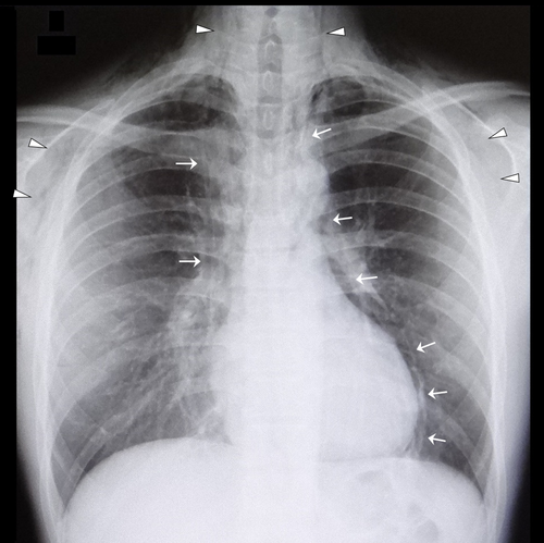
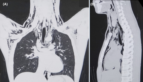
A healthy, 20-year-old man presented with a 1-day history of hoarseness, neck swelling, throat pain, chest pain, and upper back pain that had developed suddenly on the day before the presentation, while training in yelling out loud for 6 h as a member of his college cheerleading club. On arrival, his respiratory rate was 16/min, and he maintained an oxygen saturation of 97% on room air. A crackling sensation was perceived on palpation of the neck. Hamman's sign was not observed. His chest radiograph showed pneumomediastinum and diffuse subcutaneous emphysema (Figure 1). A CT scan showed widespread free air in the mediastinum and subcutaneous areas of the axilla, neck, and jaw (Figure 2). There was no evidence of pneumothorax or esophageal injury. He was kept under observation for 5 days in our hospital. A repeat radiograph obtained 12 days after the first arrival revealed complete resolution of the free air.


There have been a few reports of pneumomediastinum complicating head and neck surgeries, dental procedures, or Valsalva maneuvers.1 Spontaneous pneumomediastinum caused by simple vocal exercises is even more uncommon.2, 3 The emphysemas caused by vocal exercises typically resolve without intervention, even if extensive free air has emerged.1-3
AUTHOR CONTRIBUTIONS
YH conceived the idea for the document and contributed to the management of the patient and writing of the manuscript. TN contributed to the editing of the manuscript.
ACKNOWLEDGMENT
None.
CONFLICT OF INTEREST
All the authors have no pertinent conflict of interest to report for this manuscript.
ETHICAL APPROVAL
This report complies with the provisions of the Declaration of Helsinki.
CONSENT
Written informed consent to publish this report was obtained from the patient in accordance with the journal's patient consent policy.


