Giant multinodular goiter for 24 years; hidden in a village in Western Nepal
Abstract
Here, we present the case of a giant multinodular goiter with retrosternal extension in an old lady with dyspnea for 3 months. The patient was treated with microscopic-assisted total thyroidectomy without any postoperative complications.
1 CASE REPORT
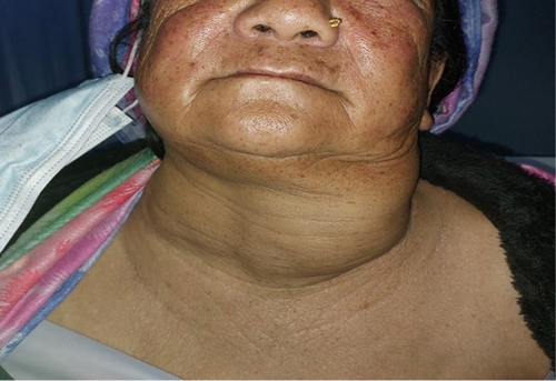
A 67-year-old woman presented at the health camp organized in the Kihun village by Bhawana Foundation, Nepal, with complaints of painless neck swelling for 24 years and shortness of breath for 3 months. Shortness of breath was gradually progressive and aggravated while sleeping in supine position. On neck examination, a large left greater than right mass was present. The mass was non-tender, non-pulsatile, and moved with deglutition (Figure 1).

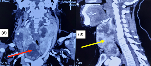
Thyroid function tests and serum calcium were within normal limits. Ultrasound of the neck showed multiple thyroid nodules and cystic lesions. The CECT neck revealed heterogeneously enhancing lesions extending retrosternally (Figure 2A,B). FNAC was suggestive of atypia of undetermined significance.

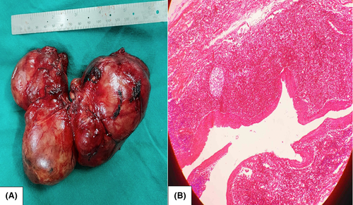
The patient underwent microscopic-assisted total thyroidectomy under general anesthesia. Her postoperative recovery was uneventful and relieved her shortness of breath. The patient was discharged on the sixth postoperative day with levothyroxine replacement therapy. The mass removed from the neck weighed 461.5 g and measured approximately 14 cm (Figure 3). Microscopic examination was consistent with multinodular goiter.

Benign multinodular goiter leading to airway compromise has become a rare clinical entity.1 Universal salt iodization, cosmetic concern, and improved surgical technique with minimal disfigurement have led to the disappearance of large goiter from modern clinical practice. The definitive management of multinodular goiter includes total thyroidectomy.2
AUTHOR CONTRIBUTIONS
BS involved in diagnosis, treatment, and conceptualization of study. BS, BN, AP, and PN involved in manuscript preparation, editing, and proofreading of final version of manuscript.
ACKNOWLEDGMENT
None.
CONFLICT OF INTEREST
We declare no competing interests.
CONSENT
Written informed consent was obtained from the patient to publish this report.
Open Research
DATA AVAILABILITY STATEMENT
Data available on request.


