

Journal list menu

 Issue
IssueActa Crystallographica Section D: Volume 78, Issue 5
542-682May 2022
Export Citations
Download PDFs
research papers
Validation analysis of EMDB entries
- Pages: 542-552
- First Published: 20 April 2022
A new and comprehensive resource is described that contains validation information for all cryo-EM structures that are available in the public archives, EMDB and PDB, based on recommendations from the cryo-EM community.
MrParse: finding homologues in the PDB and the EBI AlphaFold database for molecular replacement and more
- Pages: 553-559
- First Published: 26 April 2022

MrParse is a software package designed to aid decision making in molecular replacement (MR). It performs a sequence search to find search models, not only in the PDB, as would conventionally be performed, but also in the EBI AlphaFold database, and provides a series of analyses relevant to MR such as crystal pathology detection and sequence analysis.
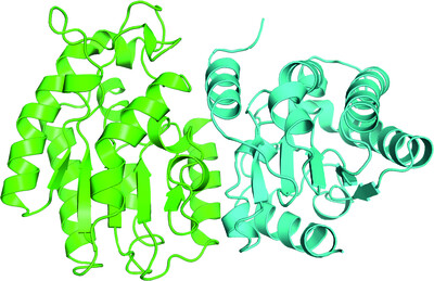
The BAM7 gene in Zea mays encodes a protein with similar structural and catalytic properties to Arabidopsis BAM2
- Pages: 560-570
- First Published: 08 April 2022
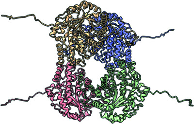
The Zea maysBAM7 gene encodes two transcripts. The shorter transcript, called ZmBAM7-S, forms a catalytically active, tetrameric structure with sigmoidal kinetics that is similar to Arabidopsis BAM2.
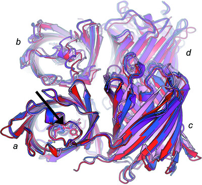
Crystal structure of the middle and C-terminal domains of Hsp90α labeled with a coumarin derivative reveals a potential allosteric binding site as a drug target
- Pages: 571-585
- First Published: 08 April 2022

Allosteric inhibitors that bind to the middle domain (MD) or C-terminal domain (CTD) of Hsp90 have become promising drug leads for the development of effective and nontoxic chemicals in anticancer drug discovery. The structure of MDCC-labeled Hsp90α MD and CTD reported here provides the first direct visual insight into allosteric binding inhibitors of Hsp90 MD or CTD and provides a basis for the design of novel drugs for the treatment of cancer and neurodegenerative diseases.
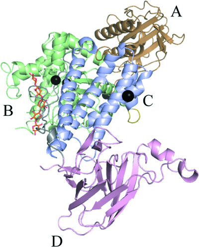
Structural studies of a novel auxiliary-domain-containing phenylalanine hydroxylase from Bacillus cereus ATCC 14579
- Pages: 586-598
- First Published: 08 April 2022
The crystal structure of phenylalanine hydroxylase from Bacillus cereus ATCC 14579 with three distinct domains is reported. The structural features and biochemical properties of the two novel auxiliary domains are investigated.
Over the rainbow: structural characterization of the chromoproteins gfasPurple, amilCP, spisPink and eforRed
- Pages: 599-612
- First Published: 08 April 2022
The structures of four coral chromoproteins reveal conserved dimer interfaces and insights into chromophore interactions that are important for colour development.
Crystal structure of the domain-swapped dimeric maltodextrin-binding protein MalE from Salmonella enterica
- Pages: 613-622
- First Published: 08 April 2022
Based on crystal structures of MalE from Salmonella enterica bound to maltopentaose, domain-swapped dimeric conformations are discussed.
Monoclonal antibody 7H2.2 binds the C-terminus of the cancer-oocyte antigen SAS1B through the hydrophilic face of a conserved amphipathic helix corresponding to one of only two regions predicted to be ordered
- Pages: 623-632
- First Published: 20 April 2022
The structure of the antigen-binding fragment of mouse monoclonal antibody 7H2.2 in complex with a 15-residue fragment from the metalloproteinase sperm acrosomal SLLP1 binding protein (SAS1B) has been determined at 2.75 Å resolution. The antigen is an amphipathic α-helix that corresponds to one of only two elements of secondary structure that are predicted to be ordered within the C-terminal region of SAS1B, which provides a basis for the targeted use of SAS1B.
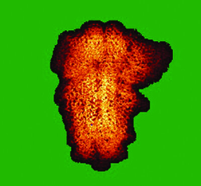
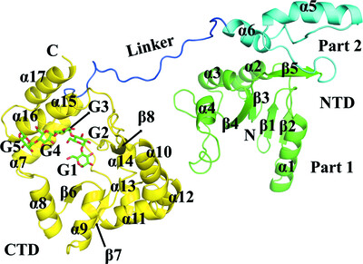
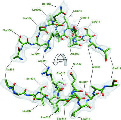
Synergic action of an inserted carbohydrate-binding module in a glycoside hydrolase family 5 endoglucanase
- Pages: 633-646
- First Published: 20 April 2022
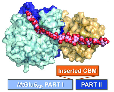
A unique endoglucanase with a carbohydrate-binding module inserted in the middle of the catalytic domain has been characterized structurally and functionally, providing insights into the mode of action responsible for its enhanced catalytic performance.
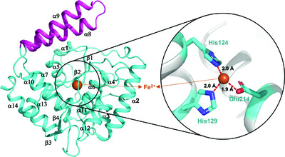
The structure of Phocaeicola vulgatus sialic acid acetylesterase
- Pages: 647-657
- First Published: 26 April 2022
The sialic acid acetylesterase from P. vulgatus was produced heterologously in Escherichia coli, purified and crystallized in two different crystal forms, from which structures at 1.44 and 2.06 Å resolution were obtained.
A GH115 α-glucuronidase structure reveals dimerization-mediated substrate binding and a proton wire potentially important for catalysis
- Pages: 658-668
- First Published: 20 April 2022
The crystal structure of a GH115 α-glucuronidase obtained in complex with xylohexaose and Ca2+ reveals that the two molecules constituting the homodimer cooperatively bind the substrate and that a divalent ion is involved in formation of the Michaelis–Menten complex and is likely to contribute to the formation of a protein wire that is essential for catalysis.
Structural insights into choline-O-sulfatase reveal the molecular determinants for ligand binding
- Pages: 669-682
- First Published: 26 April 2022
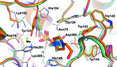
The first structures of a choline-O-sulfatase bound to different ligands are reported.
Sign up for email alerts
Tools
Distributed on behalf of the International Union of Crystallography