Journal list menu

 Issue
IssueJournal of Biophotonics: Volume 14, Issue 12
December 2021
Export Citations
Download PDFs
COVER PICTURE
Front cover
- First Published: December 2021

Despite the fact that both acute cardiac cessation and respiratory arrest are the reasons of brain ischemia, its hemodynamics, oxygenation and structural pathology differs. This article represents blood re-distribution and neuronal tissue degeneration during the first decade of minutes after the breathing and heart beating violation. The results show that cardiac cessation is much more life-threatening statement that provokes blood circulation abnormalities and causes strong signs of cerebral ischemia in compare with the respiratory arrest.
Further details can be found in the article by G. Piavchenko, I. Kozlov, V. Dremin, D. Stavtsev, E. Seryogina, K. Kandurova, V. Shupletsov, K. Lapin, A. Alekseyev, S. Kuznetsov, A. Bykov, A. Dunaev, and I. Meglinski (e202100216).
Inside Front cover
- First Published: December 2021

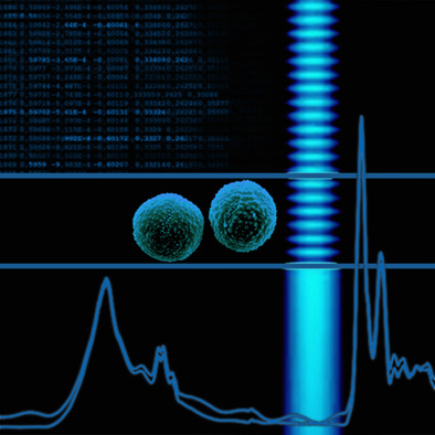
Pulmonary cancer is one of the most malignancies worldwide. We introduce a novel three-dimensional Convolutional Neural Network (3D-PulCNN) to classify three pulmonary subtypes based on the microscopic hyperspectral image because it can supply spatial information as well as spectral information. Combining the difference of spectral curves and texture feature between the three subtypes, we can classify LUAD, LUSC and SCLC more accurately in cytodiagnosis.
Further details can be found in the article by Qing Zhang | Yan Wang | Song Qiu | Jiangang Chen | Li Sun and Qingli Li (e202100142).
ISSUE INFORMATION
RESEARCH ARTICLES
Impairments of cerebral blood flow microcirculation in rats brought on by cardiac cessation and respiratory arrest
- First Published: 17 September 2021
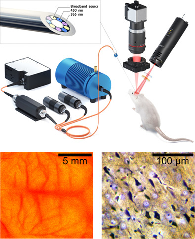
After an impair of blood and respiration functions there are significantly different changes in the structure and function of brain tissue that we present in this article.
3D-PulCNN: Pulmonary cancer classification from hyperspectral images using convolution combination unit based CNN
- First Published: 18 August 2021
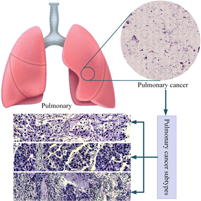
Pulmonary cancer is one of the most common malignancies worldwide. Existing algorithms for its classification are mostly based on color images, and the improvement of accuracy is quite challenging. This article proposes a new framework named as three-dimensional convolutional neural network (3D-PulCNN) for classifying pulmonary cancer based on microscopic hyperspectral image. Also, 3D-UNet is leveraged to segment cancer cells, and their morphological characteristics are calculated to help with prognosis assessment.
Deep-learning-based motion correction in optical coherence tomography angiography
- First Published: 20 July 2021
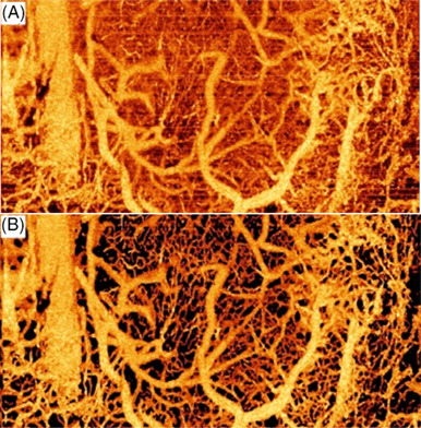
Motion artifacts significantly bias the visualization and quantification of OCT angiography (OCTA) images. Here, we present a deep-learning-based method to effectively reduce the motion artifacts, in which two deep neural networks were applied to identify artifacts and restore microvasculature, respectively. As shown in the figure, the proposed method can efficiently retrieve vasculature signal from the overwhelming motion artifacts (A) and significantly enhance OCTA image (B).
Single-shot phase contrast microscopy using polarisation-resolved differential phase contrast
- First Published: 13 August 2021
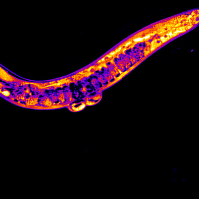
Phase contrast microscopy provides a label-free means to visualise structures in transparent samples, including cells and organisms. Implementations with low phototoxicity should enable extended imaging of live subjects that can complement fluorescence microscopy. Building on the development of more quantitative phase microscopy modalities, we present a widely applicable single-shot phase contrast technique (pDPC) that can provide semi-quantitative readouts of dynamic samples, which we have applied to Caenorhabditis Elegans.
An automated approach for fringe frequency estimation and removal in infrared spectroscopy and hyperspectral imaging of biological samples
- First Published: 01 September 2021
In infrared spectroscopy of thin film samples, multiple internal reflection causes sine wave signatures in the absorbance spectra. These signatures are referred to as fringes, and they impede the chemical analysis of the samples. In this study, an algorithm based on extended multiplicative signal correction (EMSC) is presented for estimating and removing interference fringes in infrared spectroscopy and imaging. The Fringe EMSC algorithm is made openly accessible on GitHub.
Redox ratio in the left ventricle of the growth restricted fetus is positively correlated with cardiac output
- First Published: 09 September 2021
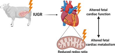
Babies born too small due to receiving insufficient nutrients and oxygen during pregnancy have an increased risk of developing cardiovascular disease in later life. We have combined two imaging modalities, two-photon microscopy and phase-contrast MRI, to assess and intrinsically link alterations in cardiac metabolism with cardiac function in normally grown and growth restricted fetuses.
Accurately calibrated frequency domain diffuse optical spectroscopy compared against chemical analysis of porcine adipose tissue
- First Published: 09 September 2021
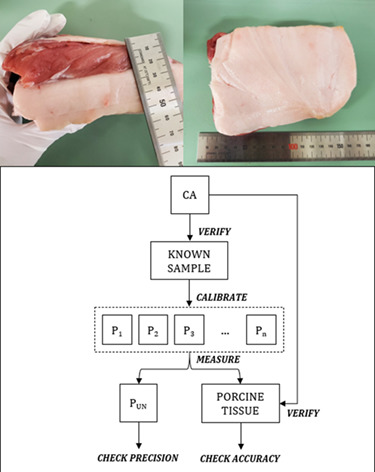
Frequency domain diffuse optical spectroscopy (fdDOS) is a technique which can non-invasively quantify the water and fat content of tissues. Decomposition of biological material into these basic components using chemical analysis (CA) is also a known technique in the nutrition industry, but has yet to be compared to fdDOS. In this study, we utilize both CA and fdDOS on porcine adipose tissue following a method to verify accurate fdDOS calibration.
Monitoring aging-associated structural alterations in Caenorhabditis elegans striated muscles via polarization-dependent second-harmonic generation measurements
- First Published: 17 August 2021
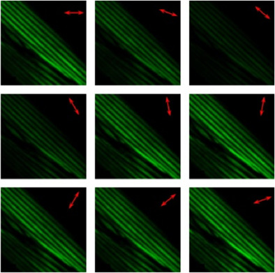
The employment of label free, non-destructive imaging techniques as novel diagnostic tools for the in-vivo monitoring of muscular changes due to aging is an important issue with high biological significance. Polarization-dependent second-harmonic generation measurements were used for age classification of the Caenorhabditis elegans samples and evaluation of structural alterations in their striated muscles during the worm lifespan. The obtained results indicate the usefulness of this technique as a potential new diagnostic tool for the investigation of muscle disorders.
Toward absolute viability measurements for bacteria
- First Published: 12 September 2021
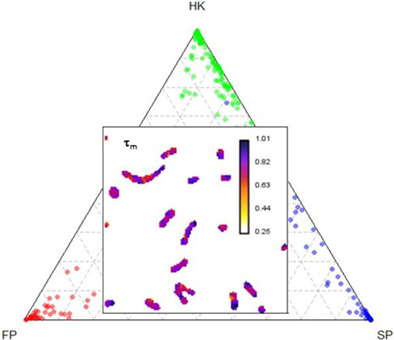
Viability of quiescent microbes is an important but elusive property to measure, and even more difficult to quantify using referenceable units for comparability. Here, we present a study using fluorescence lifetime imaging microscopy of a membrane-voltage fluorophore incubated with an oral microbe, Streptococcus mutans, and demonstrated differences in lifetime response of individual microbes in a quiescent state (SP) compared to those killed using heat (HK) or chemical fixation (FP).
Gingivitis resolution followed by optical coherence tomography and fluorescence imaging: A case study
- First Published: 28 August 2021
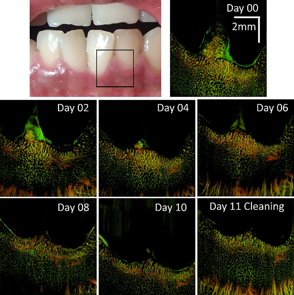
OCT angiography provides detailed information of microvascular involvement during a reversible gingivitis over a time course from Day 0 to Day 11 at which time the subject was scheduled for teeth cleaning. Shown are the vessel-depth encoded color vascular maps for better 3D appreciation.
Spectral photokinetic conversion of the fluorescent probes BSB and K114 for improved detection of amyloid assemblies
- First Published: 09 September 2021
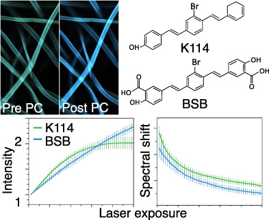
Exposure of amyloids stained with BSB and K114 to high laser power resulted in a permanent increase in fluorescence intensity, as well as a spectral blue-shift. The degree of fluorescence change over time (“photoconversion,” PC) was quantified in different amyloids. The kinetics of photoconversion-induced intensity and emission changes was found to strongly depend on such factors as dye structure, pH, presence of glycerol and the type of amyloid assembly.
Analysis of diffuse reflectance spectroscopy by means of Bayesian inference and separation of the parameters for scattering strength and spectral dependence of the scattering
- First Published: 10 September 2021
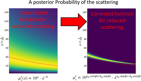
Scattering has a strong spectral dependence. While its characterization should be a simple fit, experimental results show strong fluctuations even for the same tissue type. These instabilities are analyzed by means of Bayesian Inference. By a simple coordinate transform, which separates scattering strength and scatterer size effects, a stable mathematical description of the scattering is derived. Thus, the resulting a posteriori probability of the reduced scattering coefficient narrows down significantly.
Cd-free InP/ZnSeS quantum dots for ultrahigh-resolution imaging of stimulated emission depletion
- First Published: 15 September 2021
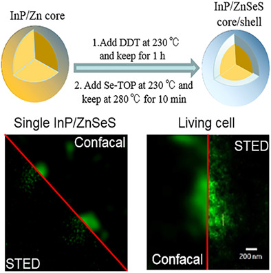
The environmentally friendly InP/ZnSeS quantum dots (InP/ZnSeS QDs) was successfully synthesized by a two-step method. The as-prepared InP/ZnSeS QDs possess smaller size and stronger photobleaching resistance. So, the InP/ZnSeS QDs as fluorescent probes are applied in cell super-resolution imaging. These results show that Cd-free InP/ZnSeS QDs have significant potential for application in fluorescent probes for stimulated emission depletion nanoscopy.
CORRIGENDUM
Corrigendum: Quantification of collagen structural changes during chick corneal development
- First Published: 08 November 2021
Sign up for email alerts
Tools
More from this journal
Related Titles
 Volume 19, Issue 14July 22, 2025
 Volume 5, Issue 3-4August-November 2023
 Volume 13, Issue 21July 25, 2025