

Journal list menu

 Issue
IssueJournal of Comparative Neurology: Volume 531, Issue 18
Special Issue:Honoring Deepak Pandya
1867-2193December 2023Guest Editor(s):
Export Citations
Download PDFs
FEATURED COVER
Cover Image, Volume 531, Issue 18
- First Published: 30 December 2023
The cover image is based on the Research Article India ink to 3D imaging: The legacy of Dr. Deepak “Dee” N. Pandya and his influence on generations of neuroanatomists by Gene J. Blatt et al., https://doi.org/10.1002/cne.25551.
ISSUE INFORMATION - TOC
The Journal of Comparative Neurology, Table of Content, Vol. 531, No. 18, December, 2023
- Pages: 1867-1868
- First Published: 30 December 2023
EDITORIAL
COMMENTARIES
In memoriam: Deepak N. Pandya (December 6, 1932 to October 4, 2020)
- Pages: 1870-1872
- First Published: 04 January 2023
RESEARCH ARTICLE
India ink to 3D imaging: The legacy of Dr. Deepak “Dee” N. Pandya and his influence on generations of neuroanatomists
- Pages: 1875-1882
- First Published: 02 November 2023

In this tribute article, we describe the legacy of Dr. Deepak Pandya on neuroanatomy and how he influenced our research. We provide examples of tracing with India ink and how methods have evolved to tracking neurons and their connections with artificial intelligence and whole brain imaging.
REVIEWS
Anatomy of the auditory cortex then and now
- Pages: 1883-1892
- First Published: 27 November 2023
Using neuroanatomical investigations in the macaque, Deepak Pandya and his colleagues have established the framework for auditory cortex organization, with subdivisions into core and belt areas. This has aided subsequent neurophysiological and imaging studies in monkeys and humans, and a nomenclature building on Pandya's work has also been adopted by the Human Connectome Project. The foundational work by Pandya and his colleagues is highlighted here in the context of subsequent and ongoing studies on the functional anatomy and physiology of auditory cortex in primates, including humans, and their relevance for understanding cognitive aspects of speech and language.
Medial–lateral organization of primary auditory cortex and the question of sound localization
- Pages: 1893-1896
- First Published: 26 June 2023
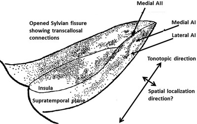
Illustration of the supratemporal plane after opening of the Sylvian fissure. AI is the primary auditory cortex; AII is association cortex. The dots indicate terminations of transcallosal connections between the two hemispheres. Note that there are connections to medial AI but not to lateral AI. In the anterior-posterior direction, the cells show a tonotopic distribution. The reason for the difference medial-lateral is unknown and was speculated in 1969 to be related to auditory localization in space.
The dorsal stream of visual processing and action-specific domains in parietal and frontal cortex in primates
- Pages: 1897-1908
- First Published: 28 April 2023
RESEARCH ARTICLE
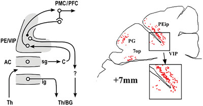
Architecture of the inferior parietal cortex in capuchin monkey
- Pages: 1909-1925
- First Published: 02 January 2023
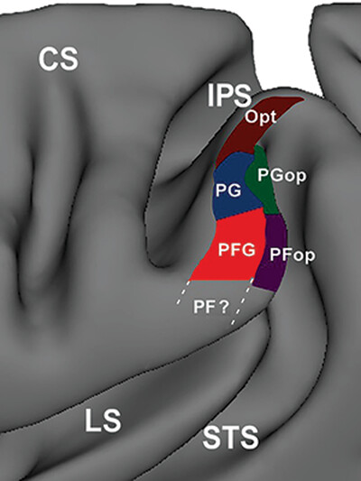
Architectural subdivision of the inferior parietal cortex (IPC) of the capuchin monkey in a 3D reconstruction of the left hemisphere. Five architectural areas are shown: three areas in the IPC convexity (PFG, in red; PG, in blue; and Opt, in brown), and two areas in the IPC operculum (PFop, in purple; and PGop, in green).
COMMENTARY
Cellular and laminar architecture: A short history and commentary
- Pages: 1926-1933
- First Published: 08 November 2023
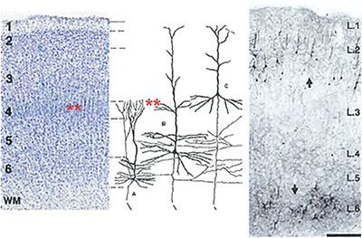
Lamination is an important classification criterion for feedforward, feedback, and other cortical connections. Lamination, however, needs to be considered with neuropil and local environments. There is substantial heterogeneity of parent cell types, within and across layers, and substantial heterogeneity of postsynaptic dendritic targets, as these cross the layers.
RESEARCH ARTICLES
Comparative features of calretinin, calbindin, and parvalbumin expressing interneurons in mouse and monkey primary visual and frontal cortices
- Pages: 1934-1962
- First Published: 26 June 2023
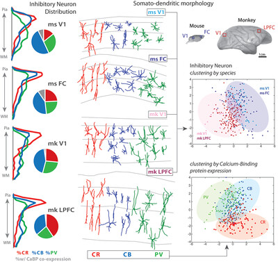
Medalla et al. quantified the density, laminar distribution, and somatodendritic morphology of inhibitory interneurons expressing one or more of the calcium-binding proteins (CaBPs) (calretinin [CR], calbindin [CB], and/or parvalbumin [PV]) in mouse versus rhesus monkey in two functionally and cytoarchitectonically distinct regions—the primary visual and frontal cortical areas. This study revealed that laminar distribution and dendritic morphology of inhibitory interneurons were largely dependent on CaBP expression. The main species difference was the significantly greater density and proportion of CR+ interneurons and lower extent of CaBP coexpression in monkey compared to mouse cortices. Cluster analyses revealed that the somatodendritic morphology of layer 2–3 inhibitory interneurons is dependent on CaBP and, to a lesser extent, species, but not cortical area. By contrast to pyramidal cells that show highly distinctive area- and species-specific features, the current study found more subtle differences in the distribution and features of interneurons across areas and species. These data yield insight into how nuanced differences in the cytoarchitecture, distribution, and properties of neurons may underlie specializations in cortical regions to confer species- and area-specific functional capacities.
Pattern of ventral temporal lobe interconnections in rhesus macaques
- Pages: 1963-1986
- First Published: 02 November 2023
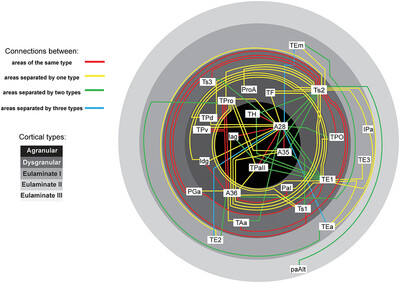
In macaques, connections are more frequent between architectonically similar ventral temporal cortices, in line with the Structural Model, which predicts density and laminar distribution of connections based on cortical type differences between linked areas. Pathways associated with memory in the entorhinal cortex are embedded in the laminar inhibitory microenvironment.
REVIEW
On the evolution of polysensory superior temporal sulcus and middle temporal gyrus: A key component of the semantic system in the human brain
- Pages: 1987-1995
- First Published: 11 July 2023
The superior temporal sulcus and middle temporal gyrus region underlies multisensory integration resulting in a key component of the semantic system in the language dominant hemisphere of the human brain
RESEARCH ARTICLES
Terminal organization of the corticospinal projection from the arm/hand region of the rostral primary motor cortex (M1r or old M1) to the cervical enlargement (C5–T1) in rhesus monkey
- Pages: 1996-2018
- First Published: 08 November 2023
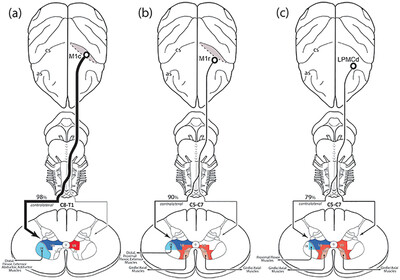
Summary diagram showing the general topography of the M1r CSP found in the present study (b) compared to the topography of the M1c CSP (a) and LPMCd CSP (c) found in our previous studies (Morecraft et al., 2013, 2019) .
Cytoarchitectonic and connection stripes in the dysgranular insular cortex in the macaque monkey
- Pages: 2019-2043
- First Published: 17 December 2023
The tight overlap of tract-tracing labeling patterns and cytoarchitectonic subareas in the macaque monkey insular cortex supports the notion that the primate insular cortex is highly organized, with separate dysgranular modules possibly integrating interoception with functionally distinct exteroceptive modalities.
Comparison of monkey and human retrosplenial neurocytology
- Pages: 2044-2061
- First Published: 07 December 2023
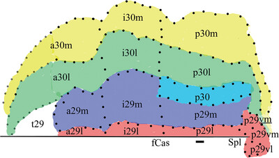
Critical to human imaging studies is the localization of cortical areas based on cytoarchitectural analyses. Retrosplenial cortex forms a complex region that is not localized only around the splenium of the corpus callosum as previously thought but it extends rostrally to abut midcingulate cortex in both monkey and human brains. This extension involves anterior and intermediate divisions and we provide a logical nomenclature for divisions of both based on the distribution of neuron-specific nuclear binding protein and intermediate neurofilament antibodies. The graphical image shows the flat map of area distributions and associated designations in human based on location and modifications to Brodmann's original map. Note that flattening distorts cortex caudal to the splenium (Spl). fCas, fundus of the callosal sulcus. For further details see Figure 2.
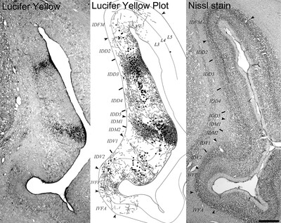
Assessing individual variability of the entorhinal subfields in health and disease
- Pages: 2062-2079
- First Published: 12 September 2023
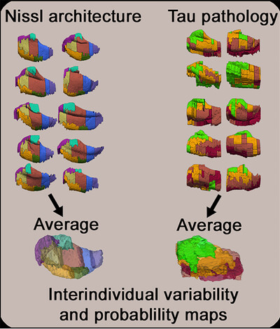
Based on histologically validated Nissl architecture delineations and tau pathology estimations, Oltmer et al. generated an average entorhinal cortex, provided probability maps, and investigated interindividual variability within the human entorhinal subfields in health and disease.
Oligomeric, phosphorylated, and truncated tau and spliceosome pathology within the entorhinal–hippocampal connectome across stages of Alzheimer's disease
- Pages: 2080-2108
- First Published: 29 March 2023
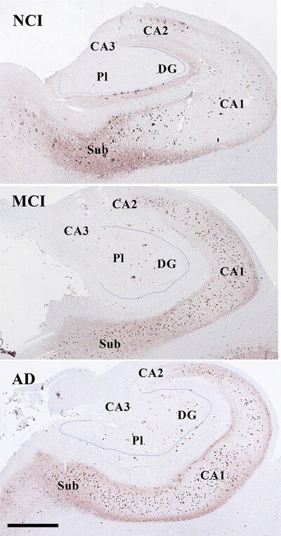
In this study, we determined the density of posttranslational oligomeric, phosphorylated, and late truncated tau epitopes within medial temporal lobe (MTL) subfields including entorhinal cortex (EC) layer II, subiculum, Cornu Ammonis (CA) subfields, and dentate gyrus (DG) in subjects who died with a clinical diagnosis of no cognitive impairment, mild cognitive impairment (MCI), and Alzheimeŕs disease (AD). We also examined whether alterations of the nuclear alternative splicing protein, SRSF2, are associated with tau pathology. Our results suggest a dual-hit process in NFT formation, with decreased nuclear SRSF2 in the presence of cytoplasmic phosphorylated within the entorhinal hippocampal connectome during the onset of AD. Although oligomeric and phosphorylated tau follow a stereotypical pattern, clinical disease stage determined density of tau deposition and not anatomic location within the entorhinal-hippocampal connectome.
Shades of gray in human white matter
- Pages: 2109-2120
- First Published: 27 June 2023
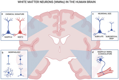
This graphic illustrates a coronal section of the human brain and highlights key characteristics of white matter neurons (WMNs). Two classes emerged, subplate and subcortical deep WMNs, which appeared to be neurochemically heterogenous; for example, image (a) demonstrates that there was no chemical overlap between NADPH-d (blue) and MAP-2 (red) positivity. Inspection of morphological features led to (b) the classification of three groups of NADPH-d+ WMNs: (1) unipolar, (2) bipolar, and (3) multipolar—the latter of which was most frequently observed. (c) In cognitively normal par, subplate WMNs were found to be significantly larger than subcortical deep WMNs. (d) NADPH-d+ subcortical neurons also tended to embrace the outer walls of microvessels, suggesting a functional role in vasodilation. Together, these observations provide a landscape for future systematic investigations of this unique cell population. Created with BioRender.com.
A novel subdivision of the bed nucleus of stria terminalis in monkey, rat, and mouse brains
- Pages: 2121-2145
- First Published: 30 December 2022
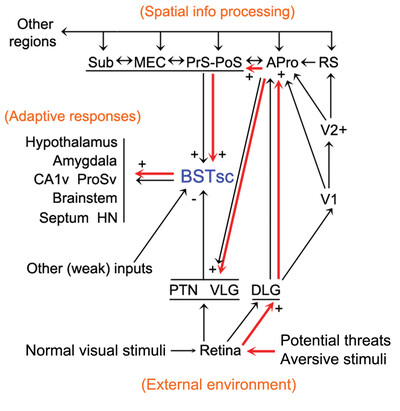
A novel subdivision of the bed nucleus of stria terminalis (BST) was identified in monkey, rat, and mouse brains based on the human equivalent. This spindle-shaped small cell subdivision (BSTsc) receives its main inputs from the earlier visual and spatial processing structures and projects to many common target regions as other BST subdivisions do (see the graph). The neurons in the BSTsc mostly expresses excitatory marker genes while those in other BST components mainly contains inhibitory marker genes. The BSTsc is poised to serve as a shortcut bridge directly linking spatial information from the environment to vigilant adaptive internal responses.
Analyzing the cortical fine structure as revealed by ex-vivo anatomical MRI
- Pages: 2146-2161
- First Published: 31 July 2023
The human cortex has a rich fiber structure, as revealed by myelin-staining of histological slices. Myelin also contributes to the image contrast in MRI. Recent advances in MR scanner and imaging technology allowed the acquisition of an ex-vivo data set at an isotropic resolution of 100 µm. We focused on a computational analysis of this data set with the aim of bridging histological knowledge with MRI-based results. As an example, the discrimination between unistriate (visual) cortex and adjacent astriate cortex is shown in the figure.
Associations between white matter integrity of the cingulum bundle, surrounding gray matter regions, and cognition across the dementia continuum
- Pages: 2162-2171
- First Published: 27 November 2023
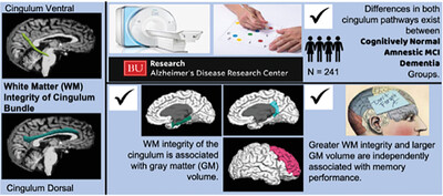
Key Findings:
- Differences exist in the white matter integrity of the cingulum bundle between participants who are on the dementia continuum.
- White matter integrity of the cingulum is associated with gray matter volume.
- White matter integrity of the cingulum ventral, independent of gray matter volume, may be predictive of memory performance.
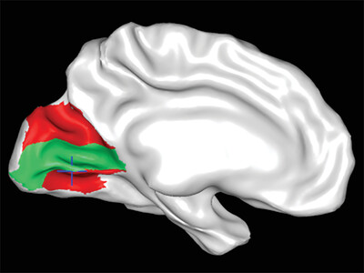
A proposed structural connectivity matrices approach for the superior fronto-occipital fascicle in the Harvard-Oxford Atlas comparative framework following the Pandya comparative extrapolation principle
- Pages: 2172-2184
- First Published: 27 November 2023
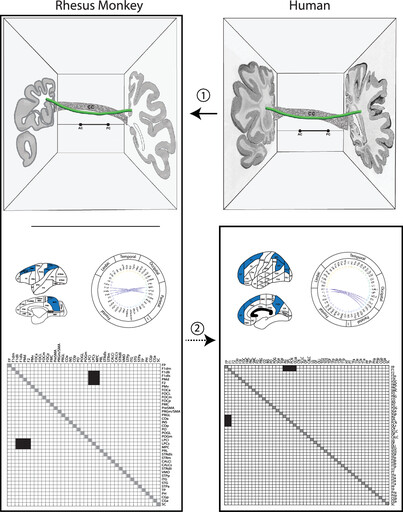
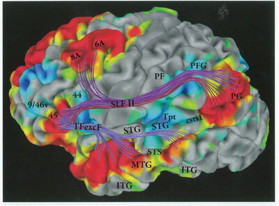
Direct information about origins and terminations of cortico-cortical long association fiber pathways in humans is extremely limited. The topographic location and trajectory of cortico-cortical association pathway stems in humans and nonhuman primates is similar (1), but only in nonhuman primates is there complete information on origins and terminations as well as stems. Thus, an extrapolation approach, from nonhuman primate to human (2), is applied in the present report to infer connectivity patterns for the superior fronto-occipital fascicle in the human brain.
Diaschisis in the human brain reveals specificity of cerebrocerebellar connections
- Pages: 2185-2193
- First Published: 23 August 2023
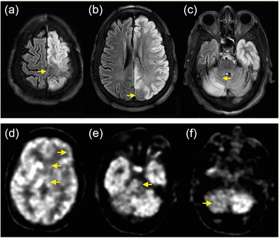
Brain magnetic resonance imaging and positron emission tomography following unilateral status epilepticus revealed abnormalities in cerebral association areas and contralateral cerebellar cognitive posterior lobe regions, with sparing of primary sensory and motor areas in cerebral cortex and cerebellum. This unique observation provides confirmation in the human brain of anatomical-functional specialization in cerebrocerebellar circuits.