

Journal list menu

 Issue
IssueJournal of Comparative Neurology: Volume 531, Issue 11
1095-1216August 2023
Export Citations
Download PDFs
FEATURED COVER
Cover Image, Volume 531, Issue 11
- First Published: 06 June 2023
The cover image is based on the Research Article Expression of tau and phosphorylated tau in the brain of normal and hemiparkinsonian rhesus macaques by Julia C. Gambardella et al., https://doi.org/10.1002/cne.25490.
ISSUE INFORMATION - TOC
The Journal of Comparative Neurology, Table of Content, Vol. 531, No. 11, August, 2023
- Page: 1095
- First Published: 06 June 2023
RESEARCH ARTICLES
Sex differences in white matter tracts of capuchin monkey brains
- Pages: 1096-1107
- First Published: 01 May 2023
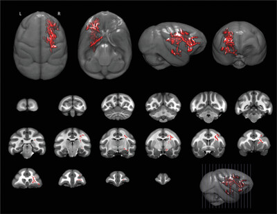
Nonhuman primates exhibit sexual dimorphism in behavior, suggesting that there could be underlying differences in brain organization and function. Females showed significantly higher fractional anisotropy than males in regions of frontal-parietal white matter in the right cerebral hemisphere. These anisotropy values were non-overlapping between males and females.
Derivation of human retinal cell densities using high-density, spatially localized optical coherence tomography data from the human retina
- Pages: 1108-1125
- First Published: 19 April 2023
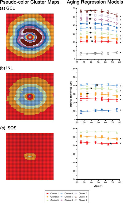
Pseudocolor cluster maps and regression models describing normal, age-related change in the ganglion cell layer, inner plexiform layer, and inner segment–outer segment region of photoreceptors, as derived from optical coherence tomography data.
Distribution of the transcription factor islet-1 in the central nervous system of nonteleost actinopterygian fish: Relationship with cholinergic and catecholaminergic systems
- Pages: 1126-1146
- First Published: 18 April 2023
The effects of voluntary running exercise on the astrocytes of the medial prefrontal cortex in APP/PS1 mice
- Pages: 1147-1162
- First Published: 05 May 2023
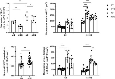
Pathological changes in the medial prefrontal cortex (mPFC) and astrocytes are closely associated with Alzheimer's disease (AD).
3D atlas of cerebral neuropils with previously unknown demarcations in the honey bee brain
- Pages: 1163-1183
- First Published: 18 April 2023
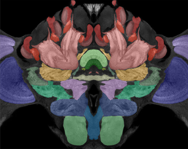
Confocal image showing the demarcation of neuropils in a layer of the central brain (cerebrum) of the honey bee. Distinct anti-synapsin immunolabeled neuropils are depicted by different colors. The study by Habenstein et al. provides a detailed 3D atlas of neuropils and tracts in the honeybee brain.
Rod bipolar cells receive cone photoreceptor inputs through both invaginating synapses and flat contacts in the mouse and guinea pig retinas
- Pages: 1184-1197
- First Published: 18 April 2023
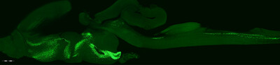
Expression of tau and phosphorylated tau in the brain of normal and hemiparkinsonian rhesus macaques
- Pages: 1198-1216
- First Published: 26 April 2023
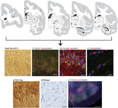
Accumulation of the neuronal protein tau had been linked to neurodegenerative disorders. Rhesus macaques are widely used for studying aging and neurodegenerative disorders, yet little is known about the natural expression of tau in their brains. Here, we demonstrate that tau is present across brain regions in normal and hemiparkinsonian rhesus macaques detected by immunohistochemical methods. Tau is found in neuronal soma and fibers of gray matter brain regions and oligodendrocytes in white matter brain regions. Both 3R and 4R tau isoforms are detected. Tau phosphorylated at Thr231, but not Ser202/Thr205 (AT8), is also present. Nigral dopaminergic (TH+) cell loss associated to neurotoxin-induced hemiparkinsonism did not affect tau expression as the protein is mainly expressed in nigral GABAergic (GAD67+) cells.