

Journal list menu

Export Citations
Download PDFs
Table of Contents
Emerging Hubs in Plant Light and Temperature Signaling
- First Published: 06 October 2015
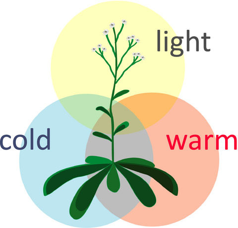
Temperature and Light Perception integration.
Green Light to Plant Responses to Pathogens: The Role of Chloroplast Light-Dependent Signaling in Biotic Stress
- First Published: 19 May 2015
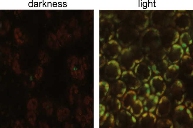
The effect of light on ROS production by chloroplasts during a non-host pathogen interaction causing LCD is illustrated by infiltrating tobacco leaves with Xanthomonas campestris (see reference [16] in the text). Leaves were incubated with a ROS-reacting probe and observed 19 h post-infiltration by confocal laser microscopy, merging bright field, chlorophyll self-fluorescence (red), and ROS fluorescence (green). In leaves kept in the dark (left), chloroplasts were visualized as a red necklace around the central vacuole. Illuminated tissue (right) shows chloroplasts in yellow due to combination of red and green fluorescence, indicating that pathogen interaction results in light-dependent plastid ROS production.
How Does Photoreceptor UVR8 Perceive a UV-B Signal?
- First Published: 21 May 2015
Light perception of UV-B photoreceptor UVR8 is triggered by light-induced molecular events originating in two epicenters located at the dimer interface. Clustered tryptophan residues serve as UV-B pigments and undergo concerted conformational changes upon absorbing UV-B photons, which disrupt the dimer interface and lead to dimer dissociation of UVR8.
The Evolution and Functional Role of Flavin-based Prokaryotic Photoreceptors
- First Published: 03 July 2015
Blue-light photosensors of the LOV type, related to plant phototropins, are wide spread among bacteria. What is their role in vivo and by which pathways did they evolve? Why to bacterial pathogens often see the same blue color as they host plants and in the same way? News and views on a burning photobiology topic.
Photoreceptors in Chemotrophic Prokaryotes: The Case of Acinetobacter spp. Revisited
- First Published: 04 July 2015
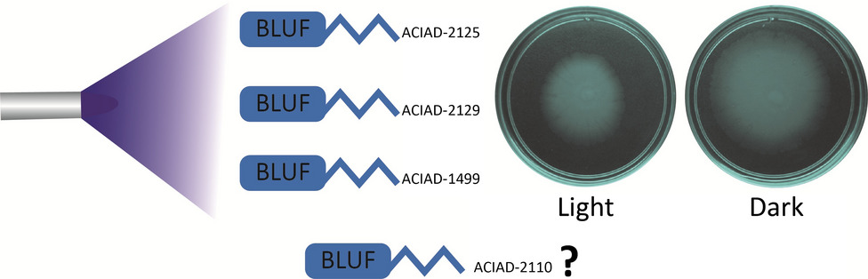
Most members of the genus Acinetobacter have at least one BLUF protein encoded in their genome. A. baylyi ADP1 has four of them, of which three are jointly involved in the inhibition of twitching motility in response to blue light at suboptimal temperatures.
Activation of the General Stress Response of Bacillus subtilis by Visible Light
- First Published: 18 July 2015
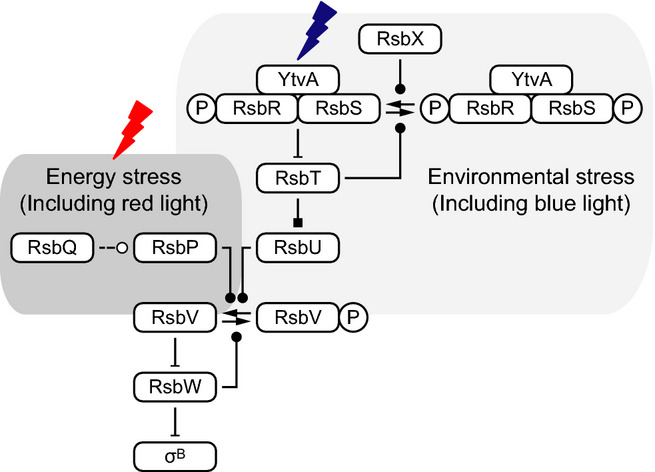
The general stress response of Bacillus subtilis can be activated with blue and red light. The mechanism of its activation is well understood, to the extent that computational analysis allows one to recognize mutationally altered properties of blue-light photoreceptor protein YtvA (one of the components of high-molecular-weight complexes called stressosomes) in the in vivo output characteristics of the stress response. This system qualifies exceptionally well as a model system for future studies of genome evolution, driven by altered stimulus regimes.
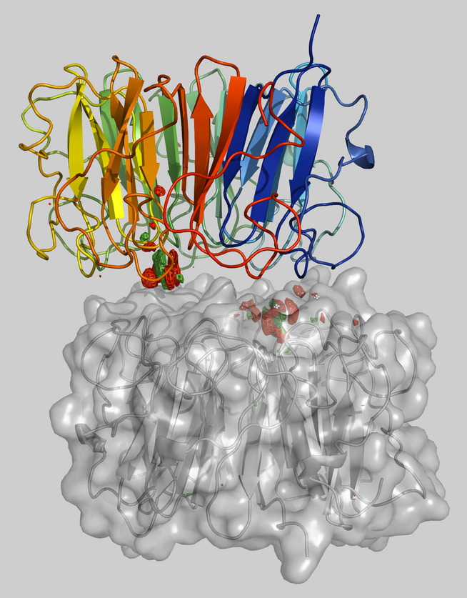
More Than Just Light: Clinical Relevance of Light Perception in the Nosocomial Pathogen Acinetobacter baumannii and Other Members of the Genus Acinetobacter
- First Published: 25 August 2015
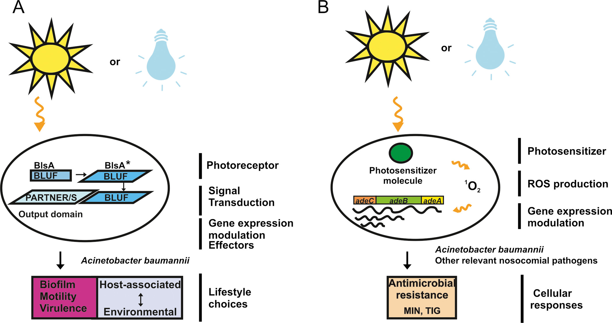
In Acinetobacter baumannii light modulates physiological aspects related to the success of the microorganism as a nosocomial pathogen. In the figure are outlined models for light perception and signal transduction in A. baumannii. (A) Light absorption induces a conformational change in the A. baumannii BLUF photoreceptor BlsA, now able to bind unknown partners that modulate the response, resulting ultimately in regulation of surface motility, biofilm formation and virulence against Candida albicans. (B) Light can also induce reduction in susceptibility to antibiotics in a photoreceptor-independent manner in A. baumannii and other clinically relevant pathogens. In this case, light application could result in the excitation of an unknown photosensitizer molecule with the concomitant production of ROS such as 1O2, leading ultimately to the induction of expression of antibiotic resistance genes such as those coding for the AdeABC pump. This results in reduction in susceptibility to the antibiotics MIN and TIG. All these phenotypes occur at 25°C.
Melanopsin and the Non-visual Photochemistry in the Inner Retina of Vertebrates
- First Published: 23 October 2015
The inner retina of most vertebrates expresses the photopigment melanopsin (Opn4). Opn4 has been involved in a number of nonimage forming tasks (synchronization of circadian rhythms, pupillary light reflexes, etc.) and although it has been characterized as a bi/tristable photopigment, little is known about the mechanism/s involved in its chromophore regeneration. Based on some molecular and biochemical similarities with rhabdomeric photoreceptors (the biochemical nature of phosphoinositol-photocascade and opsin homology), we can infer that a novel visual cycle may operate in Opn4 (+) cells further cooperating to the chromophore regeneration via a supplementary alternative process as described in Drosophila.
Nonvisual Opsins and the Regulation of Peripheral Clocks by Light and Hormones
- First Published: 14 July 2015
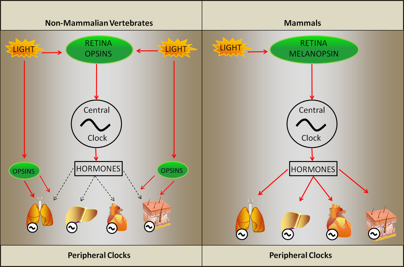
Nonvisual opsins have a wide distribution in all vertebrate classes, even though their role in the modulation of peripheral clocks remains elusive. Together with light, hormones represent an important zeitgeber in the entrainment of peripheral clocks. In this review, we focus the entrainment of peripheral clocks of nonmammalian vertebrates by light and hormones, highlighting the main differences between mammalian and nonmammalian models. In addition, the putative photopigments and the signaling pathways triggered by light in ectothermic vertebrates are also discussed.
Forged Under the Sun: Life and Art of Extremophiles from Andean Lakes
- First Published: 09 December 2015
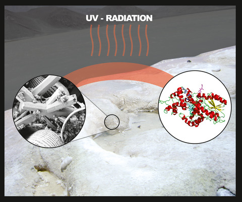
High-altitude Andean lakes are a treasure chest for microbiological research in South America. Their indigenous microbial communities including the highest modern stromatolites are exposed to outstandingly high UV irradiation and share a complex system of genetic and physiological mechanisms called as UV-resistome.
UV-induced DNA Damage: The Role of Electronic Excited States
- First Published: 05 October 2015
Electronic interactions among DNA bases give rise to excited states delocalized over two or more bases. As a result, the excited state properties depend strongly on conformational motions.
Oxidatively Generated Damage to Cellular DNA by UVB and UVA Radiation
- First Published: 18 October 2014
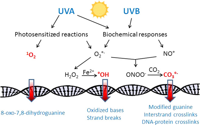
UVA predominantly generates singlet oxygen that selectively oxidize guanine leading to 8-oxo-7,8-dihydroguanine. Superoxide anion radical () is also produced as the precursor of H2O2 and subsequently of highly reactive hydroxyl radical (•OH) that induces oxidized bases and single-strand breaks. Both UVA and UVB are able to trigger delayed biochemical responses including activation of enzymes, inflammation and bystander effects. As a result and nitric oxide are released leading to the formation of •OH and peroxynitrite. The latter species is expected through reaction with CO2 to produce the carbonate anion radical, an efficient one-electron oxidant of guanine.
Photobiological Origins of the Field of Genomic Maintenance
- First Published: 20 October 2015
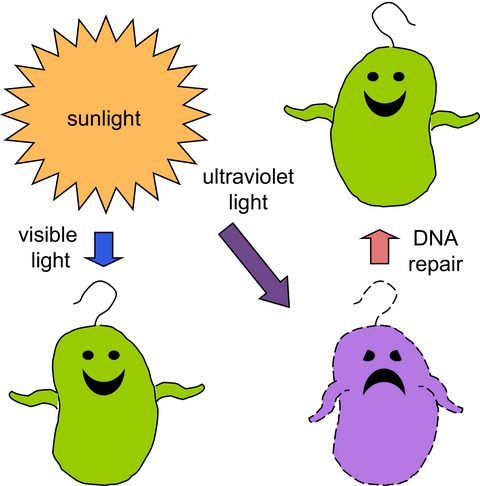
Although sunlight is essential for life, the UV wavelengths constitute a threat to life. Cellular responses evolved to deal with DNA damage inflicted by UV; the study of these responses spawned the burgeoning field of DNA repair and genomic maintenance.
UV Signature Mutations
- First Published: 29 October 2014
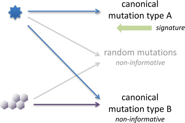
Inverse relationship of canonical mutation patterns and mutation signatures for inferring the mutagen from mutations. Two mutagens are illustrated. A mutagen's canonical mutations deviate from random base changes, establishing a pattern typical for that mutagen. Different mutagens can produce the same canonical mutations (non-informative mutations). Signature mutations are the subset of canonical mutations that, in addition, are unique to that mutagen and permit inference backward from mutations to mutagen. A mutagen therefore produces signature mutations plus non-informative mutations. The latter are real and are produced by the mutagen, but are not useful for identifying that mutagen or carcinogen.
Research Articles
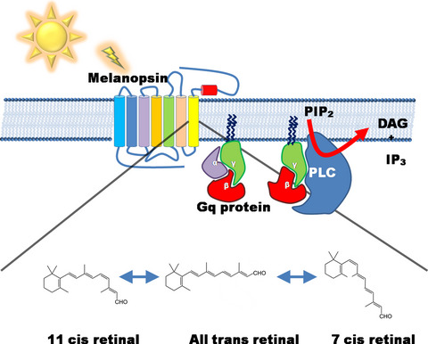
Melanopsins: Localization and Phototransduction in Xenopus laevis Melanophores
- First Published: 25 June 2015
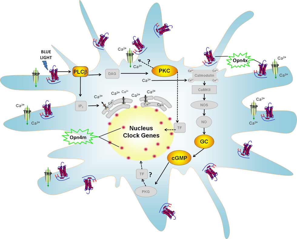
Xenopus laevis melanophores express two melanopsins, Opn4x and Opn4m. Their proteins are differentially located: Opn4x is present in the cytoplasm and the cell membrane, and Opn4m is located in the nuclear region. In this model, blue light, which maximally activates melanopsins, increases clock gene expression. Given the localization of melanopsins and our pharmacological data, the light-induced clock gene expression seems to be mediated by Opn4x through the activation of the phosphoinositide cascade. This intracellular signaling ultimately cross-talks with cGMP-producing pathway, leading to the reset of the biological clock in X. laevis melanophores.
Functional Characterization of a LOV-Histidine Kinase Photoreceptor from Xanthomonas citri subsp. citri
- First Published: 14 July 2015
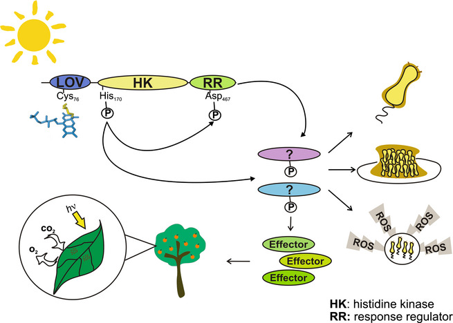
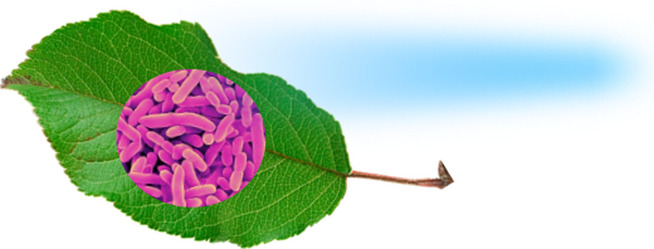
Xanthomonas citri subsp. citri (Xcc) is the bacterium responsible for citrus canker. Xcc-LOV is a blue-light (BL) sensing, hybrid histidine-kinase protein. This spectroscopic characterization confirmed that Xcc-LOV presents a canonical LOV-photochemistry, involving the BL-induced formation of a covalent cystein-chromophore (flavin) adduct, preceded by a transient flavin triplet state and followed by thermal conversion into the dark state. Moreover, Xcc-LOV showed BL-induced kinase activity, initiating a signal transduction cascade. Therefore, we correlated the previously observed regulatory effects of BL on Xcc physiology and synthesis of effectors for maintaining plant energetic metabolism and counteracting plant defense, with the molecular activation of Xcc-LOV.
Acute Effects of Light on Alternative Splicing in Light-Grown Plants
- First Published: 17 November 2015
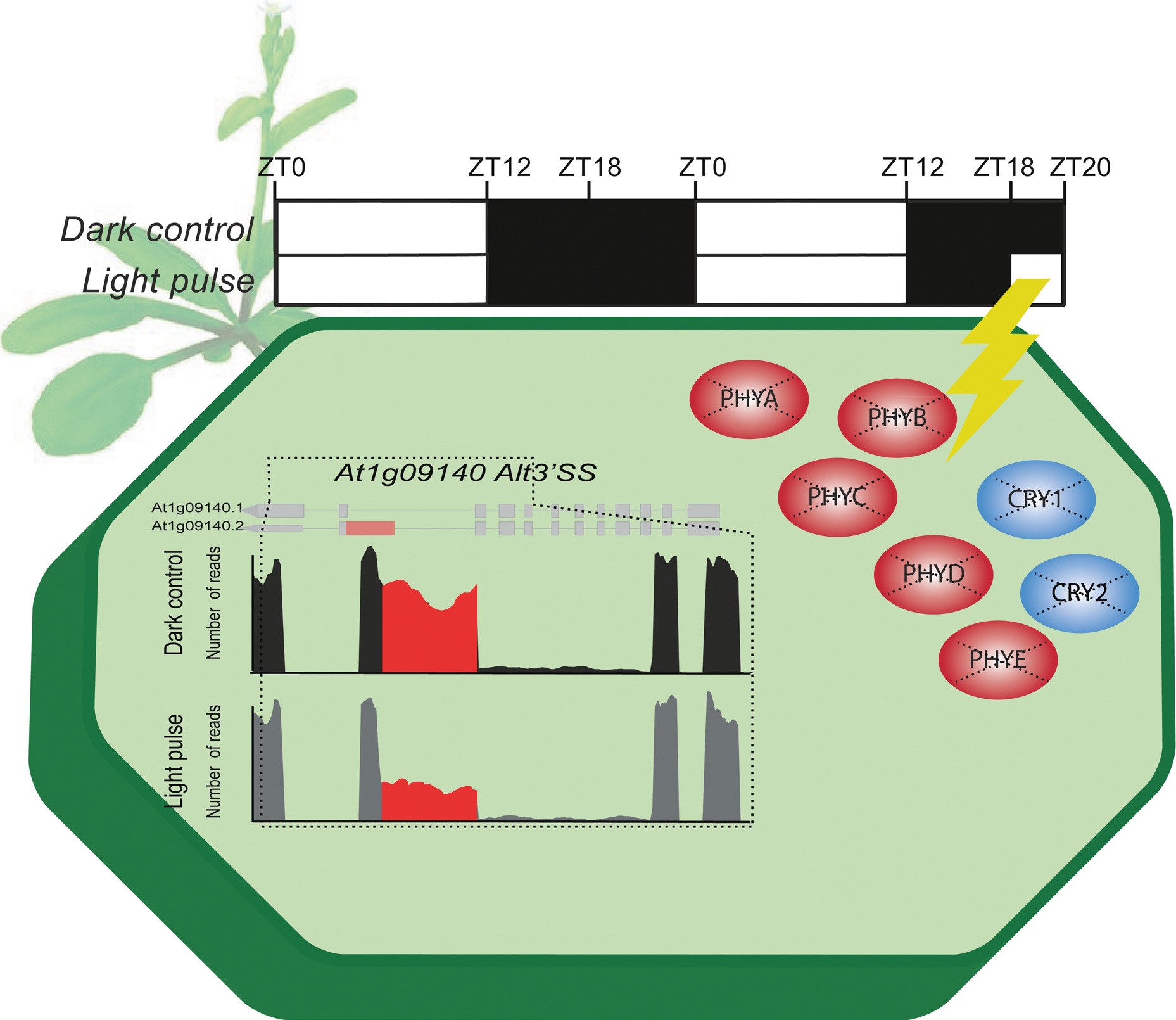
Light modulates plant growth and development to a great extent by regulating gene expression programs. Here, we evaluated the effect of light on alternative splicing (AS) in light-grown Arabidopsis thaliana plants using high-throughput RNA sequencing (RNA-seq). Interestingly, the effect of a red-light pulse on AS of a gene encoding a splicing factor was not impaired in a quintuple phytochrome mutant, providing unequivocal evidence that nonphotosensory photoreceptors control AS in light-grown plants.
Novel In Vitro Antioxidant and Photoprotection Capacity of Plants from High Altitude Ecosystems of Colombia
- First Published: 20 October 2015
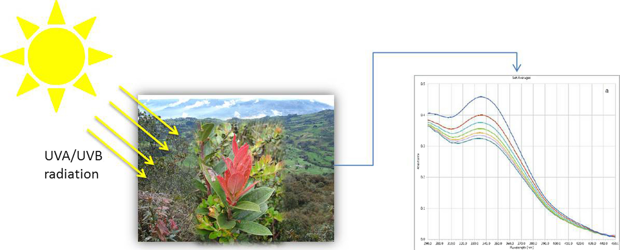
Selected plants grow on high mountains of tropical zones that are exposed to high UVR levels all year long. Their extracts showed an exceptional absorption coefficient and their sunscreen formulations displayed values of SPF, UVAPF, UVA/UVB ratio, and critical wavelength high enough to satisfy requirements for broad-spectrum UVB/UVA protection. In addition, the photostability study in formulations showed an in vitro SPF effectiveness (%SPFeff) greater than 80%. Therefore, the proposed natural formulation could be considered as a photostable product.
Photodynamic Action Mechanism Mediated by Zinc(II) 2,9,16,23-Tetrakis[4-(N-methylpyridyloxy)]phthalocyanine in Candida albicans Cells
- First Published: 24 June 2015
![Photodynamic Action Mechanism Mediated by Zinc(II) 2,9,16,23-Tetrakis[4-(N-methylpyridyloxy)]phthalocyanine in Candida albicans Cells](/cms/asset/9bafb40a-0b4a-4aec-a8a9-0f6d8c8ad2cd/php12483-toc-0001-m.jpg)
The photoreaction type I/type II pathways mediated by ZnPPc4+ was studied in Candida albicans cells. Photoinactivation was investigated in presence of specific molecular probes to detect the presence of ROS, under different experimental conditions and in presence of scavengers of ROS to find information about the photoprocesses that produces the cell death. These investigation indicates that O2(1Δg) is generated in the cells, although a minor extension other radical species can also be involved in the photoinactivation of C. albicans mediated by ZnPPc4+.
Sign up for email alerts
Tools
Published on behalf of the American Society for Photobiology