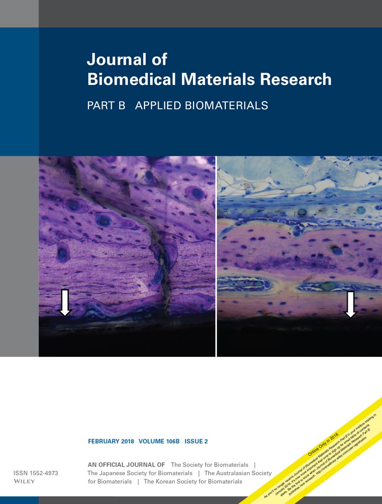
In vivo response to decellularized mesothelium scaffolds
Michael J. Cronce
Center for Regenerative Medicine, Massachusetts General Hospital, Boston, Massachusetts, 02114
Both contributed equally as joint first authors.
Search for more papers by this authorRenea A. Faulknor
Center for Regenerative Medicine, Massachusetts General Hospital, Boston, Massachusetts, 02114
Harvard Medical School, Boston, Massachusetts, 02115
Both contributed equally as joint first authors.
Search for more papers by this authorIrina Pomerantseva
Center for Regenerative Medicine, Massachusetts General Hospital, Boston, Massachusetts, 02114
Department of Surgery, Massachusetts General Hospital, Boston, Massachusetts, 02114
Harvard Medical School, Boston, Massachusetts, 02115
Search for more papers by this authorEmmanuel C. Ekwueme
Center for Regenerative Medicine, Massachusetts General Hospital, Boston, Massachusetts, 02114
Harvard Medical School, Boston, Massachusetts, 02115
Search for more papers by this authorOlive Mwizerwa
Center for Regenerative Medicine, Massachusetts General Hospital, Boston, Massachusetts, 02114
Search for more papers by this authorCraig M. Neville
Center for Regenerative Medicine, Massachusetts General Hospital, Boston, Massachusetts, 02114
Department of Surgery, Massachusetts General Hospital, Boston, Massachusetts, 02114
Harvard Medical School, Boston, Massachusetts, 02115
Search for more papers by this authorCorresponding Author
Cathryn A. Sundback
Center for Regenerative Medicine, Massachusetts General Hospital, Boston, Massachusetts, 02114
Department of Surgery, Massachusetts General Hospital, Boston, Massachusetts, 02114
Harvard Medical School, Boston, Massachusetts, 02115
Correspondence to: C. Sundback; e-mail: [email protected]Search for more papers by this authorMichael J. Cronce
Center for Regenerative Medicine, Massachusetts General Hospital, Boston, Massachusetts, 02114
Both contributed equally as joint first authors.
Search for more papers by this authorRenea A. Faulknor
Center for Regenerative Medicine, Massachusetts General Hospital, Boston, Massachusetts, 02114
Harvard Medical School, Boston, Massachusetts, 02115
Both contributed equally as joint first authors.
Search for more papers by this authorIrina Pomerantseva
Center for Regenerative Medicine, Massachusetts General Hospital, Boston, Massachusetts, 02114
Department of Surgery, Massachusetts General Hospital, Boston, Massachusetts, 02114
Harvard Medical School, Boston, Massachusetts, 02115
Search for more papers by this authorEmmanuel C. Ekwueme
Center for Regenerative Medicine, Massachusetts General Hospital, Boston, Massachusetts, 02114
Harvard Medical School, Boston, Massachusetts, 02115
Search for more papers by this authorOlive Mwizerwa
Center for Regenerative Medicine, Massachusetts General Hospital, Boston, Massachusetts, 02114
Search for more papers by this authorCraig M. Neville
Center for Regenerative Medicine, Massachusetts General Hospital, Boston, Massachusetts, 02114
Department of Surgery, Massachusetts General Hospital, Boston, Massachusetts, 02114
Harvard Medical School, Boston, Massachusetts, 02115
Search for more papers by this authorCorresponding Author
Cathryn A. Sundback
Center for Regenerative Medicine, Massachusetts General Hospital, Boston, Massachusetts, 02114
Department of Surgery, Massachusetts General Hospital, Boston, Massachusetts, 02114
Harvard Medical School, Boston, Massachusetts, 02115
Correspondence to: C. Sundback; e-mail: [email protected]Search for more papers by this authorAbstract
Biological surgical scaffolds are used in plastic and reconstructive surgery to support structural reinforcement and regeneration of soft tissue defects. Macrophage and fibroblast cell populations heavily regulate scaffold integration into host tissue following implantation. In the present study, the biological host response to a commercially available surgical scaffold (Meso BioMatrix Surgical Mesh (MBM)) was investigated for up to 9 weeks after subcutaneous implantation; this scaffold promoted superior cell migration and infiltration previously in in vitro studies relative to other commercially available scaffolds. Infiltrating macrophages and fibroblasts phenotypes were assessed for evidence of inflammation and remodeling. At week 1, macrophages were the dominant cell population, but fibroblasts were most abundant at subsequent time points. At week 4, the scaffold supported inflammation modulation as indicated by M1 to M2 macrophage polarization; the foreign body giant cell response resolved by week 9. Unexpectedly, a fibroblast subpopulation expressed macrophage phenotypic markers, following a similar trend in transitioning from a proinflammatory to anti-inflammatory phenotype. Also, α-smooth muscle actin-expressing myofibroblasts were abundant at weeks 4 and 9, mirroring collagen expression and remodeling activity. MBM supported physiologic responses observed during normal wound healing, including cellular infiltration, host tissue ingrowth, remodeling of matrix proteins, and immune modulation. © 2017 Wiley Periodicals, Inc. J Biomed Mater Res Part B: Appl Biomater, 106B: 716–725, 2018.
CONFLICT OF INTEREST
SMG is a current employee and XHL is a former employee of DSM Biomedical. Both played substantial roles providing information regarding clinical use and properties of dECM materials as well as potential issues with manufacturing methods. DSM partially funded this study but had no role in the study design; data collection, analysis, or interpretation; or manuscript preparation.
REFERENCES
- 1Wolf MT, Dearth CL, Ranallo CA, LoPresti ST, Carey LE, Daly KA, Brown BN, Badylak SF. Macrophage polarization in response to ECM coated polypropylene mesh. Biomaterials 2014; 35: 6838–6849.
- 2Laschke MW, Haufel JM, Scheuer C, Menger MD. Angiogenic and inflammatory host response to surgical meshes of different mesh architecture and polymer composition. J Biomed Mater Res B Appl Biomater 2009; 91: 497–507.
- 3Anderson JM, Rodriguez A, Chang DT. Foreign body reaction to biomaterials. Semin Immunol 2008; 20: 86–100.
- 4Arbos MA, Ferrando JM, Quiles MT, Vidal J, López-Cano M, Gil J, Manero JM, Peña J, Huguet P, Schwartz-Riera S, Reventós J, Armengol M. Improved surgical mesh integration into the rat abdominal wall with arginine administration. Biomaterials 2006; 27: 758–768.
- 5Arenas-Herrera JE, Ko IK, Atala A, Yoo JJ. Decellularization for whole organ bioengineering. Biomed Mater 2013; 8: 014106.
- 6Gilbert TW. Strategies for tissue and organ decellularization. J Cell Biochem 2012; 113: 2217–2222.
- 7Mancuso L, Gualerzi A, Boschetti F, Loy F, Cao G. Decellularized ovine arteries as small-diameter vascular grafts. Biomed Mater 2014; 9: 045011.
- 8Luo X, Kulig KM, Finkelstein EB, Nicholson MF, Liu XH, Goldman SM, Vacanti JP, Grottkau BE, Pomerantseva I, Sundback CA, Neville CM. In vitro evaluation of decellularized ECM-derived surgical scaffold biomaterials. J Biomed Mater Res B Appl Biomater 2015. doi: 10.1002/jbm.b.33572. [Epub ahead of print]
10.1002/jbm.b.33572 Google Scholar
- 9Hoganson DM, Owens GE, O'Doherty EM, Bowley CM, Goldman SM, Harilal DO, Neville CM, Kronengold RT, Vacanti JP. Preserved extracellular matrix components and retained biological activity in decellularized porcine mesothelium. Biomaterials 2010; 31: 6934–6940.
- 10Kulig KM, Luo X, Finkelstein EB, Liu XH, Goldman SM, Sundback CA, Vacanti JP, Neville CM. Biologic properties of surgical scaffold materials derived from dermal ECM. Biomaterials 2013; 34: 5776–5784.
- 11Chun SY, Lim GJ, Kwon TG, Kwak EK, Kim BW, Atala A, Yoo JJ. Identification and characterization of bioactive factors in bladder submucosa matrix. Biomaterials 2007; 28: 4251–4256.
- 12Voytik-Harbin SL, Brightman AO, Kraine MR, Waisner B, Badylak SF. Identification of extractable growth factors from small intestinal submucosa. J Cell Biochem 1997; 67: 478–491.
10.1002/(SICI)1097-4644(19971215)67:4<478::AID-JCB6>3.0.CO;2-P CAS PubMed Web of Science® Google Scholar
- 13Brown BN, Badylak SF. Extracellular matrix as an inductive scaffold for functional tissue reconstruction. Transl Res 2014; 163: 268–285.
- 14Bryers JD, Giachelli CM, Ratner BD. Engineering biomaterials to integrate and heal: The biocompatibility paradigm shifts. Biotechnol Bioeng 2012; 109: 1898–1911.
- 15Diegelmann RF, Evans MC. Wound healing: An overview of acute, fibrotic and delayed healing. Front Biosci 2004; 9: 283–289.
- 16Heng MC. Wound healing in adult skin: Aiming for perfect regeneration. Int J Dermatol 2011; 50: 1058–1066.
- 17Martin P. Wound healing–aiming for perfect skin regeneration. Science 1997; 276: 75–81.
- 18Mosser DM, Edwards JP. Exploring the full spectrum of macrophage activation. Nat Rev Immunol 2008; 8: 958–969.
- 19Saraiva M, O'Garra A. The regulation of IL-10 production by immune cells. Nat Rev Immunol 2010; 10: 170–181.
- 20Frantz C, Stewart KM, Weaver VM. The extracellular matrix at a glance. J Cell Sci 2010; 123(Pt 24): 4195–4200.
- 21Brown BN, Londono R, Tottey S, Zhang L, Kukla KA, Wolf MT, Daly KA, Reing JE, Badylak SF. Macrophage phenotype as a predictor of constructive remodeling following the implantation of biologically derived surgical mesh materials. Acta Biomater 2012; 8: 978–987.
- 22Brown BN, Valentin JE, Stewart-Akers AM, McCabe GP, Badylak SF. Macrophage phenotype and remodeling outcomes in response to biologic scaffolds with and without a cellular component. Biomaterials 2009; 30: 1482–1491.
- 23Jones KS. Effects of biomaterial-induced inflammation on fibrosis and rejection. Semin Immunol 2008; 20: 130–136.
- 24Keane TJ, Londono R, Turner NJ, Badylak SF. Consequences of ineffective decellularization of biologic scaffolds on the host response. Biomaterials 2012; 33: 1771–1781.
- 25Arlein WJ, Shearer JD, Caldwell MD. Continuity between wound macrophage and fibroblast phenotype: Analysis of wound fibroblast phagocytosis. Am J Physiol 1998; 275(4 Pt 2): R1041–R1048.
- 26Ploeger DT, Hosper NA, Schipper M, Koerts JA, de Rond S, Bank RA. Cell plasticity in wound healing: Paracrine factors of M1/M2 polarized macrophages influence the phenotypical state of dermal fibroblasts. Cell Commun Signal 2013; 11: 29.
- 27Van Linthout S, Miteva K, Tschope C. Crosstalk between fibroblasts and inflammatory cells. Cardiovasc Res 2014; 102: 258–269.
- 28Corporation KN. Meso BioMatrix Acellular Peritoneum Matrix Breast Reconstruction Feasibility Trial. clinicaltrials.gov/show/NCT01823107.
- 29 Abramoff MDM, Paulo J.; Ram, Sunanda J. Image processing with ImageJ. Biophotonics international 2004; 11: 36–42.
- 30Goodpaster T, Legesse-Miller A, Hameed MR, Aisner SC, Randolph-Habecker J, Coller HA. An immunohistochemical method for identifying fibroblasts in formalin-fixed, paraffin-embedded tissue. J Histochem Cytochem 2008; 56: 347–358.
- 31Darby IA, Laverdet B, Bonte F, Desmouliere A. Fibroblasts and myofibroblasts in wound healing. Clin Cosmet Investig Dermatol 2014; 7: 301–311.
- 32Hinz B. Formation and function of the myofibroblast during tissue repair. J Invest Dermatol 2007; 127: 526–537.
- 33Pittet P, Lee K, Kulik AJ, Meister JJ, Hinz B. Fibrogenic fibroblasts increase intercellular adhesion strength by reinforcing individual OB-cadherin bonds. J Cell Sci 2008; 121(Pt 6): 877–886.
- 34Badylak SF, Valentin JE, Ravindra AK, McCabe GP, Stewart-Akers AM. Macrophage phenotype as a determinant of biologic scaffold remodeling. Tissue Eng Part A 2008; 14: 1835–1842.
- 35Grinnell F. Fibroblast biology in three-dimensional collagen matrices. Trends Cell Biol 2003; 13: 264–269.
- 36Sandor M, Xu H, Connor J, Lombardi J, Harper JR, Silverman RP, McQuillan DJ. Host response to implanted porcine-derived biologic materials in a primate model of abdominal wall repair. Tissue Eng Part A 2008; 14: 2021–2031.
- 37Kelley P, Gordley K, Higuera S, Hicks J, Hollier LH. Assessing the long-term retention and permanency of acellular cross-linked porcine dermal collagen as a soft-tissue substitute. Plast Reconstr Surg 2005; 116: 1780–1784.
- 38Faulknor RA, Olekson MA, Nativ NI, Ghodbane M, Gray AJ, Berthiaume F. Mesenchymal stromal cells reverse hypoxia-mediated suppression of alpha-smooth muscle actin expression in human dermal fibroblasts. Biochem Biophys Res Commun 2015; 458: 8–13.
- 39Sindrilaru A1, Peters T, Wieschalka S, Baican C, Baican A, Peter H, Hainzl A, Schatz S, Qi Y, Schlecht A, Weiss JM, Wlaschek M, Sunderkötter C, Scharffetter-Kochanek K. An unrestrained proinflammatory M1 macrophage population induced by iron impairs wound healing in humans and mice. J Clin Invest 2011; 121: 985–997.
- 40Valentin JE, Stewart-Akers AM, Gilbert TW, Badylak SF. Macrophage participation in the degradation and remodeling of extracellular matrix scaffolds. Tissue Eng Part A 2009; 15: 1687–1694.
- 41Jordana M, Sarnstrand B, Sime PJ, Ramis I. Immune-inflammatory functions of fibroblasts. Eur Respir J 1994; 7: 2212–2222.
- 42Forster R, Davalos-Misslitz AC, Rot A. CCR7 and its ligands: Balancing immunity and tolerance. Nat Rev Immunol 2008; 8: 362–371.
- 43Pickens SR, Chamberlain ND, Volin MV, Pope RM, Mandelin AM, 2nd, Shahrara S. Characterization of CCL19 and CCL21 in rheumatoid arthritis. Arthritis Rheum 2011; 63: 914–922.
- 44Pickens SR, Chamberlain ND, Volin MV, Pope RM, Talarico NE, Mandelin AM, 2nd, Shahrara S. Role of the CCL21 and CCR7 pathways in rheumatoid arthritis angiogenesis. Arthritis Rheum 2012; 64: 2471–2481.
- 45Shields JD, Fleury ME, Yong C, Tomei AA, Randolph GJ, Swartz MA. Autologous chemotaxis as a mechanism of tumor cell homing to lymphatics via interstitial flow and autocrine CCR7 signaling. Cancer Cell 2007; 11: 526–538.
- 46Bruhl H, Mack M, Niedermeier M, Lochbaum D, Scholmerich J, Straub RH. Functional expression of the chemokine receptor CCR7 on fibroblast-like synoviocytes. Rheumatology (Oxford) 2008; 47: 1771–1774.
- 47Eckes B, Zigrino P, Kessler D, Holtkotter O, Shephard P, Mauch C, Krieg T. Fibroblast-matrix interactions in wound healing and fibrosis. Matrix Biol 2000; 19: 325–332.


