Tubulin Beta-3 Chain as a New Candidate Protein Biomarker of Human Skin Aging: A Preliminary Study
Abstract
Skin aging is a complex process, and a lot of efforts have been made to identify new and specific targets that could help to diagnose, prevent, and treat skin aging. Several studies concerning skin aging have analyzed the changes in gene expression, and very few investigations have been performed at the protein level. Moreover, none of these proteomic studies has used a global quantitative labeled proteomic offgel approach that allows a more accurate description of aging phenotype. We applied such an approach on human primary keratinocytes obtained from sun-nonexposed skin biopsies of young and elderly women. A total of 517 unique proteins were identified, and 58 proteins were significantly differentially expressed with 40 that were downregulated and 18 upregulated with aging. Gene ontology and pathway analysis performed on these 58 putative biomarkers of skin aging evidenced that these dysregulated proteins were mostly involved in metabolism and cellular processes such as cell cycle and signaling pathways. Change of expression of tubulin beta-3 chain was confirmed by western blot on samples originated from several donors. Thus, this study suggested the tubulin beta-3 chain has a promising biomarker in skin aging.
1. Introduction
Life expectancy in developed countries over the past two centuries has considerably increased, and if this trend continues through the 21st century, most babies born since 2000 in such countries will reach 100 years. Also, it is expected that by 2030, one in eight people worldwide will be 65 or above and the global aging of the population will lead to several societal, economical, and medical challenges [1].
Aging is a complex process influenced by multiple genetic and environmental factors and is characterized by a progressive decline in multiple physiological functions. Skin like other organs is affected by aging that can be accelerated by environmental factors such as UV radiation. Intrinsic skin aging is observed in sun-nonexposed skin and reflects the aging process of the entire organism [2]. Thereby, skin is an interesting alternative approach to decipher the intrinsic aging process as it is easily accessible compared to internal organs or tissues. Skin undergoes several morphological and physiological changes with intrinsic aging such as fine wrinkle formation, thinning of the epidermis and dermis, increased vulnerability and fragility, dryness, loss of elasticity, and disturbed barrier function [2]. The underlying mechanisms of intrinsic aging are multiple: cellular senescence and decreased proliferative capacity; shortening of the telomeres; increase in DNA damage and reduction in DNA repair processes; mitochondrial and genomic DNA mutations; hormonal decline and oxidative stress [3, 4].
Over the last decade, several transcriptomic studies have investigated the effect of aging on gene expression in several organism models and in humans [5, 6]. Concerning skin aging, only few studies have been conducted in humans. The first showed that genes differently expressed in elderly and young human male skin were involved in various cellular processes such as metabolism, signal transduction, apoptosis, and regulation of transcription [7]. More recently, a study has compared the gene expression profile from sun-nonexposed skin in both genders depending on aging. There was a significant different response in both genders with aging, with only 39 genes commonly dysregulated and 4 of them regulated in the opposite manner in both genders. From these results, the WNT signaling pathway has emerged as the major downregulated pathway with aging in both sexes [8]. And lately, 75 differentially expressed genes were identified in human epidermis according to age status [9]. Pathway analysis revealed that these genes were mainly involved in cell migration, cancer, dermatological diseases, and cell proliferation. Also, genes involved in the development of the epidermis were significantly enriched, and an overall downregulation of keratinocytes differentiation was observed.
Proteins are the workhorses of the cell and the main effectors of numerous cellular processes. Quantitative mass spectrometry-based proteomics has proven its utility for the description of protein dynamics in order to decipher complex processes and to describe normal and pathological states [10–13]. Relatively few studies have used proteomic to investigate skin aging, and they all used a two-dimensional gel electrophoresis approach which leads to a lower coverage of the proteome than gel-free approaches that could provide a proteomic signature of aging [14–16].
Skin is composed of several cell layers, and previous studies have investigated the protein expression level in whole skin or in stratum corneum samples. But the different cell layers can behave very differently and exhibit a different response to aging. Keratinocytes are the major components of the epidermis which is the most superficial and accessible layer of the skin. Focusing on keratinocytes will help to gain a deeper insight into skin aging and to have access to the proliferative compartment of the epidermis. Skin structure differs between genders, mainly due to the role of androgens in skin morphology. In humans, male skin is thicker than female skin and female subcutaneous tissues are thicker than male’s one [17]. We then investigated the changes in the protein expression profile in human primary keratinocytes derived from sun-nonexposed skin obtained from young and elderly Caucasian women (mean age 62.5 years, n = 2, and 29.5 years, n = 2, resp.). Considering that the skin undergoes significant changes during menopause [18], the age categories were chosen so that the elderly women would have more probably reached menopause. Indeed, the average age where women reached menopause was 48.8 ± 4 years in a French cohort of women [19].
Our quantitative proteomic profiling, which expression was significantly dysregulated, of young and elderly primary human keratinocytes identified 58 proteins that are putative candidate biomarkers for intrinsic skin aging. Further western blot analysis on 14 donors confirms that tubulin beta-3 chain could be a biomarker of skin aging.
2. Material and Methods
2.1. Cell Culture
Human keratinocyte cultures were established by outgrowth from skin biopsies obtained after plastic mammary surgery (Centre Hospitalier Universitaire Grenoble Alpes, France) from healthy donors with their informed consent. Donors were Caucasian women aged 57–71 years (n = 10) and 18–32 years (n = 8) classified in two age groups designed hereafter elderly and young, respectively. Skin biopsies were from sun-nonexposed skin. Isolation and culture of primary keratinocytes in KSFM medium supplemented with 25 μg/mL bovine pituitary extract (BPE), 1.5 ng/mL EGF (Life Technologies), and 75 μg/mL Primocin™ (InvivoGen) were done as previously described [20]. To avoid replicative senescence, cells were used at passage 2 or 3.
2.2. Protein Extraction for MS Analysis
Cultured keratinocytes obtained from young (27 and 32 years) and elderly (60 and 65 years) donors were cultivated up to passage 2 and harvested by trypsinization. After two washes with PBS (Life Technologies), cell pellets were kept at −80°C up to the extraction. Frozen cell pellets were lysed for 30 minutes at 4°C in a solution containing 40 mM HEPES pH 7.4, 100 mM NaCl, 1 mM EDTA, 0.02% Triton, 0.02% sodium deoxycholate, 0.2 mM TCEP, and protease and phosphatase inhibitor cocktails (PhosSTOP) from Roche. Lysis was achieved by short sonication on ice, and the lysates were cleared by centrifugation at 14,000 rpm for 20 minutes at 4°C. The concentration of the protein extract was determined using BCA protein assay kit (Thermo Fisher Scientific, IL, USA).
2.3. Protein Digestion and iTRAQ Labeling
Protein samples were labeled with iTRAQ reagents according to the manufacturer’s instructions (iTRAQ Reagents 8 plex Applications kit; Sciex, Framingham, MA, USA). Briefly, equal amount of protein extract obtained from cells originated from young donors was pooled in order to achieve a total of 100 μg of proteins. The same procedure was applied for cells from elderly donors. The samples were reduced in 20 mM of TCEP (tris-(2-carboxyethyl)phosphine) at 37°C for 1 h, and cysteine residues were blocked in 10 mM of MMTS (methyl methanethiosulfonate) at room temperature for 10 min, followed by trypsin (Promega, Lyon, France) digestion at a ratio of 1 : 10 (trypsin : protein) at 37°C overnight. Each peptide solution was labeled with one iTRAQ reagent: iTRAQ reporter ions of m/z 113.1 for young and m/z 117.1 for elderly. iTRAQ labeling was verified for all reactions, and the samples were pooled in a ratio 1 : 1 and dried by vacuum centrifugation prior to the OFFGEL peptide fractionation.
2.4. Peptide OFFGEL Isoelectrofocusing
Peptide fractionation according to their pI was performed with 3100 OFFGEL Fractionator and the OFFGEL Kit linear pH 3–10 (Agilent Technology, Les Ulis, France) in a 24-well setup following the manufacturer’s instructions. The device was set up for the 24-fraction separation by using a 24-cm-long IPG gel strip with a linear pH gradient ranging at 3–10. iTRAQ-labeled peptide mix was dried by vacuum centrifugation and resuspended in focusing OFFGEL buffer prior to loading in each of the 24 wells. Peptides were focused with a constant current of 50 μA until 50 kVh was reached. After complete fractionation, peptide samples were recovered from each well, dried in a vacuum concentrator, and then desalted using C18 ZipTips (Millipore, MA, USA).
2.5. Reversed Phase Nanoliquid Chromatography
Further peptide separation was performed on an Ultimate 3000 C18 reversed phase nanoliquid chromatography (RP-nanoLC) system (Ultimate 3000, Dionex/Thermo Scientific) controlled by Chromeleon v. 6.80 software (Dionex/Thermo Scientific/LC Packings, Amsterdam, The Netherlands) and coupled with a PROBOT MALDI spotting device controlled by the μCarrier 2.0 software (Dionex/Thermo Scientific/LC Packings, Amsterdam, The Netherlands). Vacuum dried fractions were resuspended in buffer A (98% water, 2% ACN, and 0.05% TFA) before injection on a nanotrapping column (C18, 3 μm, 100 Å pore size; LC Packing) in 2% ACN and 0.05% TFA at a flow rate of 20 μL/min for 5 min. Then, trapped peptides were separated by reversed phase chromatography (Acclaim PepMap300 75 μm, 15 cm, nanoViper C18, 3 μm, 100 Å pore size; Thermo Scientific) with a binary gradient of buffer A (2% ACN and 0.05% TFA) and buffer B (80% ACN and 0.04% TFA) at a flow rate of 0.3 μL/min. The entire run lasted 60 min, and the nanoLC gradient was set up as follows: 5–35 min, 8–42% B; 35–40 min, 42–58% B; 40–50 min, 58–90% B; and 50–60 min, 90% B. Fractions from eluted solution were collected and spotted on a MALDI sample plate (Sciex, Les Ulis, France) at a frequency of one spot per 15 seconds. The α-cyano-4-hydroxy-cinnamic acid matrix (HCCA, 2 mg/mL in 70% ACN and 0.1% TFA) was continuously added to the column effluent at a flow rate of 0.9 μL/min and, therefore, integrated in each spot of MALDI sample plate.
2.6. MALDI-TOF/TOF Analysis
MS and MS/MS analysis of nanoLC-off-line spotted peptide samples was performed using the 4800 MALDI-TOF/TOF mass spectrometer (Sciex, Les Ulis, France) controlled by the 4000 Series Explorer software v. 3.5. The mass spectrometer was operated in a positive reflector mode. Each spectrum was externally calibrated using the Peptide Calibration Standard II (Bruker Daltonics, Bremen, Germany), and the peptide mass tolerance was set to 50 ppm. MS spectra were acquired in a m/z 700–4000 range. Up to 30 of the most intense ions per spot position characterized by a S/N (signal/noise) ratio higher than 40 were chosen for MS/MS analysis. Selected ions were activated by using CID (collision-induced dissociation) in order to obtain the corresponding MS/MS spectrum.
2.7. Analysis of iTRAQ Data
MS and MS/MS spectra were used for identification and relative quantitation by using ProteinPilot™ software v 4.0 with the Paragon™ (Sciex, Les Ulis, France) and Mascot (Matrix Science, London, UK) search engines. The analysis was performed with the UniProtKB database released on June 2015, and the taxonomy was limited to Homo sapiens. Concerning Paragon search engine, the search effort was set to “Thorough ID” and the False Discovery Rate Analysis (FDR) of 1% was applied. For quantification, bias and background correction was applied and only quantified proteins with at least 1 peptide at the 95% peptide confidence level were included. For Mascot search engine, the FDR was set lower than 1% and only peptides with a score higher than 30 were considered. Data were merged at the peptide level after Paragon and Mascot analysis. In order to obtain a high-quality quantitative analysis, we analyzed our data with the R package Isobar [21] which allows the determination of statistical significance of protein/peptide regulation. A normal fit was used and only proteins which ratio had a p value ratio and a p value sample <0.05 are then considered as significantly differently expressed depending on age. For output of our quantitative iTRAQ results, all protein ratios were expressed as elderly over young (117 : 113) to present relative protein quantification ratios. A summary of the parameters applied for the mass data analysis is presented in Supplemental Data (Table S1) available online at https://doi.org/10.1155/2017/5140360.
2.8. Gene Ontology and Pathway Analysis
Gene ontology and protein function analysis were performed using PANTHER (http://www.pantherdb.org/) [22] by importing the list of dysregulated proteins. Each protein was classified into one or several categories regarding PANTHER family, protein class, GO-slim molecular function, biological process, cellular component, and pathway. This functional classification of proteins regarding biological process and protein class is graphically illustrated in a pie chart. Pathway enrichment analysis was performed with PathVisio 3.2.2 Revision: 4047 [23] by importing the list presented in Table S2. The following criteria have been used [Log10 ratio] < −0.15 OR [Log10 ratio] > 0.16 AND [is significant] = 1 and the calculation method was pathway-centric. The top enriched pathways and their Z-scores are represented.
2.9. Western Blot Analysis
Human primary keratinocytes were harvested and cultivated as described previously from skin biopsies of 8 young (age: 18, 21, 24, 26, 27 (2 donors), 30, and 32) and 10 elderly donors (age: 57, 59, 60, 62 (2), 65 (2), 66, 68, and 71). At early passages (2 or 3, when cells are still proliferating), cells were lysed by vortexing in RIPA Buffer (Sigma-Aldrich) containing protease inhibitors (Complete Mini protease inhibitor cocktail, Roche, Switzerland), 1 mM DTT and 100 μM PMSF. Samples were then centrifuged for 15 minutes at 14,000 rpm and the supernatants collected. Protein concentration was determined with MicroBC Assay (Interchim), and 20 μg of total protein was loaded on TGX Stain-Free™ FastCast™ 12% Acrylamide gels (Biorad). Proteins were transferred onto a nitrocellulose membrane using Trans-Blot® Turbo™ Transfert System (Biorad). Membranes were blocked with TBS-Tween 0.5% containing 5% nonfat milk and incubated with the tubulin beta-3 chain antibody (MA1-118; Thermoscientific) at 1/1000 dilution in TBS-Tween 0.5% containing 5% nonfat milk overnight at 4°C. After washing in TBS-Tween 0.5%, membranes were incubated with HRP conjugated secondary antibodies (Amersham ECL anti-mouse IgG HRP-linked, whole antibody, GE Healthcare) for 1 h at RT. Membranes were then washed in TBS-Tween 0.5%, and blot images were acquired on Molecular Imager Gel Doc XR+ and Chemidoc XRS+ Systems (Biorad). Specific detected bands were quantified with Image LAb 2.0 Software (Biorad), and corresponding intensities were normalized with total protein content and expressed as a ratio.
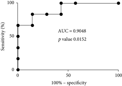
Western blot results of tubulin beta-3 chain were illustrated by box plots, and receiver operating characteristic curve (ROC curve) was created by using GraphPad Prism version 7.00 for Windows (GraphPad Software, La Jolla California USA, http://www.graphpad.com).
3. Results
3.1. Identification of Fifty-Eight Proteins Differentially Expressed with Aging by Proteomic Analysis
In order to obtain a quantitative proteomic map of elderly and young donor-derived keratinocyte cells, we used an iTRAQ labeling coupled with OFFGEL fractionation and off-line nanoLC/MS/MS as previously described [24]. The bioinformatics analysis with Paragon and Mascot search engines resulted in the identification of 517 unique proteins using a 1% FDR and considering only proteins with at least 1 peptide with confidence level ≥ 95% and score > 30. We performed a statistical analysis with the isobar package and quantified 446 proteins. Elderly keratinocytes were labeled with iTRAQ m/z 117 tag and young keratinocytes with iTRAQ m/z 113 tag. Thus, the ratio 117 : 113 (Elderly : Young) indicates the relative protein abundance between elderly and young cell samples. The complete list of identified proteins, including the UniProtKB accession number, ID, protein and gene name, peptide count, spectral count, sequence coverage, iTRAQ ratios with corresponding p value ratio and p value sample for elderly versus young cells are provided in Supplemental Data (Table S2). When the p value ratio and the p value sample were both <0.05, proteins were considered significantly differently expressed. Applying these criteria, we identified 58 proteins significantly differentially expressed depending on age status. From them, 40 were downregulated and 18 were upregulated with aging (Tables 1 and 2).
| Accession/variants | ID | Description | Gene | Peptide count | Spectral count | Sequence coverage (%) | Ratio [elderly/young] | p value rat | p value sample | Log10 ratio |
|---|---|---|---|---|---|---|---|---|---|---|
| O60814 | H2B1K_HUMAN | Histone H2B type 1-K | HIST1H2BK | 9 | 131 | 4.76 | 0.170 | 6.27E−05 | 4.36E−19 | −0.769 |
| O75334-[2-6] | LIPA2_HUMAN | Liprin-alpha-2 | PPFIA2 | 2 | 2 | 0.64 | 0.295 | 3.83E−02 | 5.16E−10 | −0.531 |
| P20674 | COX5A_HUMAN | Cytochrome c oxidase subunit 5A, mitochondrial | COX5A | 2 | 3 | 9.33 | 0.309 | 1.56E−02 | 2.23E−09 | −0.510 |
| P04732 | MT1E_HUMAN | Metallothionein-1E | MT1E | 1 | 9 | 16.39 | 0.310 | 7.22E−13 | 2.40E−09 | −0.509 |
| P05204 | HMGN2_HUMAN | Nonhistone chromosomal protein HMG-17 | HMGN2 | 4 | 11 | 8.89 | 0.330 | 1.89E−02 | 1.50E−08 | −0.482 |
| Q15075 | EEA1_HUMAN | Early endosome antigen 1 | EEA1 | 2 | 3 | 0.64 | 0.331 | 1.71E−02 | 1.61E−08 | −0.481 |
| Q9UKY7-[2] | CDV3_HUMAN | Protein CDV3 homolog | CDV3 | 3 | 16 | 11.63 | 0.342 | 1.18E−03 | 4.11E−08 | −0.466 |
| O75152 | ZC11A_HUMAN | Zinc finger CCCH domain-containing protein 11A | ZC3H11A | 1 | 1 | 1.48 | 0.349 | 2.87E−02 | 7.18E−08 | −0.457 |
| P17096 | HMGA1_HUMAN |
|
HMGA1 | 1 | 19 | 7.48 | 0.352 | 1.56E−02 | 8.83E−08 | −0.454 |
| P06454-[2] | PTMA_HUMAN |
|
PTMA | 5 | 24 | 12.61 | 0.357 | 4.01E−02 | 1.34E−07 | −0.447 |
| Q8NC51-[3] | PAIRB_HUMAN |
|
SERBP1 | 11 | 90 | 4.66 | 0.361 | 1.56E−02 | 1.82E−07 | −0.442 |
| Q13442 | HAP28_HUMAN | 28 kDa heat- and acid-stable phosphoprotein | PDAP1 | 1 | 1 | 7.18 | 0.363 | 1.23E−02 | 2.09E−07 | −0.440 |
| P63313 | TYB10_HUMAN | Thymosin beta-10 | TMSB10 | 2 | 35 | 13.64 | 0.364 | 1.30E−02 | 2.21E−07 | −0.439 |
| O00233 | PSMD9_HUMAN |
|
PSMD9 | 1 | 6 | 5.38 | 0.383 | 2.34E−04 | 8.33E−07 | −0.416 |
| P05114 | HMGN1_HUMAN | Nonhistone chromosomal protein HMG-14 | HMGN1 | 3 | 13 | 8.00 | 0.384 | 1.20E−02 | 8.80E−07 | −0.415 |
| P62158 | CALM_HUMAN | Calmodulin | CALM1 | 10 | 147 | 8.72 | 0.385 | 2.89E−02 | 9.13E−07 | −0.415 |
| P02795, P13640-[2], P80297 | MT1G_HUMAN, MT1X_HUMAN, MT2_HUMAN | Metallothionein-1G, Metallothionein-1X, Metallothionein-2 | MT1G, MT1X, MT2A | 1 | 20 | 16.29 | 0.392 | 3.61E−03 | 1.47E−06 | −0.406 |
| P67936 | TPM4_HUMAN | Tropomyosin alpha-4 chain | TPM4 | 5 | 48 | 3.23 | 0.401 | 3.64E−02 | 2.51E−06 | −0.397 |
| P22528 | SPR1B_HUMAN | Cornifin-B | SPRR1B | 4 | 56 | 8.99 | 0.405 | 2.54E−02 | 3.18E−06 | −0.392 |
| Q92538-[2,3] | GBF1_HUMAN | Golgi-specific brefeldin A-resistance guanine nucleotide exchange factor 1 | GBF1 | 1 | 1 | 0.32 | 0.406 | 4.48E−02 | 3.34E−06 | −0.391 |
| P51858 | HDGF_HUMAN | Hepatoma-derived growth factor | HDGF | 4 | 12 | 3.75 | 0.429 | 2.37E−02 | 1.17E−05 | −0.368 |
| P61604 | CH10_HUMAN | 10 kDa heat shock protein, mitochondrial | HSPE1 | 12 | 121 | 7.84 | 0.455 | 2.20E−02 | 4.16E−05 | −0.342 |
| P07108-[2-5] | ACBP_HUMAN | Acyl-CoA-binding protein | DBI | 4 | 50 | 9.20 | 0.460 | 1.86E−02 | 5.21E−05 | −0.337 |
| Q9C030-[2] | TRIM6_HUMAN | Tripartite motif-containing protein 6 | TRIM6 | 2 | 8 | 1.23 | 0.463 | 1.10E−02 | 6.03E−05 | −0.334 |
| P20962 | PTMS_HUMAN | Parathymosin | PTMS | 4 | 11 | 8.82 | 0.468 | 1.44E−02 | 7.30E−05 | −0.330 |
| Q9GZP8 | IMUP_HUMAN | Immortalization upregulated protein | IMUP | 3 | 5 | 9.43 | 0.476 | 8.05E−03 | 1.05E−04 | −0.322 |
| Q9H299 | SH3L3_HUMAN | SH3 domain-binding glutamic acid-rich-like protein 3 | SH3BGRL3 | 3 | 36 | 10.75 | 0.490 | 1.62E−02 | 1.82E−04 | −0.310 |
| P62857 | RS28_HUMAN | 40S ribosomal protein S28 | RPS28 | 3 | 20 | 17.39 | 0.491 | 9.21E−03 | 1.93E−04 | −0.309 |
| P16949-[2] | STMN1_HUMAN | Stathmin | STMN1 | 3 | 24 | 8.72 | 0.492 | 2.51E−02 | 2.00E−04 | −0.308 |
| P02765 | FETUA_HUMAN | Alpha-2-HS-glycoprotein | AHSG | 4 | 61 | 3.27 | 0.507 | 4.77E−02 | 3.38E−04 | −0.295 |
| O15212 | PFD6_HUMAN | Prefoldin subunit 6 | PFDN6 | 1 | 9 | 9.30 | 0.519 | 1.72E−02 | 5.20E−04 | −0.285 |
| P52926 | HMGA2_HUMAN | High mobility group protein HMGI-C | HMGA2 | 3 | 8 | 11.93 | 0.528 | 1.02E−04 | 7.07E−04 | −0.277 |
| P61956 | SUMO2_HUMAN | Small ubiquitin-related modifier 2 | SUMO2 | 1 | 7 | 12.63 | 0.534 | 1.89E−02 | 8.65E−04 | −0.272 |
| Q9UHV9 | PFD2_HUMAN | Prefoldin subunit 2 | PFDN2 | 1 | 5 | 9.09 | 0.544 | 1.81E−02 | 1.16E−03 | −0.265 |
| P20929-[2,3] | NEBU_HUMAN | Nebulin | NEB | 4 | 4 | 0.13 | 0.563 | 5.23E−06 | 2.03E−03 | −0.250 |
| P09429 | HMGB1_HUMAN | High mobility group protein B1 | HMGB1 | 5 | 33 | 5.58 | 0.563 | 9.96E−07 | 2.07E−03 | −0.249 |
| P62328 | TYB4_HUMAN | Thymosin beta-4 | TMSB4X | 1 | 16 | 15.91 | 0.582 | 4.60E−03 | 3.41E−03 | −0.235 |
| P09497-[2] | CLCB_HUMAN | Clathrin light chain B | CLTB | 7 | 24 | 3.49 | 0.593 | 1.87E−03 | 4.46E−03 | −0.227 |
| P35749-[2-4] | MYH11_HUMAN | Myosin-11 | MYH11 | 1 | 5 | 0.56 | 0.649 | 4.13E−02 | 1.53E−02 | −0.188 |
| Q16629-[2-4] | SRSF7_HUMAN | Serine/arginine-rich splicing factor 7 | SRSF7 | 2 | 13 | 3.78 | 0.651 | 3.09E−02 | 1.59E−02 | −0.187 |
| Accession/variants | ID | Description | Gene | Peptide count | Spectral count | Sequence coverage (%) |
Ratio [elderly/young] | p value ratio | p value sample | Log10 ratio |
|---|---|---|---|---|---|---|---|---|---|---|
| P26373 | RL13_HUMAN | 60S ribosomal protein L13 | RPL13 | 4 | 15 | 4.27 | 1.455 | 2.54E−03 | 3.04E−02 | 0.163 |
| P01861 | IGHG4_HUMAN | Ig gamma-4 chain C region | IGHG4 | 1 | 4 | 4.89 | 1.482 | 2.00E−02 | 2.46E−02 | 0.171 |
| Q13200 | PSMD2_HUMAN |
|
PSMD2 | 1 | 5 | 1.65 | 1.511 | 4.13E−02 | 1.96E−02 | 0.179 |
| P13797 | PLST_HUMAN | Plastin-3 | PLS3 | 5 | 13 | 1.90 | 1.584 | 2.13E−02 | 1.08E−02 | 0.200 |
| P49721 | PSB2_HUMAN | Proteasome subunit beta type-2 | PSMB2 | 1 | 22 | 5.47 | 1.635 | 1.82E−03 | 7.01E−03 | 0.214 |
| P48643 | TCPE_HUMAN | T-complex protein 1 subunit epsilon | CCT5 | 6 | 35 | 1.29 | 1.696 | 2.17E−03 | 4.15E−03 | 0.229 |
| P11166 | GTR1_HUMAN | Solute carrier family 2, facilitated glucose transporter member 1 | SLC2A1 | 4 | 46 | 2.03 | 1.891 | 2.60E−02 | 7.28E−04 | 0.277 |
| P61158 | ARP3_HUMAN | Actin-related protein 3 | ACTR3 | 6 | 26 | 2.63 | 2.040 | 3.21E−02 | 1.84E−04 | 0.310 |
| P09211 | GSTP1_HUMAN | Glutathione S-transferase P | GSTP1 | 10 | 117 | 7.62 | 2.308 | 3.65E−02 | 1.47E−05 | 0.363 |
| P78417 | GSTO1_HUMAN | Glutathione S-transferase omega-1 | GSTO1 | 4 | 25 | 5.81 | 2.482 | 2.89E−10 | 2.77E−06 | 0.395 |
| P13667 | PDIA4_HUMAN | Protein disulfide-isomerase A4 | PDIA4 | 3 | 4 | 1.09 | 2.633 | 2.99E−02 | 6.60E−07 | 0.420 |
| P62277 | RS13_HUMAN | 40S ribosomal protein S13 | RPS13 | 1 | 1 | 7.95 | 2.638 | 2.88E−02 | 6.27E−07 | 0.421 |
| Q13509 | TBB3_HUMAN | Tubulin beta-3 chain | TUBB3 | 4 | 19 | 4.00 | 2.664 | 4.21E−04 | 4.87E−07 | 0.426 |
| P30048 | PRDX3_HUMAN | Thioredoxin-dependent peroxide reductase, mitochondrial | PRDX3 | 1 | 1 | 5.47 | 2.769 | 3.86E−02 | 1.80E−07 | 0.442 |
| Q01813-[2] | PFKAP_HUMAN |
|
PFKP | 2 | 3 | 2.68 | 3.116 | 3.23E−02 | 6.75E−09 | 0.494 |
| P50213 | IDH3A_HUMAN | Isocitrate dehydrogenase [NAD] subunit alpha, mitochondrial | IDH3A | 3 | 3 | 2.19 | 3.239 | 6.67E−03 | 2.14E−09 | 0.510 |
| Q9Y6N5 | SQRD_HUMAN | Sulfide:quinone oxidoreductase, mitochondrial | SQRDL | 1 | 2 | 2.22 | 3.487 | 8.97E−03 | 2.17E−10 | 0.543 |
| P38606-[2] | VATA_HUMAN | V-type proton ATPase catalytic subunit A | ATP6V1A | 2 | 2 | 2.43 | 4.833 | 1.38E−02 | 1.75E−15 | 0.684 |
3.2. Gene Ontology Analysis
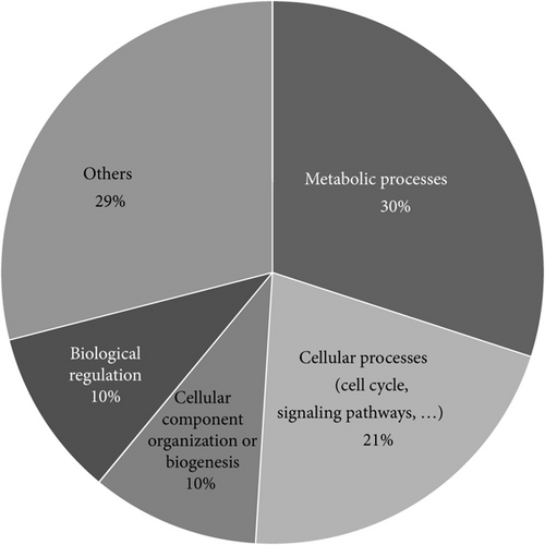
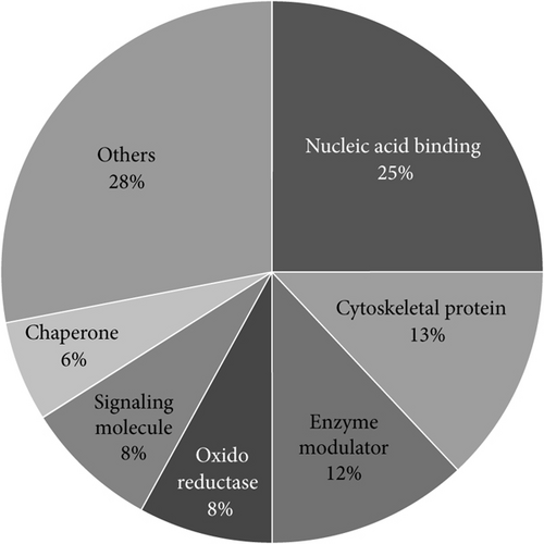
The 58 proteins previously identified were analyzed using PANTHER [22]. Their classifications into gene ontology and PANTHER categories are as follows: protein family, protein class, molecular function, biological process, cellular component, and pathway is listed in Table 3. This functional classification of biological process and PANTHER protein class is graphically illustrated in Figure 1. The main represented biological process categories are metabolism (30%); cellular process including cell cycle/cell signaling pathways/cell component movement (21%); cellular component organization/biological regulation (10%); localization/developmental process (8%); response to stimuli (4%); multicellular organismal process/immune system process (3%); and biological adhesion (1%). Concerning protein class, dysregulated proteins belong to the main following protein classes: nucleic acid binding (25%); cytoskeletal protein (13%); enzyme modulator (12%); oxidoreductase and signaling molecules (8%); chaperone (6%); transferase/transcription factor (4%); and extracellular matrix protein/hydrolase/carrier protein/membrane traffic protein/cell junction protein/kinase/isomerase/receptor (2%).
| Accession | ID | Description | Gene | PANTHER family/subfamily |
PANTHER protein class | PANTHER GO-slim molecular function |
PANTHER GO-slim biological process | PANTHER GO-slim cellular component |
Pathway |
|---|---|---|---|---|---|---|---|---|---|
| P61158 |
|
|
ACTR3 |
|
|
|
|
|
|
| P02765 |
|
Alpha-2-HS-glycoprotein | AHSG |
|
|
|
|
|
|
| P38606 |
|
|
ATP6V1A |
|
|
|
|
|
|
| P62158 |
|
Calmodulin | CALM1 |
|
Calmodulin (PC00060) |
|
|
|
|
| P48643 |
|
|
CCT5 |
|
Chaperonin (PC00072) |
|
|||
| Q9UKY7 |
|
|
CDV3 | Protein CDV3 homolog (PTHR16284:SF13) | |||||
| P09497 |
|
|
CLTB |
|
|
|
|
|
|
| P20674 |
|
|
COX5A |
|
Oxidase(PC00176) |
|
|
||
| P07108 |
|
|
DBI |
|
|
|
|
||
| Q15075 |
|
|
EEA1 |
|
|||||
| Q92538 |
|
|
GBF1 |
|
|
|
|
|
|
| P78417 |
|
|
GSTO1 |
|
|
|
|
|
|
| P09211 |
|
|
GSTP1 |
|
|||||
| P51858 |
|
|
HDGF |
|
|
|
|
||
| O60814 |
|
|
HIST1H2BK |
|
Histone (PC00171) |
|
|
|
|
| P17096 |
|
|
HMGA1 |
|
|
|
|
|
|
| P52926 |
|
|
HMGA2 |
|
|
|
|
|
|
| P09429 |
|
|
HMGB1 |
|
|
|
|||
| P05114 |
|
|
HMGN1 |
|
|
|
|
||
| P05204 |
|
|
HMGN2 |
|
|
|
|
||
| P61604 |
|
|
HSPE1 |
|
Chaperonin (PC00072) |
|
|||
| P50213 |
|
|
IDH3A |
|
|
|
|
|
|
| P01861 |
|
|
IGHG4 |
|
|||||
| Q9GZP8 |
|
|
IMUP |
|
|||||
| P04732 |
|
Metallothionein-1E | MT1E | Metallothionein-1E (PTHR23299:SF2) | |||||
| P13640 |
|
|
MT1G | Metallothionein-1G (PTHR23299:SF3) | |||||
| P35749 |
|
Myosin-11 | MYH11 |
|
|
|
|
|
|
| P20929 |
|
Nebulin | NEB | Nebulin (PTHR11039:SF37) | |||||
| Q13442 |
|
|
PDAP1 |
|
|||||
| P13667 |
|
|
PDIA4 |
|
|
|
|
||
| Q9UHV9 |
|
Prefoldin subunit 2 | PFDN2 |
|
Chaperone (PC00072) |
|
|||
| O15212 |
|
Prefoldin subunit 6 | PFDN6 | Prefoldin subunit 6 (PTHR21431:SF0) |
|
|
|
||
| Q01813 |
|
|
PFKP |
|
|
|
|
||
| P13797 |
|
Plastin-3 | PLS3 | Plastin-3 (PTHR19961:SF32) | Nonmotor actin-binding protein (PC00085) |
|
|
|
|
| O75334 |
|
Liprin-alpha-2 | PPFIA2 | Liprin-alpha-2 (PTHR12587:SF6) |
|
||||
| P30048 |
|
|
PRDX3 |
|
Peroxidase (PC00176) |
|
|
||
| P49721 |
|
|
PSMB2 |
|
|||||
| Q13200 |
|
|
PSMD2 |
|
Enzyme modulator (PC00095) |
|
|
|
|
| O00233 |
|
|
PSMD9 |
|
Enzyme modulator (PC00095) |
|
|
|
|
| P06454 |
|
|
PTMA | Prothymosin alpha (PTHR22745:SF0) |
|
||||
| P20962 |
|
Parathymosin | PTMS |
|
|
||||
| P26373 |
|
|
RPL13 |
|
Ribosomal protein (PC00171) |
|
Translation (GO:0008152) | ||
| P62277 |
|
|
RPS13 |
|
Ribosomal protein (PC00171) |
|
|
||
| P62857 |
|
|
RPS28 |
|
Ribosomal protein (PC00171) |
|
|
|
|
| Q8NC51 |
|
|
SERBP1 |
|
RNA-binding protein (PC00171) |
|
|
||
| Q9H299 |
|
|
SH3BGRL3 |
|
|||||
| P11166 |
|
|
SLC2A1 |
|
|
||||
| P22528 |
|
Cornifin-B | SPRR1B |
|
|||||
| Q9Y6N5 |
|
|
SQRDL |
|
|||||
| Q16629 |
|
|
SRSF7 | ||||||
| P16949 |
|
Stathmin | STMN1 |
|
|
|
|
|
|
| P61956 |
|
|
SUMO2 |
|
|
||||
| P63313 |
|
Thymosin beta-10 | TMSB10 |
|
|||||
| P62328 |
|
Thymosin beta-4 | TMSB4X |
|
|||||
| P67936 |
|
|
TPM4 |
|
Actin-binding motor protein (PC00085) |
|
|
|
|
| Q9C030 |
|
|
TRIM6 |
|
|||||
| Q13509 |
|
|
TUBB3 | Tubulin beta-3 chain (PTHR11588:SF43) | Tubulin (PC00085) |
|
|
|
|
| O75152 |
|
|
ZC3H11A |
|
Nucleic acid binding (PC00171) |
|
|||
Pathway enrichment analysis was performed using PathVisio [23], and the top enriched pathways are listed in Table 4. Metal homeostasis is in the top enriched pathways, as well as histone modifications, DNA replication, oxidative stress, and electron transport chain pathways.
| Pathway | Positive (r) | Measured (n) | Total | % | Z-score | p value (permuted) |
|---|---|---|---|---|---|---|
| Zinc homeostasis | 4 | 4 | 39 | 100.00% | 3.30 | 0.000 |
| Copper homeostasis | 5 | 6 | 58 | 83.33% | 3.13 | 0.000 |
| Arachidonate epoxygenase/epoxide hydrolase | 2 | 2 | 17 | 100.00% | 2.33 | 0.001 |
| DNA replication | 2 | 2 | 50 | 100.00% | 2.33 | 0.009 |
| Retinoblastoma (RB) in cancer | 2 | 2 | 98 | 100.00% | 2.33 | 0.012 |
| Histone modifications | 11 | 25 | 69 | 44.00% | 1.98 | 0.027 |
| Aryl hydrocarbon receptor | 1 | 1 | 51 | 100.00% | 1.65 | 0.048 |
| Cardiac hypertrophic response | 1 | 1 | 60 | 100.00% | 1.65 | 0.063 |
| Constitutive androstane receptor pathway | 1 | 1 | 34 | 100.00% | 1.65 | 0.029 |
| Dual hijack model of Vif in HIV infection | 1 | 1 | 9 | 100.00% | 1.65 | 0.007 |
| Endochondral ossification | 1 | 1 | 69 | 100.00% | 1.65 | 0.072 |
| Endothelin pathways | 1 | 1 | 47 | 100.00% | 1.65 | 0.047 |
| Gastric cancer network 1 | 1 | 1 | 32 | 100.00% | 1.65 | 0.027 |
| Melatonin metabolism and effects | 1 | 1 | 55 | 100.00% | 1.65 | 0.057 |
| NOTCH1 regulation of human endothelial cell calcification | 1 | 1 | 18 | 100.00% | 1.65 | 0.014 |
| Notch signaling pathway | 1 | 1 | 62 | 100.00% | 1.65 | 0.058 |
| RalA downstream regulated genes | 1 | 1 | 13 | 100.00% | 1.65 | 0.015 |
| T-cell receptor and costimulatory signaling | 1 | 1 | 45 | 100.00% | 1.65 | 0.050 |
| TarBasePathway | 1 | 1 | 19 | 100.00% | 1.65 | 0.019 |
| Type II interferon signaling (IFNG) | 1 | 1 | 38 | 100.00% | 1.65 | 0.043 |
| Metapathway biotransformation | 2 | 3 | 189 | 66.67% | 1.55 | 0.120 |
| Cytoplasmic ribosomal proteins | 13 | 34 | 89 | 38.24% | 1.54 | 0.121 |
| Circadian rythm-related genes | 5 | 11 | 210 | 45.45% | 1.40 | 0.146 |
| Apoptosis modulation and signaling | 3 | 6 | 97 | 50.00% | 1.28 | 0.190 |
| Alzheimers disease | 4 | 9 | 163 | 44.44% | 1.19 | 0.259 |
| Electron transport chain | 4 | 9 | 118 | 44.44% | 1.19 | 0.252 |
| Oxidative stress | 2 | 4 | 32 | 50.00% | 1.04 | 0.232 |
| Preimplantation embryo | 2 | 4 | 60 | 50.00% | 1.04 | 0.299 |
| Vitamin B12 metabolism | 2 | 4 | 118 | 50.00% | 1.04 | 0.337 |
| TNF alpha signaling pathway | 3 | 7 | 97 | 42.86% | 0.95 | 0.340 |
3.3. Western Blot Analysis of Tubulin Beta-3 Chain, a Promising Candidate Protein Biomarker of Aging
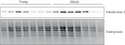
In proteomic quantitation analysis, tubulin beta-3 chain was evidenced as a promising candidate protein biomarker of aging. Indeed, tubulin beta-3 chain was upregulated in elderly donors with an iTRAQ ratio of protein expression level of 2.66 (elderly versus young) and significant p values (p value ratio of 4.21 × 10⁻⁴ and a p value sample of 4.87 × 10⁻⁷). Thus, tubulin beta-3 chain was further analyzed by western blot on human primary keratinocyte cells from the same donors but also from at least fourteen other donors in order to exclude the interindividual variability and to figure out whether or not these proteins were dysregulated in other donors. For this western blot analysis, total protein quantification was used as a control of gel loading and relative intensity of the specific antibodies on the membrane versus the amount of protein loaded on the gel was calculated and represented on box-plot diagram. Representative western blot images and results of quantification are shown in Figure 2. The trend of dysregulation of tubulin beta-3 chain was confirmed, and a similar ratio was obtained by western blot quantification and with our proteomic workflow. Moreover, receiver operating characteristic curve (ROC curve) confirmed the specificity and the sensitivity of tubulin beta-3 chain expression level for determination of age status with an area under the ROC curve of 0.9048 and a p value of 0.0152.

4. Discussion
Skin aging is a complex process with multifactorial origins that can decipher using new technological approach such as global quantitative proteomics. We carried out an iTRAQ-MALDI-TOF/TOF MS and MS/MS analysis to identify and quantify changes in human primary keratinocyte proteomes from young and elderly donors. 517 proteins were identified including proteins found mainly in keratinocytes such as cornifin-B and keratin-2E which are associated with keratinocyte activation, proliferation, and keratinization [25]. After applying robust statistical analysis, 58 proteins were found significantly differentially expressed depending on age status with 40 that were downregulated and 18 upregulated with aging.
Comparison of our results with previous gene and protein expression studies of skin aging shows some similarities. We found that more proteins are downregulated (40) than upregulated (18) with aging which is consistent with the previous results from a gene expression study in women [8]. The majority of proteins which expression is affected by age are nucleic acid-binding proteins (25%), cytoskeletal proteins (13%), and enzyme modulators (12%). These proteins are mostly involved in metabolism (30%) or cellular process (21%) such as cell cycle and cell signaling pathways. Similar results have been observed in a previous transcriptomic study [7]. Pathway enrichment analysis reveals metal (zinc and copper) homeostasis as one of the most enriched pathways which is consistent with the observed dysregulation of several metallothionein proteins that bind diverse types of metal and also participated in oxidative stress response [26]. Oxidative stress and electron transport chain pathways are also in the enriched pathways’ list, and this is consistent with the increase of reactive oxygen species (ROS) production with aging [3].
In this work, tubulin beta-3 chain expression is upregulated with aging in our proteomic experiment and also in western blot analyses on samples from several donors. Statistical analysis of these data has shown that tubulin beta-3 chain may discriminate age status. Tubulin beta-3 chain is a component of the microtubules that are complex polymers composed of tandem repeats of α- and β-tubulin heterodimers. In humans, six β-tubulin isoforms have been described and the isoform composition of the microtubule is determining its behavior. For example, it has been shown that in vitro purified microtubules enriched in tubulin beta-3 chain are more dynamic compared to microtubules containing other β-tubulin isoforms [27]. Tubulin beta-3 chain mutations have been linked in humans to different types of neurological disorders with abnormal axon guidance [28, 29]. Tubulin beta-3 was also reported as a biomarker for melanocyte lineage and as involved in melanocyte differentiation and melanogenesis [30], and it has been shown that tubulin beta-3 expression is decreased in senescent melanocytes [31]. Locher et al. [30] also reported that tubulin beta-3 may play a substantial role in melanosome transport which is regulated by proteins (such as kinesin and dynein) using microtubule tracks. Moreover, melanosomes may be transferred from melanocytes to keratinocytes through the shedding vesicle system [32], but the cellular process is still unclear and no data was reported concerning ageing aspects. Interestingly, tubulin beta-3 was identified as a direct downstream protein of human melanocortin 1 receptor (MC1R) [33], a protein associated to skin pigmentation, ultraviolet radiation, and to other aspects such as skin cancers [34]. High level of tubulin beta-3 chain has been linked to resistance to antitubulin agents such as taxanes and vinorelbine and lower overall survival in nonsmall cell lung cancer (NSCLC) [35–37] and prostate tumor patients [38]. But the prognostic value of tubulin beta-3 chain is still debated in cutaneous malignant melanoma prognosis [39]. The high expression of tubulin beta-3 chain in NSCLC has been shown to be regulated by ras, PI3/akt, and MAP kinase-ERK signaling [40]. Tubulin beta-3 chain has also been described to be a prognostic marker for bladder urothelial carcinoma with patients showing a higher level of tubulin beta-3 chain presenting a shorter disease-free survival [41]. To date, no publication reported tubulin beta-3 chain protein as a candidate biomarker for aging and there is no known mechanism of tubulin beta-3 chain dysregulation in skin aging. But it is interesting to notice that alterations of the cytoskeleton have been reported with aging [7] and that mutations in lamin genes (type V intermediate filaments) are responsible for some premature aging diseases such as the Hutchinson-Gilford progeria syndrome (HGPS) [42].
In our study, we identified other dysregulated proteins such as peroxiredoxin 3, 6-phosphofructokinase, platelet type, and cornifin-B. Thioredoxin-dependent peroxide reductase, mitochondrial, also known as peroxiredoxin 3 (PrxIII), a mitochondrial member of the antioxidant family of thioredoxin (Trx) peroxidases, was found upregulated with aging in our study. Two other family members, peroxiredoxins 1 and 2, were also upregulated in a previous report [14]. Peroxiredoxins are important cellular antioxidant; indeed, they act as hydrogen peroxide and organic hydroperoxide scavengers [43]. It is well established that with age, there is an increase in reactive oxygen species (ROS) production and a decrease in antioxidant activity both contributing to chronological aging [3]. In oxidative stress conditions, PrxIII undergoes overoxidation and subsequent irreversible inactivation. And it has been shown that in rats, this modified PrxIII form accumulates with aging [44]. In our analysis, we did not identify the peptide containing the cysteine that is overoxidized to sulfonic acid and we then cannot discriminate between the two forms, explaining why, in consequence, we observed a global upregulation of the protein. Concerning 6-phosphofructokinase, platelet type (ATP-PFK) which is upregulated with aging in our study, this enzyme catalyzes the phosphorylation of D-fructose 6-phosphate to fructose 1,6-bisphosphate by ATP, the first committing step of glycolysis. It has been shown that human primary keratinocytes derived from elderly donors showed higher glucose uptake and increased lactate production, which are the indicators of a shift in metabolism towards increased glycolysis [45]. Thus, the observed upregulation of ATP-PFK is correlated with the increased glycolysis in primary keratinocytes. Interestingly, cornifin-B is a marker of keratinocyte differentiation [46] and two transcriptomic studies reported that cornifin-B was downregulated with aging using women epidermis [9] and skin biopsies [47]. Our study is in concordance with this view as our proteomic study showed a downregulation of cornifin-B expression with aging (iTRAQ ratio erlderly versus young of 0.405 with a p value ratio of 2.54 × 10⁻² and a p value sample of 3.18 × 10⁻⁶).
Comparing our results with other studies aimed at identifying biomarkers of skin aging shows some differences that may be explained by different type/origin of skin samples, gender, difference in sample processing all along the workflow, and the variable correlation between mRNA and protein expression levels [48]. But using different and complementary approaches is interesting as it can lead to the identification of even more candidate biomarkers than with a single method. Also, considering the occurrence of oxidative stress during aging and the subsequent induced altered posttranslational modifications (PTMs) of proteins such as carbonylation, 3-nitrityrosilation, emphasis must be placed on the study of PTMs with aging in future proteomic experiment concerning skin aging [49].
5. Conclusions
Defining the differential protein signature with aging even if these changes could be initiating adaptive or compensatory events is crucial to further increase our knowledge of skin aging. Our aim was to identify some biomarker candidates for skin aging from human primary keratinocyte culture. 58 proteins were dysregulated with aging, and from this, tubulin beta-3 chain was also observed dysregulated in western blot analysis of keratinocyte extracts isolated from multiple donors. Statistical analysis has confirmed especially that an increase in tubulin beta-3 chain is associated with aging. Further studies will be needed in order to evaluate the effect of this change of expression on the complex process of aging.
This study brings a new effort to reach a better understanding of the biology of skin aging and to identify new and specific targets that could help to diagnose, prevent, and treat skin aging. Indeed, emerging diagnostic tools now require a combination of multiple biomarkers to achieve a better accuracy, and we propose that tubulin beta-3 chain could be one of these.
Conflicts of Interest
The authors declare that there is no conflict of interest regarding the publication of this paper.
Acknowledgments
The authors would like to thank Valérie Cunin, Sylvie Michelland, and David Beal for providing their help and technical assistance and Friederike Ehrhart for the help with PathVisio.


