Characterization of Lumpy skin disease virus isolated from a giraffe in Vietnam
Tung Duy Dao and Long Hoang Tran contributed equally to this work.
Abstract
While investigating a giraffe's death in a Vietnamese zoo, we successfully identified and isolated Lumpy skin disease virus (LSDV) from skin nodule biopsies and ruptured nodule wound swab samples. Phylogenetic analysis indicated that the isolate obtained in this study was closely related to the previous Vietnamese and Chinese LSDV strains from cattle. This is the first report on the genome detection and isolation of LSDV in a diseased giraffe in Vietnam. Further study is needed to better understand the epidemiology of this disease in wildlife.
Lumpy skin disease (LSD) is a disease of cattle caused by a DNA virus that belongs to the Capripoxvirus genus within the Poxviridae family (Buller et al., 2005). LSD is the most economically significant virus in the Poxviridae family as it affects domestic ruminants, leading to a reduction in milk yield, abortion, infertility in cows, a decline of the growth rate in beef cattle and death (Gari et al., 2011; Tuppurainen & Oura, 2012). LSD was first reported in sick cattle in Zambia in 1929 (MacDonald, 1931). From there, the disease spread across countries in sub-Saharan Africa (Mercier et al., 2018). In 1989, LSD was detected outside the African continent for the first time in Israel (Davies, 1980). Since then, LSD outbreaks have been frequently documented in Middle Eastern nations including Kuwait, Oman, Lebanon and Jordan (Davies, 1980; Wainwright, 2013). LSD was reported in Europe in 2015 (Agianniotaki et al., 2017; Mercier et al., 2018). It was detected in Southeast Asia (Nepal, China and India) in 2019 (Acharya & Subedi, 2020; Calistri et al., 2020). In Vietnam, the first outbreak of the disease was reported in Lang Son province in 2020 and quickly spread to 32 provinces (Personal communication). Domestic ruminants, including cattle and buffalos, are mainly affected by Lumpy skin disease virus (LSDV), but there are only a few records of infection in wildlife. In this study, we report the identification and characterization of the LSDV isolated from a giraffe (Giraffa camelopardalis) in Vietnam in 2021.
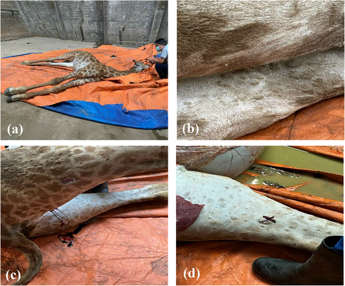
In early May 2021, a sick giraffe showing depletion, in-appetence, nasal discharge, salivation and lachrymation was reported in a zoo in a large city in northern Vietnam. The giraffe had circumscribed skin nodules that were 2–7 cm in diameter, primarily on the neck and legs (Figure 1), and died on day 9 of illness. An autopsy conducted by a veterinarian revealed many skin lesions but few lesions on its internal organs. Blood, skin nodule biopsies and ruptured nodule wound swab samples were collected from the carcass and the samples were sent to Vietnam National Institute of Veterinary Research (NIVR) for further analysis. Upon arrival at NIVR, samples were titurated to make a 10% suspension in the Minimum Essential Medium.

DNA was extracted using DNeasy Blood & Tissue Kits (Qiagen, Valencia, CA, USA) according to the manufacturer's instructions. The samples were screened by real-time PCR (rt-PCR) for detection of the LSDV genome as previously reported (Bowden et al., 2008). rt-PCR was conducted with primers CaPV-074F1, CaPV074R1 and probe CaPV-074P1 (Bowden et al., 2008). The result of rt-PCR are considered positive when the cycle threshold (Ct) value was less than 40. The results indicated that three samples, including blood, nodule biopsies and nodule swabs, were positive for the LSDV genome with Ct values at 38.20, 21.38 and 27.50, respectively (Table 1). To obtain the viral isolate, we inoculated the samples into Chorioallantoic membrane (CAM) of embryonated eggs, following previously described methods (Hierholzer & Killington, 1996). After 3 days, the virus was successfully recovered from nodule swabs and skin nodule biopsies (Table 1). Recovered viruses from CAMs were confirmed as LSDV by rt-PCR (Table 1). The virus was temporarily designated as Giraffe/Vietnam/2021.
| Real-time PCR | Isolation | |||
|---|---|---|---|---|
| Sample | Ct value | Egg | Real-time PCR confirm | PCR for sequencing |
| Blood | 38.20 | NA | NA | NA |
| Nodule swab | 27.50 | Pos | 35.47 | Pos |
| Homo. of biopsies | 21.38 | Pos | 36.86 | Pos |
- NA, not applicable; Pos, positive.
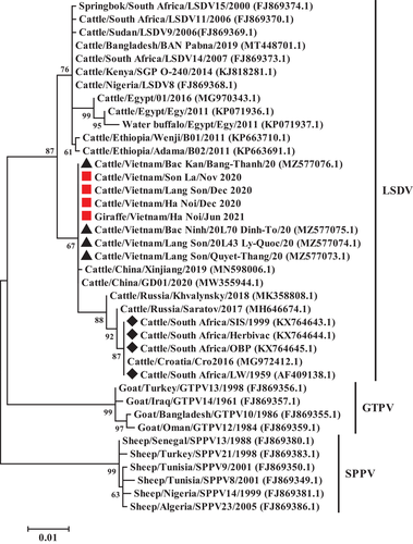
To further characterize the virus isolate from the giraffe, we compared the isolate to three LSDV isolates recovered from cattle in Hanoi, Lang Son and Son La during 2020. A PCR kit using the GoTaq Green Master Mix (Promega, Madison, WI, USA) was used to amplify the full-length GPCR gene sequence of LSDV. Two pair of primers, LSD-F1/R1 and LSD-F2/R2 (Le Goff et al., 2009), were used to amplify 1146 bp of the full-length LSDV GPCR gene sequence. Two PCR products were applied to gel-purified kit using the QIAquick Gel Extraction Kit (Qiagen, Hilden, Germany). The purification PCR products were sent to first the Base company in Singapore to sequence. All raw sequencing data were obtained from both strands of each PCR product for verification and multiply aligned by sequence analysis program Genetyx version 13.0.3 (Genetyx Corp., Tokyo, Japan). The consensus of complete GPCR gene sequences were generated from four bidirectional repeats of the two PCR products. The obtained sequences were submitted to GenBank with accession number of MZ966323 to MZ966326. Four Vietnamese LSDV GPCR gene sequences, together with representative nucleotide sequences of LSDV, sheep-pox virus (SPPV) and goat-pox virus (GTPV) available in GenBank were aligned using Cluster W (Thompson et al., 1994) in BioEdit version 7.2.5 (Hall, 1999). Phylogenetic analysis was carried out by MEGA X using maximum likelihood method with the best-fit model T92+G and 1000 bootstrap replicates (Kumar et al., 2018).
Phylogenetic analysis indicated that the sequence obtained from giraffe was closely related to other LSDV strains in Vietnam and in Xinjiang city of China. Four LSDVs isolated in Vietnam, one LSDV isolated in China together with the LSDV isolated in Russia were clustered into the same group with LSDV vaccine strains (Figure 2). The Vietnamese and Chinese LSDV strains showed a close relationship, indicating that the LSDVs circulating in Vietnam and China have the same origin. LDSVs were reported in Vietnam in 2019, however, information of GPCR gene nucleotide sequence of the LSDVs in Vietnam is very limited. Our study provided preliminary information of GPCR gene of Vietnamese LSDVs, and further contributed the data to better understand the situation of LSD in Vietnam.

It is known that LSDV has a narrow host range (Haller et al., 2014). Although the disease is endemic in most African countries and has spread to Europe, Middle East and Asia, including Vietnam (Calistri et al., 2020; Tran et al., 2021), cattle, have been primarily reported as hosts during LSD outbreaks. There are few reports of LSDV infection in wildlife but the susceptibility of wildlife was documented by detection of neutralizing antibodies (Davies, 1980; Hedger & Hamblin, 1983). Specific antibodies to LSDV were reported previously in wildlife, such as African buffalo (Syncerus caffer), blue wildebeest (Connochaetes taurinus), eland (Taurotragus oryx), Arabian oryx (Oryx leucoryx), giraffe, impala and greater kudu (Barnard, 1997; Fagbo et al., 2014; Greth et al., 1992; Hedger & Hamblin, 1983; Molini et al., 2021). These studies also suggested that wild animals may play a role in the epidemiology of LSD. Regarding LSDV infection in giraffes, experimental infection has demonstrated the susceptibility of the giraffe with LDSV (Young et al., 1970). However, susceptibility of wildlife has been relatively poorly studied and very little is known about natural infection of wildlife, especially in giraffe as mild clinical cases. The disease is easily missed because it can be difficult to monitor skin lesions. Moreover, monitoring of diseases in free-ranging wildlife populations is challenging. To our knowledge, this is the first report on a natural LSDV infection detected in giraffes. The diseased animal showed typical clinical signs of LSD, such as skin lesions, nasal and ocular discharge and salivation, which suggests that giraffes may play a role in the epidemiology of LSD. In this study, LSD-specific antibodies were not assessed due to the death of the animal and histopathological diagnosis was also not performed by the local veterinarians. Therefore, further studies should be conducted to clarify whether the disease can be induced and even lead to death in giraffes or not. This finding has also highlighted the fact that monitoring this disease in wildlife is important for determining the role of wildlife as a potential reservoir of the virus.
Recombination is one of the evolutionary processes which has been reported in LSDV from Russia (Sprygin et al., 2018) and China (Wang et al., 2020) based on sequence data of complete genomes. A previous study reported that the four Vietnamese LSDVs were most closely related to putative recombinative isolates China/GD01/2020 (MW355944), and Russia/Saratov/2017 (MH646674) based on the viral complete genome sequences (Mathijs et al., 2021). The author also suggested that the four Vietnamese LSDVs were likely generated from recombination events. Kononova et al. (2021) demonstrated that the recombinant LSDV isolate (Saratov/2017) exhibited more aggressive replication properties. In the present study, the LSDV obtained from a diseased giraffe and the four published Vietnamese recombination LSDVs belonged to a single cluster, together with Saratov/2017 and China/GD01/2020 isolates based on the full-length GPCR gene sequence. To further clarify whether the giraffe LSDV strain was generated from recombination events or not, the complete genome of the isolate would need to be sequenced and analyzed.
In summary, the present study demonstrated that LSDV was successfully recovered from samples of the giraffe in a zoo in Vietnam. This is the first report demonstrating that, in addition to experimental infection (Young et al., 1970), giraffes can be naturally infected by LSDV. Genetic analysis of the LSDV in the giraffe showed a close relationship with Vietnamese LSDVs in cattle, and highly related to LSDV isolates in China, Russia and vaccine strains. These data implied that this virus may has been circulating in domestic cattle then introduced to the zoo and threaten other animals such as giraffe. Further research on infectious disease in wildlife is needed to better understand the epidemiology of the disease and protect wildlife from the disease.
ACKNOWLEDGMENTS
We thank Ngo Thi Minh Quyen and other members of our laboratory in the Virology Department of NIVR for technical support. We also thank Dr. Kristen Kelli Coleman at Duke University and Dr. Amanda Farrell at Harvard School of Medicine for English edits and proofreading of our manuscript.
CONFLICT OF INTEREST
The authors declare that they have no conflict of interest.
Open Research
DATA AVAILABILITY STATEMENT
The data that support the findings of this study are available from the corresponding author upon reasonable request.


