Peering deeper into asthmatic lungs
Abstract
See related Article
Treating patients with asthma forms a major part of practice for most respirologists; as such, they are well acquainted with symptoms due to the underlying pathological disease drivers in asthma, a combination of airway inflammation and smooth muscle constriction leading to bronchoconstriction. Targeting airway inflammation and bronchoconstriction has been the mainstay of treatment for asthma since the 1980s, and this continues with the development of monoclonal antibodies directed against specific molecules in the inflammatory cascade, as well as the use of anti-inflammatory antibiotics.1, 2 Despite ongoing anti-inflammatory and bronchodilator therapy, there remains a significant worldwide disease burden, with over 339 million people affected, suggesting that we are still not getting to the heart of understanding asthma.
One aspect of asthma that has been modelled extensively in silico and in vitro is the proposed buckling of the internal surface of the airway during bronchoconstriction.3, 4 This anticipated buckling, especially when combined with a thickened subepithelial collagen layer in the ‘remodelled’ airways seen in asthma, is proposed to contribute to early airway closure, as well as the generation of mechanical forces when epithelial cells come into contact within airway folds. Previously, we and others demonstrated that mechanical forces induced by this projected airway folding induce airway remodelling in the absence of inflammation.5, 6 The application of compressive mechanical stress in vitro on cultured epithelial cells and ex vivo in precision-cut lung slices induces collagen production, an increase in goblet cell number and changes in smooth muscle contractility and proliferation.5, 7, 8 Many of these changes are also seen in asthmatic patients who undergo repeated induced bronchoconstriction. These findings imply a model of asthma where bronchoconstriction induces airway folding, which in turn leads to downstream changes and more difficult asthma control.
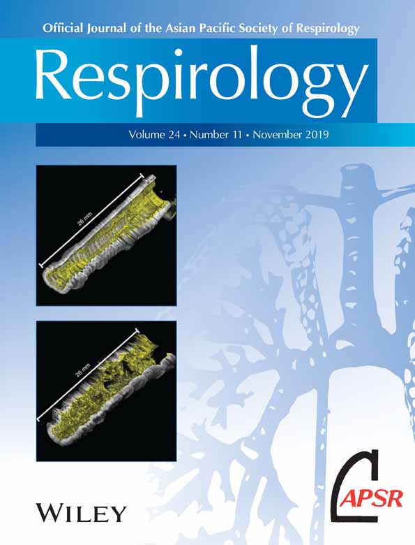
Despite these models recapitulating many of the findings seen in chronic asthma, the fundamental premise on which they are based, that small airways buckle during bronchoconstriction, has until now not been directly observed in humans; high-resolution computed tomography (CT) scanning has demonstrated irregularities only in medium-to-large airways.9 In a recent publication in Respirology, Adams et al. demonstrate mucosal buckling in the airways of allergic asthmatic patients following segmental allergen challenge using optical coherence tomography (OCT) via a bronchoscope10 OCT is routine in retinal imaging and uses light to image a few millimetres into living tissue without causing damage. The investigating team performed endobronchial OCT on a 3-cm length of the airway in the right middle and upper lobes of the study subjects, before and following segmental allergen challenge. Using this novel approach, they imaged the airway structure and quantified epithelial and mucosal thickness, as well as assessing mucosal buckling both at baseline and post-challenge. At baseline, allergic asthmatic lungs had airway structural changes when compared to allergic non-asthmatic lungs, with increases in mucosal thickness, epithelial thickness and perhaps, most intriguingly, mucosal buckling. Until now, it had been assumed that mucosal buckling, and hence mechanical force generation, on the airway epithelium only occurred during significant bronchoconstriction. The subjects in this study had near-normal baseline spirometry (mean: 90% predicted) and only withheld bronchodilators for 24 h, but despite this, mucosal buckling was present at initial bronchoscopy. The degree of baseline mucosal buckling strongly correlated with airway obstruction as measured by forced expiratory volume in 1 s/forced vital capacity (FEV1/FVC) ratio. Segmental allergen challenge resulted in an increase in mucosal and epithelial thickness, as well as inducing more mucosal buckling; this increase was still detectable 24 h after allergen challenge.10 This is the first directly observed mucosal buckling in human airways at baseline or following allergen challenge.
This study importantly demonstrates that repeatable OCT imaging of the airways via bronchoscopy is feasible and achievable, which opens the door to using this imaging modality as a marker of disease improvement following experimental early-phase interventions. In addition, the impact of therapy on airway remodelling in asthma has always been challenging to assess, but now, this may be possible longitudinally, allowing at least a correlation between asthma control, quality of life and airway remodelling. Not surprisingly, the study raises other questions. First, how good does lung function have to be in our asthmatic patients to eliminate mucosal folding? Given that it has been clearly demonstrated that the mechanical forces associated with airway buckling lead to deleterious downstream effects, the elimination of buckled airways seems a reasonable goal. Performing OCT in all patients is unfeasible, but it might be possible to assess lung function in conjunction with OCT to find the point at which mucosal buckling is eliminated. How long mucosal buckling lasts after an acute episode of bronchoconstriction is another interesting issue, and answering this question would allow more accurate in vitro modelling of airway mechanical stressors and allow us to model the impact of longer-term airway folding. Finally, although this study does not allow us to differentiate between the role of inflammation and mechanical bronchoconstriction in the generation of mucosal buckling, mucus hypersecretion or airway remodelling, it demonstrates a method through which this question might be answered. A future study might compare inflammation alone (allergen administered with bronchodilators via the bronchoscope) to inflammation plus bronchoconstriction (allergen challenge) to bronchoconstriction alone (methacholine alone), although the impact of diluent in the asthmatic group in this study may make interpreting results difficult.
OCT at bronchoscopy has been in development for many years and is now beginning to deliver exciting and translatable results. Using these and other data obtained via this technique, we should be able to model, investigate and treat asthma more effectively in the future, an outcome all respirologists and their patients will appreciate.