Influence of Signalment Variables on Body Weight-Normalized Echocardiographic Measurements of Heart Size in 56 169 Adult Unsedated Normal Pure-Bred Cats
Funding: The authors received no specific funding for this work.
ABSTRACT
Background
Echocardiography is widely used to breed-screen cats for the presence of heart disease. Left-sided cardiac dimensions are non-linearly related to body weight (BW), but the association with signalment variables is incompletely evaluated.
Objective
To validate previously published prediction equations (PE) and 95% prediction intervals (PI) and study the effects of breed, age, sex, and neutering on BW-normalized aortic (Ao), left atrial (LA) and ventricular (LV) dimensions.
Animals
56 169 pure-bred adult cats.
Methods
Data from heart screens conducted between 1999 and 2023 were included. Body-weight-(BW)-based PE and 95% PIs were obtained by allometric scaling including only cats considered normal. The effects of signalment variables on BW-normalized cardiac dimensions were examined using group-wise comparisons and uni- and multivariable analyses.
Results
The PE and PI changed marginally from those previously reported. The BW-normalized measurements showed greater variation for LV systolic than diastolic measurements (p < 0.001), and LA showed greater variation than Ao measurements. All signalment variables had small but significant effects on BW-normalized variables (p < 0.001), where the effect of breed was most prominent. None of the breeds had a variable median measurement > 10% above or below the PE, or > 10% of cats outside the PI. Signalment main effects persisted after adjusting for examiner and year of examination.
Conclusions and Clinical Relevance
Breed, age, sex, and neutering status had small and mostly clinically irrelevant effects on BW-normalized Ao, LA, and LV linear dimensions. The PE and PI intervals are valid in adult pure-bred cats across many breeds, different ages, sexes, and neutering status.
Abbreviations
-
- Ao
-
- aorta
-
- BW
-
- body weight
-
- FCM
-
- feline cardiomyopathy
-
- FS%
-
- fractional shortening
-
- GDPR
-
- general data protection regulation
-
- HCM
-
- hypertrophic cardiomyopathy
-
- HR
-
- heart rate
-
- IQD
-
- interquartile distance
-
- IQR
-
- interquartile range
-
- IVS
-
- interventricular septum systole
-
- IVSd
-
- interventricular septum diastole
-
- LA
-
- left atrium
-
- LA:Ao
-
- left atrial to aortic root diameter ratio
-
- LPI
-
- lower prediction interval
-
- LSM
-
- least square mean
-
- LV
-
- left ventricle
-
- LVFWd
-
- left ventricular free wall diastole
-
- LVFWs
-
- left ventricular free wall systole
-
- LVIDd
-
- left ventricular internal diameter diastole
-
- LVIDs
-
- left ventricular internal diameter systole
-
- PE
-
- prediction equation
-
- PI
-
- prediction interval
-
- RCM
-
- restrictive cardiomyopathy
-
- SE
-
- standard error
-
- UPI
-
- upper prediction interval
1 Introduction
Pure-bred cats are breed screened by echocardiography to ensure that breeding animals with cardiomyopathy (CM) and other heart diseases are not used for breeding [1], as these conditions are known or in other cases presumed to be inherited traits [2-8]. Although genetic tests have become available to the public, echocardiography is likely to remain a breed screen test in the foreseen future because there are currently only a few breed specific tests available [8-10]. Furthermore, they do not identify all cases with CM, even within breeds with known disease-causing genotypes, such as the Maine Coon and Ragdoll [8-10]. Thus, echocardiography remains the only viable means to breed screen cats, regardless of breed, for the presence of CM and other types of heart disease.
Diagnosis of heart disease is partly based on size assessment of different cardiac chambers and wall thickness [1, 11, 12]. Assessment may be based on subjective impression or measurements, and the current recommendation is that measurements alone are not diagnostic, but should be used to support subjective impression and the presence of other supportive findings on the echocardiogram [1, 11].
We have previously published predicted cardiac dimensions and 95% prediction intervals (PI) in cats using allometric scaling in a large cohort of cats [13]. However, in addition to BW, there are other factors that potentially might influence echocardiographic dimensions in cats that were not investigated in our previous study. These include breed, sex, neutering status, and age. Although breed-specific reference intervals have been suggested in cats, previous studies have shown conflicting results regarding the effect of breed, sex, neutering status, and age on BW-normalized echocardiographic dimensions [3, 17-19].
The aims of our study were to investigate if the prediction equations and 95% PIs are different from those previously published when including an even larger cohort of adult pure-bred cats, and to investigate the independent effects of breed, sex, neutering status, and age on BW-normalized aortic (Ao), left atrial (LA) and left ventricular (LV) linear dimensions.
2 Material and Methods
2.1 Cats
In this cross-sectional study, results from breed heart screens conducted in Europe, Australia, New Zealand, North America, and Asia between January 1st, 1999 and December 31st, 2023 within the PawPeds screening program (www.pawpeds.com) were entered into a database after an initial plausibility check of entries. In this health program, owners and screeners are instructed to only screen cats that are apparently healthy, non-pregnant, and non-lactating. Prior to the breed-screen examination, owners are required to sign an informed consent where they allow the results to be entered into the PawPeds database and that the overall result of the screen will be available to the public on the PawPeds homepage, regardless of the outcome of the screen. Because this was a retrospective study, no ethical approval was required according to Swedish Animal Welfare Legislation. Registration and protection of owner data in the database align with General Data Protection Regulation (GDPR) legislation within the European Union [20]. Cat characteristics, including BW, and results from physical and echocardiographic examinations (including use of a sedative), were included in the database. There was no specification of the type of scale used for measuring BW. Heart rate was obtained at the physical examination by cardiac auscultation. Cats were classified according to previously published recommendations [1, 11, 13] at the discretion of each examiner into diagnostic groups: normal, equivocal for LV hypertrophy and/or other findings as previously described [1, 11, 13, 21], hypertrophic cardiomyopathy (HCM), restrictive cardiomyopathy (RCM), and other cardiac diagnoses. For the purpose of this study, only unsedated cats with unremarkable echocardiograms (cats in the cardiac healthy group) were included in the database used for analyses, as was done in a previous study [13]. In cases where a cat had been subjected to repeated screens, only the most recent screen report was included in the database to obtain the most recent evaluation. Thus, details from only one screening report per cat were included in the study data set.
2.2 Echocardiography
The echocardiographic examinations were performed by veterinary screeners cooperating with the PawPeds organization and have sufficient theoretical and practical training according to published criteria (https://www.pawpeds.com/cms/index.php/en/health-programmes/hcm/vets-join-programme) and perform cat screens using ultrasound systems and transducers with acceptable resolution and frame rate for the purpose. Standard echocardiographic examinations were performed as previously described [13, 21] using criteria for diagnosis and reference ranges that agree with published guidelines [1, 11, 13]. In brief, screeners in the PawPeds program are instructed that cats should be scanned from beneath while in right lateral recumbency. The LV should be examined in 3 different two-dimensional (2D) echocardiographic views: a right parasternal long-axis LV outflow view; a right parasternal long-axis LV inflow view; and a right parasternal LV short-axis view at the level of the papillary muscles. Left ventricular dimensions should be measured from M-mode images recorded using 2D guidance from a right parasternal short-axis view at the level of the papillary muscles according to guidelines or in the same plane using 2D images [1, 11, 13]. End-diastolic measurements of LV diameter should be made at the end of diastole, just before the onset of systole, and end-systolic LV diameter measured at the nadir of septal motion, from leading edge to leading edge [22]. Simultaneous electrocardiographic (ECG) monitoring is recommended, but not mandatory. The left atrial-to-aortic root diameter ratio (LA/Ao) should be obtained from a right parasternal short-axis view in early diastole at the first frame after aortic valve closure or time the measurement to the ECG (if monitored) [1, 11, 23, 24]. M-mode and/or 2D images of the LV outflow tract should be used to identify the presence of systolic anterior motion of the mitral valve, color Doppler echocardiography should be used to interrogate for LV outflow tract obstruction, and spectral Doppler should be used to assess maximal velocities of the left and right ventricular outflow tracts.
2.3 Cat Breed Grouping
Cats were grouped into 12 breed groups: Bengal, Birman, British Longhair/Shorthair & Scottish Fold, Cornish Rex, Devon Rex, Maine Coon, Norwegian Forest Cat, Persian/Exotic, Ragdoll, Siberian/Neva Masquerade, Sphynx, and Other cats; which consisted of all other breeds with < 300 cats in each breed. Except for the Other breed group, the decisions to combine breeds were based on how closely related the breeds were [25, 26].
2.4 Statistical Methods
All statistical calculations were made using a commercially available computerized program.1 A value of p < 0.05 was considered statistically significant. Medians and percentiles were used to provide group-wise descriptive statistics for continuous variables. Differences in variables between two groups were tested using the Mann–Whitney test, or, in the case of comparisons between > 2 groups, the Kruskal–Wallis test followed by the Steel–Dwass test if the Kruskal–Wallis test was statistically significant. The Chi-squared test was used for testing the distribution of categorical variables.
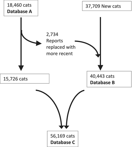
Allometric scaling was used to evaluate associations between BW and echocardiographic measurements using double-logarithmic transformations and linear curve fitting, as previously described [13]. Prediction equations (PE) and intervals (PI) of echocardiographic dimensions by BW and 95% PI by BW were created. Equations for the upper and lower PI limits were similarly estimated by double-logarithmic transformation of upper and lower PI and linear curve fitting. These calculations were made using 3 (Table S1) datasets (Figure 1): A; 1 dataset previously described including 18 460 cats [13], B; 1 dataset consisting of new reports only, and C; 1 dataset combining dataset A and B, but where reports for cats in dataset A were replaced by more recent reports from dataset B, if such existed. Measurements were normalized for BW by equations generated using dataset C and were expressed as percentage deviation from predicted value as previously described [13]. The distributions of all BW-normalized measurements were compared with regards to bias and unequal variances using the Levene test with Bonferroni correction for multiple comparisons. For each variable, the proportions of cats in each breed with BW-normalized values above the upper or below the lower PI limits, respectively, were calculated.
Curves for probability density estimation for values of BW-normalized echocardiographic variables in different groups were created by kernel smoothing, which is a non-parametric method to estimate the probability density function (Figure 2).

Hierarchical multivariable regression analysis (mixed models) was used to assess the contribution of the included signalment variables on BW-normalized variables and to address possible confounding. Two sets of analyses were done: one including sex, neutering status, age, and breed, and one including sex, age, and breed, excluding neutering status owing to a low information rate of that variable. Each BW-normalized measurement was included separately as an outcome variable in mixed models including the signalment variables breed, sex (male/female), neutering (yes/no), and age (years) modeled as fixed variables, and examiner and year of examination modeled as random variables to address possible confounding and shift over time that may occur as a consequence of improved technology and training. The distribution of the residuals from the models was investigated by normal quantile plots. The -logP value was used to assess the strength of association of each independent variable on the BW-normalized echocardiographic variables. Adjusted values for each level within each signalment variable were obtained from each model and reported as least square means (LSM) and standard error (SE) and the difference between levels was tested by the Student's t-test in case of 2 levels or the Tukey Kramer test in case of > 2 levels.
3 Results
Screen reports from 56 169 cats were included in the dataset (dataset C). This database included 15 726 reports from a previous study (database A) extended with 40 443 new reports (database B), of which 2734 were more recent reports of cats participating in the previous study (Figure 1). The cats had a median age of 1.6 years (IQR, 1.1–2.9 years) and a median BW of 4.2 kg (IQR, 3.5–5.1 kg). Female cats were more frequently represented than male (37 597 vs. 18 572 cats, p < 0.001). Information concerning sexual status was available for 68% of cats and intact cats were more common than neutered (34 771 vs. 3657 cats, p < 0.001). The database consisted of 14 151 Maine Coon, 9488 British Shorthair/Longhair/Scottish Fold, 7341 Siberian/Neva Masquerade, 6717 Norwegian Forest Cat, 5211 Ragdoll, 3484 Bengal, 2860 Sphynx, 2115 Birman, 1545 Devon Rex, 1430 Cornish Rex, 355 Persian/Exotic, and 1472 cats of other breeds. The latter group consisted of 52 different breeds of which European, Ocicat, LaPerm, and Selkirk Rex were the most commonly represented breeds (> 100 cats per breed). A total number of 151 examiners (37 board-certified cardiologists in the American or European colleges of veterinary internal medicine and 114 examiners with other cardiology training) had screened cats with a median contribution of 112 reports (IQR 39–345 reports).
3.1 Normal Prediction Formulas and 95% Prediction Intervals
Using the datasets B or C changed the constants of the PEs mainly by increasing the value of the constant b (scaling exponent) leading to small but statistically significant increases in R2 values of the models (Table 1). Furthermore, models based on datasets B or C had narrower 95% PIs as indicated by a smaller distance between upper and lower values of the constant a. Numerical values of estimates and 95% PIs by BW are presented in Table 2.
| Allometric scaling (Y = aBWb) | ||||||||||||
|---|---|---|---|---|---|---|---|---|---|---|---|---|
| Variable | Database A: Häggström et al. 2016 (n = 18 460) | Database B: new reports (n = 40 443) | Database C: combined database (n = 56 169) | |||||||||
| Prediction formula | a 95% LPI | a 95% UPI | R 2 | Prediction formula | a 95% LPI | a 95% UPI | R 2 | Prediction formula | a 95% LPI | a 95% UPI | R 2 | |
| IVSd (mm) | 2.83 × BW0.204 | 2.80 | 3.76 | 0.13 | 2.74 × BW0.223 | 2.09 | 3.58 | 0.17 | 2.77 × BW0.218 | 2.11 | 3.63 | 0.16 |
| LVIDd (mm) | 10.7 × BW0.262 | 8.45 | 13.3 | 0.28 | 10.5 × BW0.271 | 8.46 | 13.1 | 0.31 | 10.6 × BW0.270 | 8.48 | 13.2 | 0.30 |
| LVFWd (mm) | 2.63 × BW0.244 | 1.99 | 3.48 | 0.18 | 2.61 × BW0.253 | 2.02 | 3.37 | 0.22 | 2.62 × BW0.250 | 2.01 | 3.40 | 0.21 |
| IVSs (mm) | 4.41 × BW0.227 | 3.16 | 6.16 | 0.12 | 4.32 × BW0.235 | 3.16 | 5.92 | 0.13 | 4.36 × BW0.232 | 3.17 | 5.99 | 0.13 |
| LVIDs (mm) | 5.79 × BW0.261 | 3.82 | 8.80 | 0.10 | 5.55 × BW0.283 | 3.75 | 8.22 | 0.13 | 5.57 × BW0.281 | 3.76 | 8.26 | 0.13 |
| LVFWs (mm) | 4.34 × BW0.263 | 3.22 | 5.88 | 0.18 | 4.35 × BW0.262 | 3.27 | 5.79 | 0.20 | 4.36 × BW0.261 | 3.27 | 5.82 | 0.19 |
| Ao (mm) | 6.22 × BW0.277 | 4.92 | 7.87 | 0.29 | 6.25 × BW0.287 | 4.99 | 7.82 | 0.32 | 6.24 × BW0.284 | 4.97 | 7.93 | 0.31 |
| LA (mm) | 6.82 × BW0.310 | 5.12 | 9.07 | 0.25 | 6.89 × BW0.315 | 5.29 | 9.08 | 0.29 | 6.89 × BW0.313 | 5.24 | 9.08 | 0.27 |
- Note: Prediction formulas and 95% prediction intervals were calculated by allometric scaling including only cats with unremarkable echocardiograms using a dataset of 18 460 cats as previously presented (n = 18 460), a database consisting of new reports (n = 40 433) including 2734 more recent reports of cats participating in database A and using a combined dataset (n = 56 169) where the more recent reports from cats participating in database A replaced the earlier (see Figure 2). Values for the constants a and b changed slightly using the database consisting of new reports or the combined database leading to a slightly increased R2 values.
- Abbreviations: Ao, aortic diameter; IVSd, interventricular septum diastole; IVSs, interventricular septum systole; LA, left atrial diameter; LPI, lower prediction interval; LVFWd, left ventricular free wall diastole; LVFWs, left ventricular free wall systole; LVIDd, left ventricular internal diameter diastole; LVIDs, left ventricular internal diameter systole; UPI, upper prediction interval.
| Weight (kg) | IVSd (mm) | LVIDd (mm) | LVFWd (mm) | IVSs (mm) | LVIDs (mm) | LVFWs (mm) | LA (mm) | Ao (mm) | LA:Ao |
|---|---|---|---|---|---|---|---|---|---|
| 1.5 | 3.0 (2.3–4.0) | 11.8 (9.5–14.7) | 2.9 (2.2–3.8) | 4.8 (3.5–6.6) | 6.2 (4.2–9.3) | 4.9 (3.6–6.5) | 7.8 (5.9–10.3) | 7.0 (5.6–8.8) | 1.14 (8.87–1.41) |
| 2.0 | 3.2 (2.5–4.2) | 12.7 (10.2–15.9) | 3.1 (2.4–4) | 5.1 (3.7–7) | 6.8 (4.6–10.0) | 5.2 (3.9–7.0) | 8.6 (6.5–11.3) | 7.6 6.1–9.5) | 1.14 (0.87–1.42) |
| 2.5 | 3.4 (2.6–4.4) | 13.5 (10.8–16.8) | 3.3 (2.5–4.3) | 5.4 (3.9–7.4) | 7.2 (4.9–10.7) | 5.5 (4.2–7.4) | 9.2 (7.0–12.1) | 8.1 (6.5–10.2) | 1.15 (0.87–1.42) |
| 3.0 | 3.5 (2.7–4.6) | 14.2 (11.4–17.7) | 3.4 (2.6–4.5) | 5.6 (4.1–7.7) | 7.6 (5.1–11.3) | 5.8 (4.4–7.8) | 9.7 (7.4–12.8) | 8.5 (6.8–10.7) | 1.15 (0.88–1.43) |
| 3.5 | 3.6 (2.8–4.8) | 14.8 (11.9–18.4) | 3.6 (2.8–4.7) | 5.8 (4.2–8) | 7.9 (5.3–11.8) | 6.1 (4.5–8.1) | 10.2 7.8–13.4) | 8.9 (7.1–11.2) | 1.15 (0.88–1.43) |
| 4.0 | 3.7 (2.9–4.9) | 15.3 (12.3–19.1) | 3.7 (2.8–4.8) | 6.0 (4.4–8.3) | 8.2 (5.5–12.2) | 6.2 (4.7–8.4) | 10.7 (8.1–14.0) | 9.3 (7.4–11.6) | 1.16 (0.88–1.43) |
| 4.5 | 3.8 (2.9–5.0) | 15.8 (12.3–19.7) | 3.8 (2.9–5) | 6.2 (4.5–8.5) | 8.5 (5.7–12.6) | 6.5 (4.8–8.6) | 11.1 (8.4–14.6) | 9.6 (7.6–12.0) | 1.16 (0.89–1.44) |
| 5.0 | 3.9 (3.9–5.2) | 16.3 (13.1–20.3) | 3.9 (3–5.1) | 6.3 (4.6–8.7) | 8.8 (5.9–13.0) | 6.6 (5.0–8.9) | 11.4 (8.7–15.0) | 9.9 (7.9–12.4) | 1.17 (0.89–1.44) |
| 5.5 | 4.0 (3.1–5.3) | 16.7 (13.4–20.8) | 4 (3.1–5.2) | 6.5 (6.5–8.9) | 9.0 (6.1–13.3) | 6.8 (5.1–9.1) | 11.8 (8.9–15.5) | 10.1 (8.1–12.7) | 1.17 (0.89–1.44) |
| 6.0 | 4.1 (3.1–5.4) | 17.1 (13.7–21.3) | 4.1 (3.2–5.3) | 6.6 (4.8–9.1) | 9.2 (6.2–13.7) | 7.0 (5.2–9.3) | 12.1 (9.2–15.9) | 10.4 (8.3–13.0) | 1.17 (0.90–1.45) |
| 6.5 | 4.2 (3.2–5.5) | 17.5 (14–21.8) | 4.2 (3.2–5.4) | 6.7 (4.9–9.2) | 9.4 (6.4–14.0) | 7.1 (5.3–9.5) | 12.4 (9.4–16.3) | 10.6 (8.5–13.3) | 1.18 (0.90–1.45) |
| 7.0 | 4.2 (3.2–5.5) | 17.8 (14.3–22.2) | 4.3 (3.3–5.5) | 6.8 (5.0–9.4) | 9.6 (6.5–14.3) | 7.2 (5.4–9.7) | 12.7 (9.6–16.7) | 10.9 (8.7–13.6) | 1.18 (0.91–1.46) |
| 7.5 | 4.3 (3.3–5.6) | 18.2 (14.6–22.6) | 4.3 (3.3–5.6) | 7.0 (5.1–9.6) | 9.8 (6.6–14.6) | 7.4 (5.5–9.9) | 13.0 (9.9–17.1) | 11.1 (8.8–13.8) | 1.18 (0.91–1.46) |
| 8.0 | 4.3 (3.3–5.7) | 18.5 (14.8–23) | 4.4 (3.4–5.7) | 7.1 (5.1–9.7) | 10.0 (6.7–14.8) | 7.5 (5.6–10.0) | 13.2 (10.1–17.4) | 11.3 (9.0–14.1) | 1.19 (0.91–1.46) |
| 8.5 | 4.4 (3.4–5.8) | 18.8 (15.1–23.4) | 4.5 (3.4–5.8) | 7.2 (5.2–9.8) | 10.2 (6.9–15.1) | 7.6 (5.7–10.2) | 13.5 (12.3–17.8) | 11.5 (9.1–14.4) | 1.19 (0.92–1.47) |
| 9.0 | 4.5 (3.4–5.9) | 19.1 (15.3–23.8) | 4.5 (3.5–5.9) | 7.3 (5.3–10.0) | 10.3 (7.0–15.3) | 7.7 (5.8–10.3) | 13.7 (10.4–18.1) | 11.7 (9.3–14.6) | 1.20 (0.92–1.47) |
| 9.5 | 4.5 (3.5–5.9) | 19.4 (15.5–24.1) | 4.6 (3.5–6) | 7.3 (5.3–10.1) | 10.5 (7.1–15.6) | 7.9 (5.9–10.5) | 14.0 (10.6–18.4) | 11.8 (9.4–14.8) | 1.20 (0.92–1.47) |
| 10.0 | 4.6 (3.5–6.0) | 19.6 (15.8–24.5) | 4.7 (3.6–6.1) | 7.4 (5.4–10.2) | 10.6 (7.2–15.8) | 8.0 (6.0–10.6) | 14.2 (10.8–18.7) | 12.0 (9.6–15.1) | 1.20 (0.93–1.48) |
| 10.5 | 4.6 (3.5–6.1) | 19.9 (16–24.8) | 4.7 (3.6–6.1) | 7.5 (5.5–10.3) | 10.8 (7.3–16.0) | 8.1 (6.0–10.8) | 14.4 (11.0–19.0) | 12.2 (9.7–15.3) | 1.21 (0.93–1.48) |
| 11.0 | 4.7 (3.6–6.1) | 20.1 (16.2–25.1) | 4.8 (3.7–6.2) | 7.6 (5.5–10.5) | 10.9 (7.4–16.2) | 8.2 (6.1–10.9) | 14.6 (11.1–19.3) | 12.3 (9.8–15.5) | 1.21 (0.94–1.48) |
| PI as % deviation from predicted | −24 to 31 | −20 to 25 | −23 to 30 | −27 to 37 | −33 to 48 | −25 to 33 | −24 to 32 | −20% to 25% | NA |
- Abbreviations: Ao, aortic diameter; FS%, fractional shortening; IVSd, interventricular septum diastole; IVSs, interventricular septum systole; LA, left atrial diameter; LA/Ao, left atrial to aortic root ratio; LVFWd, left ventricular free wall diastole; LVFWs, left ventricular free wall systole; LVIDd, left ventricular internal diameter diastole; LVIDs, left ventricular internal diameter systole.
3.2 Variation of Body-Weight Normalized Measurements
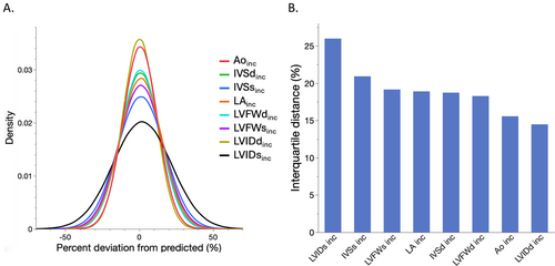
Using the formulas in Table 1 of dataset C, measurements in all cats were BW-normalized. The median deviation from predicted value ranged between the variables from −0.09% (LVIDdinc) to 1.5% (LVIDsinc). The variation differed between the variables in all comparisons (p < 0.001) and was lowest in LVIDdinc (IQR −7.2% to 7.3%) and highest in LVIDsinc (IQR −11.3% to 14.7%; Figure 3). The LV systolic measurements had generally a greater variation than diastolic measurements, and LA measurements had greater variation than Ao measurements.
3.3 Effects of Measuring on 2D-Images and Type of Cardiology Training
Left ventricular dimensions were measured using 2D images in 4496 cats. The wall dimensions were greater in diastole compared to expected values median (IQR) IVSdinc 4.1% (−4.5% to 13.7%) and LVFWdinc 5.3% (−3.4% to 14.1%), but smaller in systole median (IQR) IVSsinc −0.1% (−9.6% to 19.5%) and LVFWsinc −2.3% (−10.7% to 8.6%). The LV diameter was smaller in diastole compared to expected values median (IQR) LVIDdinc −3.0 (−10.2% to 4.0%) but greater in systole median (IQR) LVIDsinc 1.6% (−11.0% to 14.4%; p < 0.001 for all comparisons). The proportions of cats measured by 2D images above or below the PI ranged between 0.1% (IVSd) and 7.5% (LVFWs).
The distribution of examiner median values and interquartile distances (IQD) for each BW-normalized measurement revealed only minor differences between ACVIM/ECVIM board-certified cardiologists and examiners with other cardiology training. However, board-certified cardiologists demonstrated examiner median values that were slightly, yet statistically significantly, shifted towards higher median values of BW-normalized Ao and LA diameters (p < 0.05). Additionally, a broader range of examiner IQD was observed for BW-normalized values of LA (Figure S1).
3.4 Effect of Sex and Neutering
Male cats had a higher BW and were slightly younger than female cats (median (IQR) female vs. male BW and age, respectively, 3.9 kg (3.3–4.5 kg) vs. 5.2 kg (4.4–6.2 kg), and 1.64 years. (1.1–2.9 years) vs. 1.49 years. (1.0–2.8 years), both p < 0.0001), and were more commonly neutered (female vs. male: 6.7% vs. 15.1%). Male cats had slightly greater LV BW-normalized measurements, but the Ao diameters were similar, and males had smaller LA diameters (Table 3).
| Variable | Sex | Neutering | ||
|---|---|---|---|---|
| Male (N = 12 915) | Female (N = 25 513) | Intact (N = 34 771) | Altered (N = 3657) | |
| IVSd inc (%) | 3.5 (−6.0 to 12.3)a | 0.15 (−9.3 to 9.2)b | 1.4 (−8.1 to 10.4)a | 4.8 (−4.0 to 14.0)b |
| LVIDd inc (%) | 0.85 (−6.1 to 8.1)a | −0.62 (−7.8 to 6.8)b | 0.01 (−7.1 to 7.3)a | −2.3 (−9.6 to 4.5)b |
| LVFWd inc (%) | 2.7 (−6.4 to 11.3)a | −0.41 (−9.4 to 8.8)b | 0.62 (−8.3 to 9.6)a | 3.4 (−6.2 to 12.3)b |
| IVSs inc (%) | 2.5 (−8.0 to 13.3)a | 0.15 (−10 to 10.6)b | 0.64 (−9.3 to 11.1a | 4.2 (−6.1 to 15.1)b |
| LVIDs inc (%) | 2.7 (−10.2 to 15.6)a | 0.99 (−11.8 to 14.2)b | 1.9 (−10.8 to 14.8a | −4.5 (−16.8 to 8.5)b |
| LVFWs inc (%) | 2.0 (−7.5 to 12.0)a | −0.31 (−17.4 to 9.5)b | 0.06 (−9.0 to 10.0)a | 3.7 (−6.3 to 9.6)b |
| LA inc (%) | 0.35 (−8.4 to 10.7)a | 0.25 (−7.4 to 8.3)a | 0.62 (−8.5 to 10.5)a | −1.1 (−10.3 to 8.3)b |
| AO inc (%) | 0.17 (−8.8 to 9.9)a | −0.85 (−8.4 to 10.7)b | 0.37 (−7.1 to 8.3)a | −0.28 (−7.7 to 7.4)b |
- Note: Within each row in the sex respectively neutering column, values with the same superscript letter did not differ significantly.
- Abbreviations: Ao, aortic diameter; IVSd, interventricular septum diastole; IVSs, interventricular septum systole; LA, left atrial diameter; LVFWd, left ventricular free wall diastole; LVFWs, left ventricular free wall systole; LVIDd, left ventricular internal diameter diastole; LVIDs, left ventricular internal diameter systole.
Neutered cats were older and had a higher BW than intact cats (median (IQR) neutered vs. intact 3.8 years. (2.2–5.8 years) vs. 1.5 years. (1.1–2.7 years) and 4.1 kg (3.5–5.0 kg) vs. 3.5 kg (4.1–6.0 kg), p < 0.0001). Neutered cats had slightly greater LV wall thicknesses, but had smaller BW-normalized LV, aortic, and LA diameters (Table 3).
Similar statistically significant differences between the sexes and neutering status persisted in all comparisons over the different cat breed groups.
3.5 Effect of Age
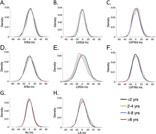
Older cats were heavier than younger cats, where the BW increased from a median (IQR) of 4.1 kg (3.5–4.9 kg) in cats < 2 years to 4.8 kg (3.8–6.0) kg in cats > 10 years, and, comparing the same age groups, were furthermore more likely to be neutered with proportions increasing from 3.6% to 75% (p < 0.001 for all comparisons). Age had a small effect on BW-normalized echocardiographic measurements, where measurements of systolic and diastolic LV wall thickness, Ao, and LA diameters increased with increasing age, and LV systolic and diastolic diameters decreased with increasing age (all p < 0.0001; Figure 4). The constants (i.e., change per year) for age when modeled as a continuous variable in the univariate models ranged from 0.53 (LAinc) to −1.0 (LVIDsinc) and had an R2 ranging from 0.0018 (IVSsinc) to 0.0076 (LVIDsinc).
3.6 Effect of Breed
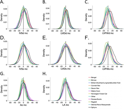
Breed affected all examined echocardiographic variables (p < 0.001), but the differences between breeds were comparably small (Table 4 and Figure 5). The following extremes (median deviating ±10% from the median for all breeds) could be identified in relation to the median for all cats: Persian/Exotic, Birman, Sphynx, Norwegian Forest Cat, and Other breeds were older, and Devon Rex and Ragdoll were younger. Maine Coon cats had a higher BW, and Sphynx, Persian, Birman, Cornish and Devon Rex, and Other cat breeds had lower BW. Sphynx cats had higher HR. Except for the Sphynx cats, none of the breeds had any median value in LV diastolic or systolic wall thickness deviating ±10% from predicted values. In the Sphynx cats, the median IVSdinc, LVFWdinc, and IVSsinc were slightly below 10%, and LVFWsinc was slightly above 10%. The Persian and Bengal cats each had one median measurement > 5% of predicted values (IVSdinc and LVWdinc, respectively). Left ventricular diameter in diastole was within ±5% of predicted values for all breeds, whereas the median systolic diameter was > 5% higher than predicted values in Norwegian Forest Cat, Maine Coon, and Devon Rex cats. Ragdoll cats had median Aoinc and LAinc < −5%, and Maine Coon cats had a median LAinc > 5%. None of the breeds deviated more than ±5% in median values from the overall median for FS and LA/Ao. The proportions of cats within each breed above or below the PI (Table S1) were generally < 5% with the following exceptions: Ragdoll cats had LVIDd, LVFWs, Ao, and LA measurements lower than the lower PI limit in proportions ranging from 5.1% to 5.6%. Maine Coon and British Shorthair/Longhair/Scottish Fold cats had 6.1% and 5.8% cats with lower than the lower PI limit concerning IVSd and LVIDs, respectively. In Sphynx cats, the proportions of cats above the upper PI limit were found to be 6.2% and 8.9% concerning LVFWd and LVFWs, respectively.
| Variable | Bengal (N = 3484) | Birman (N = 2115) | BSH/BLH and Scottish fold (N = 9488) | Cornish Rex (N = 1430) | Devon Rex (N = 1545) | Maine Coon (N = 14 151) | Norwegian Forest Cat (N = 6717) | Persian/Exotic (N = 355) | Ragdoll (N = 5211) | Sibirian/Neva Masquerade (N = 7341) | Sphynx (N = 2862) | Other (N = 1472) |
|---|---|---|---|---|---|---|---|---|---|---|---|---|
| Age (years) | 1.6 (1.1 to 2.7)a | 2.0 (1.2 to 3.4)b,c | 1.6 (1.1 to 2.9)d,e | 1.7 (1.1 to 2.9)a,d,e | 1.2 (1.0 to 2.0)f | 1.5 (1.1 to 2.8)a.e | 1.9 (1.2 to 3.3)c | 2.4 (1.5 to 3.9)b | 1.2 (1.0 to 2.0)f | 1.5 (1.1 to 2.9)a.e | 2.0 (1.2 to 3.4)c,f | 1.8 (1.2 to 3.0)d,f |
| Body weight (kg) | 4.0 (3.5 to 4.9)a | 3.4 (3.0 to 3.9)b | 4.0 (3.5 to 4.9)a | 3.1 (2.6 to 3.6)c | 3.0 (2.6 to 3.6)c | 5.1 (4.4 to 6.3)d | 4.5 (3.9 to 5.4)e | 3.4 (3.0 to 3.9)b | 4.0 (3.5 to 4.7)a | 4.1 (3.5 to 4.9)a | 3.7 (3.1 to 4.4)f | 3.7 (3.2 to 4.3)f |
| Heart rate (BPM) | 176 (160 to 189)a | 180 (165 to 192)b | 180 (165 to 198)c | 190 (170 to 210)d | 190 (170 to 210)d | 180 (162 to 200)c | 180 (160 to 200)e | 180 (160 to 200)b,c | 178 (160 to 189)a | 170 (156 to 188)e | 200 (180 to 226)f | 180 (163 to 200)c |
| IVSd (mm) | 3.9 (3.5 to 4.3)a | 3.6 (3.3 to 4.1)b,c | 3.7 (3.3 to 4.1)d | 3.6 (3.3 to 4.0)b,e | 3.5 (3.0 to 3.5)c | 4.0 (3.5 to 4.4)a | 4.0 (3.6 to 4.3)a | 3.8 (3.4 to 4.2)d,f | 3.7 (3.4 to 4.0)e,f | 3.8 (3.5 to 4.2)f | 4.0 (3.7 to 4.4)g | 3.7 (3.4 to 4.1)d,f |
| IVSd inc (%) | 4.6 (−4.7 to 14.1)a | −0.49 (−9.1 to 8-0)b,c | −0.4 (−10.4 to 9.3)c | 2.8 (−5.8 to 11.6)d | 1.0 (−7.1 to 9.2)d,e,f | 0.3 (−10.2 to 9.7)c | 2.4 (−7.0 to 11.2)d,e | 6.2 (−3.7 to 15.7)a | −1.6 (−10.0 to 6.6)g | 1.1 (−7.1 to 9.2)f | 9.5 (0.6 to 18.1)h | 1.2 (−8.8 to 10.0)b,e,f |
| LVIDd (mm) | 15.3 (14.1 to 16.7)a | 14.3 (13.3 to 15.5)b | 15.0 (13.9 to 16.3)c | 14.4 (13.2 to 15.7)b,d | 14.9 (13.8 to 16.1)c,e | 17.0 (15.6 to 18.4)f | 16.3 (15.0 to 17.6)g | 14.8 (13.4 to 16.0)d,e | 14.8 (13.8 to 16.0)e | 15.2 (14.0 to 16.4)f | 15.8 (14.6 to 17.0)g | 15.1 (14.0 to 16.5)c,f |
| LVIDd inc (%) | −0.6 (−7.5 to 6.4)a | −3.2 (−9.7 to 3.4)b | −2.6 (−9.2 to 4.2)b,c | −0.5 (−6.8 to 7.8)a | 4.6 (−1.9 to 11.6)d | 2.5 (−4.6 to 9.8)e | 2.2 (−4.9 to 9.3)e | −0.6 (−8.8 to 7.2)a,f | −4.4 (−10.5 to 2.2)g | −2.2 (−9.0 to 4.9)c,f | 4.5 (−2.6 to 11.8)d | −0.01 (−7.7 to 7.6)a |
| LVFWd (mm) | 3.9 (3.5 to 4.3)a | 3.5 (3.2 to 3.9)b | 3.7 (3.3 to 4.1)c | 3.6 (3.2 to 3.9)b | 3.4 (3.1 to 3.7)d | 4.0 (3.6 to 4.4)e | 3.9 (3.5 to 4.3)a | 3.6 (3.2 to 4.0)b,c | 3.5 (3.2 to 3.9)b | 3.8 (3.4 to 4.1f) | 4.0 (3.6 to 4.3)e | 3.6 (3.3 to 4.0)c |
| LVFWd inc (%) | 5.2 (−4.0 to 13.6)a | −0.9 (−8.7 to 8.2)b,c,d | 0.5 (−9.9 to 8.4)c,d | 2.5 (−6.4 to 12.8)e | −1.7 (−9.0 to 6.2)d | 0.9 (−9.0 to 9.9)b | 1.3 (−7.8 to 10.3)f | 1.8 (−9.3 to 11.2)b,c,e,f | −4.7 (−12.1 to 4.2)g | 0.5 (−7.9 to 8.9)b,f | 8.8 (0.1 to 18.2)h | 0–5 (−8.2 to 9.7)b,f |
| IVSs (mm) | 6.2 (5.6 to 7.0)a | 5.8 (5.1 to 6.4)b | 6.1 (5.5 to 6.8)c | 5.8 (5.2 to 6.5)b,d,e | 5.8 (5.1 to 6.4)b.e | 6.4 (5.7 to 7.2)f | 6.3 (5.7 to 7.0)g | 6.0 (5.4 to 6.7)c.d,h | 5.8 (5.4 to 6.7)d.e | 6.0 (5.4 to 6.7)h | 6.4 (5.8 to 7.1)f | 6.0 (5.3 to 6.7)h |
| IVSs inc (%) | 3.1 (−6.9 to 13.7)a | −1.0 (−10.8 to 9.3)b | 1.2 (−8.5 to 11.2)c | 3.0 (−7.6 to 11.2)a | 2.1 (−7.9 to 11.2)a,c | 0.4 (−10.2 to 11.6)d | 2.2 (−8.1 to 12.6)a,c | 4.4 (−7.2 to 14.7)a,c | −3.5 (12.5 to 6.2)e | −0.5 (−10.4 to 9.5)b | 8.2 (−2.1 to 20.0)f | 1.2 (−9.4 to 11.7)c,d |
| LVIDs (mm) | 8.4 (7.2 to 9.5)a | 7.8 (6.8 to 8.8)b | 7.9 (6.9 to 8.9)c | 7.6 (6.6 to 8.6)a.b,d | 8.0 (7.0 to 9.1)e,f | 9.3 (8.1 to 10.6)e | 9.1 (8.0 to 10.4)g | 8.0 (7.0 to 9.0)a,b,d | 8.2 (7.2 to 9.2)b | 8.2 (7.2 to 9.4)b,d | 8,4 (7.3 to 9.6)f | 8.2 (7.0 to 9.3)a,d |
| LVIDs inc (%) | 1.6 (−11.5 to 13.7)a | −1.9 (−14.0 to 11.3)b | −4.6 (−16.5 to 7.4)c | −1.2 (−14.0 to 12.4)a,b,d | 5.1 (−7.4 to 19.4)e,f | 5.5 (−7.9 to 18.5)f | 7.7 (−6.0 to 20.8)e | 0.8 (−12.1 to 12.8)a,b,d | −1.1 (−12.4 to 10.6)b | −0.4 (−12.7 to 12.2)b.d | 4.0 (−9.4 to 18.3)f | 1.0 (−12.5 to 14.0)a.d |
| LVFWs (mm) | 6.3 (5.7 to 7.0)a | 6.0 (5.4 to 6.7)b | 6.4 (5.7 to 7.0)c | 6.1 (5.5 to 6.7)b,d | 5–9 (5.2 to 6.5)e | 6.8 (6.1 to 7.5)f | 6.5 (5.9 to 7.2)g | 6.0 (5.4 to 6.7)b,d,e | 5.9 (5.4 to 6.5)e | 6.2 (5.4 to 6.5)a,h | 6.9 (6.2 to 7.5)f | 6.2 (5.6 to 6.9)d,h |
| LVFWs inc (%) | −0.4 (−9.1 to 9.2)a | −0.1 (−8.8 to 9.0)a,b,c | 0.9 (−7.6 to 10.8)c,d | 4.3 (−5.9 to 14.1)e | 0.9 (−7.9 to 10.7)b,c,d | 1.6 (−8.0 to 11.4)d | 0.3 (−9.0 to 9.7)a,b | 0.0 (−9.6 to 10.0)a,b,c,d,f | −5.8 (−13.8 to 2.9)g | −1.5 (−10.3 to 7.9)f | 11.6 (1.4 to 22.4)g | 0.8 (−8.4 to 10−4)a,b,c,d |
| FS (%) | 45 (40 to 51)a | 46 (40 to 51)a | 47 (42 to 53)b | 47 (42 to 53)b,c | 46 (42 to 52)a,c | 46 (41 to 51)d | 43 (38 to 49)e | 46 (40 to 51)a,c,d | 45 (40 to 50)d | 45 (40 to 51)a | 47 (41 to 52)c | 46 (40 to 51)a,c |
| LA (mm) | 10.8 (9.8 to 11.9)a | 9.9 (9.0 to 10.7)b | 10.9 (9.9 to 12.0)a | 9.9 (9.0 to 10.9)b | 9.4 (8.6 to 10.3)c | 12.1 (11.0 to 13.5)d | 11.1 (10.0 to 12.3)e | 10.3 (9.4 to 11.3)f,g | 10.0 (9.1 to 11.0)g | 10.4 (9.4 to 11.4)f | 10.6 (9.7 to 11.8)h | 10.4 (9.5 to 11.6)f |
| LA inc (%) | 0.4 (−8.3 to 9.9)a | −3.0 (−11.0 to 4.5)b | 1.8 (−6.9 to 11.1)c | 0.2 (−8.4 to 9.5)a | −2-1 (−10.9 to 6.0)b | 5.3 (−4.2 to 15.0)d | 0.9 (−8.5 to 10.5)a | 1.2 (−8.5 to 10.8)a,c | −6.3 (−14.0 to 2.4)e | −3.1 (−11.5 to 5.6)a | 2.4 (−7.1 to 12.9)c | −0.1 (−9.2 to 10.4)a |
| Ao (mm) | 9.1 (8.5 to 10.0)a | 8.6 (8.0 to 9.2)b | 9.6 (8.7 to 10.4)c | 8.9 (8.1 to 9.5)d | 8.4 (7.8 to 9.0)e | 10.4 (9.5 to 11.3)f | 9.5 (8.8 to 10.3)c | 9.2 (8.3 to 9.9)a.g,h | 8.8 (8.1 to 9.5)d | 9.0 (8.3 to 9.5)g | 9.0 (8.4 to 9.9)a,h | 9.0 (8.2 to 9.9)g,h |
| AO inc(%) | −1.1 (−7.5 to 5.6)a | −3.4 (−9.1 to 3.5) b | 3.2 (−4.9 to 11.2)c,d | 2.5 (−4.7 to 9.0)c | −1.8 (−8.2 to 5.3)a | 4.5 (−3.4 to 12.3)e | −0.4 (−7.9 to 6.9)a | 4.6 (−4.8 to 14.1)d,e | −5.5 (−11.9 to 1.1)f | −3.4 (−10.2 to 3.7)b | 1.8 (−5.2 to 9.5)g | 0.5 (−6.5 to 8.1)a |
| LA:Ao | 1.16 (1.07 to 1.26)a | 1.12 (1.05 to 1.21)b,c | 1.12 (1.04 to 1.22)b,c | 1.11 (1.02 to 1.20)d | 1.11 (1.03 to 1.20)c.d | 1.16 (1.06 to 1.27)a | 1.16 (1.07 to 1.26)a | 1.11 (1.00 to 1.20)c,d | 1.12 (1.04 to 1.22)b | 1.13 (1.05 to 1.24)e | 1.14 (1.06 to 1.25)a | 1.14 (1.05 to 1.24)a,e |
- Note: Within each row, values with the same superscript letter did not differ significantly.
- Abbreviations: Ao, aortic diameter; BLH, British Longhair; BSH, British Shorthair; IVSd, interventricular septum diastole; IVSs, interventricular septum systole; LA, left atrial diameter; LVFWd, left ventricular free wall diastole; LVFWs, left ventricular free wall systole; LVIDd, left ventricular internal diameter diastole; LVIDs, left ventricular internal diameter systole.
3.7 Multivariable Analysis
Hierarchical multivariable mixed models showed that all included signalment variables remained statistically significant in the analyses (Tables S2a,b). However, breed was the signalment variable with the strongest association to the BW-normalized values, whereas the associations of sex, neutering status, and age were considerably less pronounced. The LSMs from the multivariable analyses adjusting for examiner and year of examination showed a similar pattern to the univariable analyses. Concerning the BW-normalized LV wall measurements in systole and diastole (IVSdinc, LVFWdinc, IVSsinc and LVFWdinc), Sphynx cats had the greatest wall thicknesses compared to any of the other breeds, with LSM slightly above or close to 10% from predicted values (Tables S3a and S3b), regardless of whether neutering status was included in the model or not. The LSMs of LV diameter in diastole or systole, aortic, and LA diameters were within ±10% of predicted values for all breeds, regardless of whether neutering was included in the models or not. Male cats and neutered cats had thicker LV walls and greater LV diameters in diastole and systole compared to female and intact cats, respectively (Table S4), but differences were small. Similar to the univariate analyses, LV wall thickness increased and LV diameters decreased with increasing age (Table S5). The LSM for the model constants indicated that the effect of age per year was generally smaller compared to the univariate analyses and did not change substantially if neutering status was excluded.
4 Discussion
The present study is the largest published study of standard echocardiographic measurements in cats. The study showed that expanding the database with > 40 000 cat screens only marginally changed the prediction equations and marginally decreased the 95% PI. Breed was the signalment variable exhibiting the strongest effect on BW-normalized dimensions, but this effect was comparably small. Although normal reference ranges have been published for specific breeds separately [3, 17, 18, 27-31], a similar interbreed comparison has, to the authors knowledge, not been previously published. Sex, neutering, and age had a clinically unimportant effect on BW-normalized echocardiographic measurements. The PEs and 95%PIs are therefore valid in the hands of many examiners for adult pure-bred cats of different breeds, regardless of sex, neutering status, and age.
Some of the previous studies of normal standard echocardiographic measurements in cats have included a considerably smaller study group than the present and included scans performed by one or a few examiners, often working at single centers [3, 17, 18, 27-32]. However, reference intervals need to be valid in the hands of many and reflect measurements obtained under clinical conditions. Including measurements obtained by many examiners from many centers is beneficial to achieve large samples and increase generalizability, but may also increase the variation because of interobserver variation and bias and may introduce other random variation (noise) influencing the results [33]. Creating PEs and reliable PIs, therefore, requires large study groups and examinations performed by many examiners to be valid in the hands of many [33]. According to the Law of Large Numbers, the predicted value and PIs will converge towards true values, if they exist, provided independent and identical random samples [34]. Although the database of the present study is unlikely to completely fulfill these criteria, it can be assumed that the predicted values and PIs of the present study are likely to be close to true values in a cohort similar to the present. Furthermore, regardless of many sources of variation in the present study, the PIs of the present study are narrower than any previous study, again an effect of the number of included cats.
Using the PE, the distance between observed and predicted values could be calculated for each included cat and expressed as percentage deviation. The distribution of such LV BW-normalized systolic measurements showed systematically a greater variation compared to diastolic. In particular, the LVIDsinc showed the greatest variation. This is in agreement with some previous studies in cats [17, 32]. However, in these studies, the intra- and interobserver variations have been expressed as coefficients of variation (CV). The present database includes single measurements performed at one time by a single observer, which does not allow such detailed characterization of the variation. During the LV contraction, the heart base is pulled towards the apex as a consequence of the longitudinal contraction. This means that the imaging plane shifts towards the base during LV contraction, and this motion may cause more variable measurements in systole.
While this study was not designed to study the agreement between M-mode and 2D-obtained measurements of the LV walls, it showed that measuring the LV dimensions from 2D-echocardiographic images did not substantially alter the results. In other words, none of the LV dimensions, other than LVFWdinc, had median values that deviated > 5% from the expected values, and the proportions of observations outside the PI intervals were comparably small (all < 5% except for LFWsinc). Likewise, this study was not designed to study the agreement between board-certified cardiologists and examiners with other cardiology training. However, it indicated that, given the many sources of variation, the two groups provided measurements in normal cats that had similar distributions of examiner median values and IQDs, with the exception of BW-normalized Ao and LA diameters where board-certified cardiologists measured greater values and had a broader range of IQDs regarding LA diameter measurements.
The present study showed that the median percentage deviation for all echocardiographic measurements was < 10% or close to 10% (in Sphynx cats), and < 10% of the cats were either above or below the upper or lower PI limits in each breed. The breeds that presented the highest median values of LV wall thickness in diastole included the Sphynx and the Bengals. Although the PIs were valid for the Sphynx cats, the breed stands out by having slightly thicker BW-normalized diastolic walls (medians approximately 10% above predicted values) compared to other breeds. Echocardiographic dimensions have previously been studied in this breed and shown to be comparable to those found in domestic cats [27]. Although we currently have no explanation for the findings in Sphynx cats in the present study, it can be speculated that the hypotrichia and the body conformation with large ears and long legs may have indirectly an effect on the heart by leading to comparably high heat dissipation from the body surface, as shown in dogs [35]. Theoretically, this could lead to a compensatory increased metabolism to maintain normal body temperature. However, the absence of findings similar to those in the Sphynx breed in Devon Rex and Cornish Rex, that is, breeds with similar body conformation and little fur, speaks against this theory.
Differences were found for male versus female cats and for neutered versus intact cats for all BW-normalized echocardiographic variables, except for Ao diameter. Males had greater LV diameters, but smaller LAs, although the differences were numerically small and differences were < 5% between the sexes for all variables. The findings concerning LV dimensions are in agreement with previous studies showing that male cats have slightly bigger hearts after BW-normalization [17]. We have no obvious explanation for the smaller BW-normalized LA measurements found in male versus female cats, but one potential explanation could be a slight overcorrection of BW in male cats that had a higher BW. The argument against this is that BW-normalized Ao diameters were not different between male and female cats. Neutered cats had smaller BW-normalized LV diameters, Ao and LA diameters, but thicker LV walls compared to intact cats. Again, the differences were numerically small and the differences generally < 5%. Neutered cats were older and heavier than intact cats, which suggests that these differences could be caused by overcorrection for BW. However, the fact that LV wall thicknesses were greater in neutered cats speaks against the idea that the differences were entirely caused by overcorrection.
In addition to BW, one other clinical variable that differed between the neutering status groups was age; neutered cats were older than intact cats. The BW was found to increase with increasing age; age had small, but comparable effects on BW-normalized LV measurements as neutering, where LV cavity measurements became smaller and LV wall thicknesses greater with advancing age. However, BW-normalized Ao and LA measurements increased with increasing age, which means that the differences found between neutered versus intact cats cannot be explained by the age difference. Surprisingly little information is available in the literature concerning age-related changes of cardiac dimensions in normal cats, although there are Doppler echocardiographic studies suggesting that LV diastolic function declines with age in normal cats [36]. It can be speculated that the findings of increasing Ao and LV diameters with increasing age to some extent might be explained by subclinical age-related cardiovascular and renal changes causing increased systolic arterial blood pressure and increased ventricular stiffness.
A multicenter study, such as ours, in the field of echocardiographic measurements of cardiac size aims to improve the generalizability and external validity of findings by pooling data from multiple institutions. However, although the present study included a large cohort of cats examined by 151 examiners, this study, like any other similarly designed study, was susceptible to confounding factors that can influence the accuracy and reliability of the results and impact estimates of the effects of the signalment variables [37, 38]. Factors that could have introduced confounding in our study include variation in equipment, examiner skill and practices, cat characteristics, and the comparably long time period between the first cat examined and the last (1999–2023) [38]. There are several ways to address the problem with confounding, and in the present study, we chose hierarchical multivariable regression analyses adjusting for examiner and year of examination. This approach is often preferred over other statistical methods in studies including many variables in the model, or many levels (strata), or both, within variables [38], the latter being the case in the present study. The outcomes of these analyses were that the results remained comparably unchanged.
In a massive study group, as in the present study, small clinically irrelevant differences can become statistically significant. Therefore, it is more meaningful to interpret the findings by quantifying them and evaluating them with regard to their clinical importance [39]. Many of the effects of the signalment variables on BW-normalized measurements were small and are clinically irrelevant. For example, the differences in medians between the sexes or neutering groups were < 5% of predicted values for all BW-normalized measurements. If we put < 5% deviation from predicted value into actual numbers for a cat weighing 4 kg with a predicted IVSd and LVIDd of 3.7 and 15.3 mm, it equals approximately < 0.2 and < 0.8 mm, respectively. These differences are so small that they are very difficult to identify in the individual cat, and accordingly drown in the overall variation incurred with imaging and measuring the cardiac dimension. Likewise, with the exception of Sphynx cats, the majority of median BW-normalized measurements were < 5%, which means that the effect of breed is not a major contributor to the overall variation. Applying the example above of a 4 kg Sphynx cat with values deviating < 10% from predicted values, the deviation for IVSd equals < 0.4 mm and for LVIDd < 1.6 mm, respectively. These numbers are also comparably small, and it can be argued that they are clinically irrelevant. However, in some situations, systematic differences may become more relevant; for example, when the measurements are in the vicinity of upper or lower PI. In these situations, it is relevant to reassess the measurement and put its value into the context of subjective impression, supportive findings, and quality of the acquisition.
5 Limitations
This study comprises pure-bred cats examined at the will of their owner for breeding purposes. Most of the cats were comparably young at the time of examination, which means that the examined cohort of cats was skewed towards comparably young cats. The body condition score was not included in the analyses mainly because this variable was not included in the screen report form until comparably recently. Therefore, some overweight or lean cats were likely included, which might impact the results. The study was based on many echocardiographic examinations performed by many examiners with different cardiology training using different echocardiographic systems, which is both a strength and a weakness. The present study provides estimates of overall variation, but does not allow in-depth characterization of the variation, such as inter-nd intraobserver variation. Screeners provided different numbers of screen reports, which means the contribution from each examiner was different to the overall result. However, owing to the sheer number of cats, the contribution of each examiner was small and the main findings regarding the effects of the signalment variables remained after having adjusted for the effect of examiner and year of examination. Some cats were examined without ECG guidance for timing of measurements, which may contribute to increased variation of measurements. Sedatives and tranquilizers are allowed to be administered prior to scans in the Pawpeds screening program, but cats receiving such drugs were excluded in the present study owing to their different impact on the cardiovascular system.
6 Conclusions
Increasing the study group considerably only changed the BW-based PE and PI marginally from those previously reported. The BW-normalized measurements showed a greater variation for LV systolic than diastolic measurements, and LA greater variation than Ao measurements. Breed, age, sex, and neutering status had small and mostly clinically irrelevant effects on BW-normalized Ao, LA, and LV linear dimensions. The PE and PI intervals are valid in adult pure-bred cats across many breeds, different ages, sexes, and neutering status.
Acknowledgments
The authors thank all colleagues associated to the PawPeds health program for their contribution to the heart screening, which made this study possible. We also thank all the participating cat owners and the people who voluntary work on a non-profit basis registering the screening results.
Disclosure
Authors declare no off-label use of antimicrobials.
Ethics Statement
Authors declare no institutional animal care and use committee or other approval was needed. Authors declare human ethics approval was not needed.
Conflicts of Interest
The authors declare no conflicts of interest.


