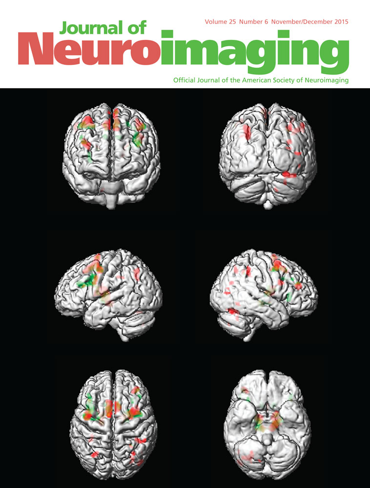
Statistical Instability of TBSS Analysis Based on DTI Fitting Algorithm
Corresponding Author
Ivan I. Maximov
Institute of Neuroscience and Medicine—4, Forschungszentrum Jülich GmbH, 52425 Jülich, Germany
Present address: Experimental Physics III, TU Dortmund University, 44221 Dortmund, Germany.
Correspondence: Address correspondence to Ivan I. Maximov, Institute of Neuroscience and Medicine—4, Forschungszentrum Jülich GmbH, 52425 Jülich, Germany. E-mail: [email protected]Search for more papers by this authorHeike Thönneßen
Institute of Neuroscience and Medicine—4, Forschungszentrum Jülich GmbH, 52425 Jülich, Germany
Department of Child and Adolescent Psychiatry and Psychotherapy, RWTH Aachen University, 52074 Aachen, Germany
Search for more papers by this authorKerstin Konrad
Department of Child and Adolescent Psychiatry and Psychotherapy, RWTH Aachen University, 52074 Aachen, Germany
Institute of Neuroscience and Medicine—3, Forschungszentrum Jülich GmbH, 52425 Jülich, Germany
JARA–BRAIN-Translational Medicine, RWTH Aachen University, 52074 Aachen, Germany
Search for more papers by this authorLaura Amort
Institute of Neuroscience and Medicine—4, Forschungszentrum Jülich GmbH, 52425 Jülich, Germany
Department of Psychiatry, Psychotherapy and Psychosomatics, RWTH Aachen University, 52074 Aachen, Germany
Search for more papers by this authorIrene Neuner
Institute of Neuroscience and Medicine—4, Forschungszentrum Jülich GmbH, 52425 Jülich, Germany
Department of Psychiatry, Psychotherapy and Psychosomatics, RWTH Aachen University, 52074 Aachen, Germany
JARA–BRAIN-Translational Medicine, RWTH Aachen University, 52074 Aachen, Germany
Search for more papers by this authorN. Jon Shah
Institute of Neuroscience and Medicine—4, Forschungszentrum Jülich GmbH, 52425 Jülich, Germany
Department of Neurology, RWTH Aachen University, 52074 Aachen, Germany
JARA–BRAIN-Translational Medicine, RWTH Aachen University, 52074 Aachen, Germany
Search for more papers by this authorCorresponding Author
Ivan I. Maximov
Institute of Neuroscience and Medicine—4, Forschungszentrum Jülich GmbH, 52425 Jülich, Germany
Present address: Experimental Physics III, TU Dortmund University, 44221 Dortmund, Germany.
Correspondence: Address correspondence to Ivan I. Maximov, Institute of Neuroscience and Medicine—4, Forschungszentrum Jülich GmbH, 52425 Jülich, Germany. E-mail: [email protected]Search for more papers by this authorHeike Thönneßen
Institute of Neuroscience and Medicine—4, Forschungszentrum Jülich GmbH, 52425 Jülich, Germany
Department of Child and Adolescent Psychiatry and Psychotherapy, RWTH Aachen University, 52074 Aachen, Germany
Search for more papers by this authorKerstin Konrad
Department of Child and Adolescent Psychiatry and Psychotherapy, RWTH Aachen University, 52074 Aachen, Germany
Institute of Neuroscience and Medicine—3, Forschungszentrum Jülich GmbH, 52425 Jülich, Germany
JARA–BRAIN-Translational Medicine, RWTH Aachen University, 52074 Aachen, Germany
Search for more papers by this authorLaura Amort
Institute of Neuroscience and Medicine—4, Forschungszentrum Jülich GmbH, 52425 Jülich, Germany
Department of Psychiatry, Psychotherapy and Psychosomatics, RWTH Aachen University, 52074 Aachen, Germany
Search for more papers by this authorIrene Neuner
Institute of Neuroscience and Medicine—4, Forschungszentrum Jülich GmbH, 52425 Jülich, Germany
Department of Psychiatry, Psychotherapy and Psychosomatics, RWTH Aachen University, 52074 Aachen, Germany
JARA–BRAIN-Translational Medicine, RWTH Aachen University, 52074 Aachen, Germany
Search for more papers by this authorN. Jon Shah
Institute of Neuroscience and Medicine—4, Forschungszentrum Jülich GmbH, 52425 Jülich, Germany
Department of Neurology, RWTH Aachen University, 52074 Aachen, Germany
JARA–BRAIN-Translational Medicine, RWTH Aachen University, 52074 Aachen, Germany
Search for more papers by this authorABSTRACT
Voxel-based DTI analysis is an important approach in the comparison of subject groups by detecting and localizing gray and white matter changes in the brain. One of the principal problems for intersubject comparison is the absence of a “gold standard” processing pipeline. As a result, contradictory results may be obtained from identical data using different data processing pipelines, for example, in the data normalization or smoothing procedures. Tract-based spatial statistics (TBSS) shows potential to overcome this problem by automatic detection of white matter changes and decreasing variation in the performed analysis. However, skeleton projection approaches, such as TBSS, critically depend on the accuracy of the diffusion scalar metric estimations. In this work, we demonstrate that the agreement and reliability of TBSS results depend on the applied DTI data processing algorithm. Statistical tests have been performed using two in vivo measured datasets and compared with different implementations of the least squares algorithm. As a result, we recommend repeating TBSS analysis using different fitting algorithms, in particular, using on iteratively-assessed robust estimators, as accurate and more reliable approach in voxel-based analysis, particularly, for TBSS. Repeating TBSS analysis allows one to detect and localize suspicious regions in white matter which were estimated as the regions with significant difference. Finally, we did not find a favorite fitting algorithm (or class of them) which can be marked as more reliable for group comparison.
Supporting Information
Disclaimer: Supplementary materials have been peer-reviewed but not copyedited.
| Filename | Description |
|---|---|
| jon12215-sup-0001-Supp.doc2.3 MB | Fig S1. Visualization of regions obtained by common TBSS results based on different fitting algorithms: (a) only least squares approaches were used; (b) only robust approaches were used. In (a) and (b) the red isosurface corresponds to the overlapping region, the green isosurface represents regions not overlapping but considered by at least one of the algorithms as an area with significant differences, and the partly transparent blue isosurface is the mean FA skeleton. (c) Shows a schematic explanation of the color coding for (a) and (b). (d) Shows the overlapping regions from (a) (red isosurface) and (b) (blue isosurface) together. An animation of all 3-dimensional objects is available in the online Supplementary Material. Fig S2. Comparing the results of TBSS analysis based on MD images obtained by the 10 fitting algorithms for Dataset II. The regions of decreased MD in the adult group are marked in blue-light blue. The grayscale background is an MNI152 T1-weighted image. The mean skeleton is marked in green. The coordinates are presented in MNI152 space and equal to X = 21, Y = −25, and Z = 44. Fig S3. Visualization of regions obtained by common TBSS results based on different fitting algorithms: (a) only least squares approaches were used; (b) only robust approaches were used. In (a) and (b) the red isosurface corresponds to overlapping region, green isosurface presents regions not overlapping. In (c), overlapping regions from (a) (red isosurface) and (b) (blue isosurface) are shown together. An animation of all3-dimensional objects is available in the online Supplementary Material. Table S1. Volumetric Comparison and Reliability Estimations Based on ICC of Mean Skeletons for Dataset I and II. |
Please note: The publisher is not responsible for the content or functionality of any supporting information supplied by the authors. Any queries (other than missing content) should be directed to the corresponding author for the article.
References
- 1Johansen-Berg H, Behrens TEJ. Diffusion MRI. From quantitative measurement to in vivo neuroanatomy. London, Burlington, San Diego: Elsevier, 2009.
- 2Jones DK. Diffusion MRI. Theory, methods, and applications. Oxford, New York: Oxford University Press, 2011.
- 3Basser PJ, Mattiello J, Le Bihan D. MR diffusion tensor spectroscopy and imaging. Biophys J 1994; 66: 259-67.
- 4Basser PJ, Mattiello J, Le Bihan D. Estimation of effective self-diffusion tensor from the NMR spin echo. J Magn Reson B 1994; 103: 247-54.
- 5Le Bihan D. Molecular diffusion, tissue microdynamics and microstructure. NMR Biomed 1995; 8: 375-86.
- 6Pierpaoli C, Basser PJ. Toward a quantitative assessment of diffusion anisotropy. Magn Reson Med 1996; 36: 893-906.
- 7Ennis DB, Kindlmann G, Rodriguez I, et al. Visualization of tensor fields using superquadric glyphs. Magn Reson Med 2005; 53: 169-76.
- 8Kindlmann G, Westin CF. Diffusion tensor visualization with glyph packing. IEEE Trans Vis Comp Graphics 2006; 12: 1329-35.
- 9Westin CF, Maier SE, Mamata H, et al. Processing and visualization of diffusion tensor MRI. Med Image Anal 2002; 6: 93-108.
- 10Kingsley PB. Introduction to diffusion tensor imaging mathematics: part I. Tensors, rotations, and eigenvectors. Conc Magn Reson 2006; 28A: 101-22.
- 11Kingsley PB. Introduction to diffusion tensor imaging mathematics: Part II. Anisotropy, diffusion-weighted factors, and gradient encoding schemes. Conc Magn Reson 2006; 28A: 123-54.
- 12Westin CF, Peled S, Gudbjartsson H, et al. Geometrical diffusion measures for MRI from tensor basis analysis. Proc Intl Soc Magn Reson Med 1997; 5.
- 13Assaf Y. Can we use diffusion MRI as a bio-marker of neurodegenerative processes? Bioessay 2008; 30: 1235-45.
- 14Beaulieu C. The basis of the anisotropic water diffusion in the nervous system—a technical review. NMR Biomed 2002; 15: 435-55.
- 15Hasan KM, Sankar A, Halphen C, et al. Development and organization of the human brain tissue compartments across the lifespan using diffusion tensor imaging. Neuroreport 2007; 18: 1735-9.
- 16Lebel C, Walker L, Leemans A, et al. Microstructural maturation of the human brain from childhood to adulthood. NeuroImage 2008; 40: 1044-55.
- 17Lebel C, Gee M, Camicioli R, et al. Diffusion tensor imaging of white matter tract evolution over the lifespan. NeuroImage 2012; 60: 340-52.
- 18Le Bihan D. The “wet mind”: water and functional neuroimaging. Phys Med Biol 2007; 52: R57-R90.
- 19Thomason ME, Thompson PM. Diffusion imaging, white matter, and psychopathology. Ann Rev Clin Psychol 2011; 7: 63-85.
- 20Cercignani M, Inglese M, Pagani E, et al. Mean diffusivity and fractional anisotropy histograms of patients with multiple sclerosis. Am J Neuroradiol 2001; 22: 952-8.
- 21Cercignani M, Bammer R, Sormani M, et al. Inter-sequence and inter-imaging unit variability of diffusion tensor MR imaging histogram-derived metrics of the brain in healthy volunteers. Am J Neuroradiol 2003; 24: 638-43.
- 22Ashburner J, Friston KJ. Voxel-based morphometry—the methods. NeuroImage 2000; 11: 805-21.
- 23Foong J, Symms MR, Barker GJ, et al. Investigating regional white matter in schizophrenia using diffusion tensor imaging. Neuroreport 2002; 13: 333-6.
- 24Buchel C, Raedler T, Sommer M, et al. White matter asymmetry in the human brain: a diffusion tensor MRI study. Cereb. Cortex 2004; 14: 945-51.
- 25Ashburner J, Friston KJ. Generative and recognition models for neuroanatomy. NeuroImage 2004; 23: 21-4.
- 26Jones DK, Cercignani M. Twety-five pitfalls in the analysis of diffusion MRI data. NMR Biomed. 2010; 23: 803-20.
- 27Van Hecke W, Leemans A, Sage CA, et al. The effect of template selection on diffusion tensor voxel-based analysis results. NeuroImage 2011; 55: 566-73.
- 28Jones DK, Symms M, Cercignani M, et al. The effect of filter size on VBM analyses of DT-MRI data. NeuroImage 2005; 26: 546-54.
- 29Smith SM, Jenkinson M, Johansen-Berg H, et al. Track-based spatial statistics: voxelwise analysis of multi-subject diffusion data. NeuroImage 2006; 31: 1487-1505.
- 30Moraschi M, Hagberg GE, Paola MD, et al. Smoothing that does not blur: effects of the anistropic approach for evaluating diffusion tensor imaging data in the clinic. J Magn Reson Imaging 2010; 31: 690-7.
- 31Van Hecke W, Leemans A, Backer SD, et al. Comparing isotropic and anisotropic smoothing for voxel-based DTI analyses: a simulation study. Human Brain Mapping 2010; 31: 98-114.
- 32Jones DK, Chitnis XA, Job D, et al. What happens when nine different groups analyze the same DT-MRI data set using voxel-based methods? Proc Intl Magn Reson Med 2007; 15: 74.
- 33Zhang H, Avants BB, Yushkevich PA, et al. High-dimensional spatial normalization of diffusion tensor images improves the detection of white matter differences: an example study using amyotrophic lateral sclerosis. IEEE Trans Med Imaging 2007; 26: 1585-97.
- 34Melonakos ED, Shenton ME, Rathi Y, et al. Can whole brain voxel-based morphometry studies applied to DTI data localize white matter changes in schizophrenia? Schizophrenia Bull 2009; 35: 202-3.
- 35Melonakos ED, Shenton ME, Rathi Y, et al. Voxel-based morphometry (VBM) studies in schizophrenia—can white matter changes be reliably detected with VBM? Psychiatr Res: Neuroimaging 2011; 193: 65-70.
- 36Van Hecke W, Sijbers J, Backer SD, et al. On the construction of a ground truth framework for evaluating voxel-based diffusion tensor MRI analysis methods. NeuroImage 2009; 46: 692-707.
- 37Smith SM, Johanse-Berg H, Jenkinson M, et al. Acquisition and voxelwise analysis of multi-subject diffusion data with track-based spatial statistics. Nature Protocols 2007; 2: 499-503.
- 38Smith SM, Nichols TE. Threshold-free cluster enhancement: addressing problems of smoothing, threshold dependence and localization in cluster inference. NeuroImage 2009; 44: 83-98.
- 39Edden RA, Jones DK. Spatial and orientational heterogeneity in statistical sensitivity of skeleton-based analysis of diffusion tensor MR imaging data. J Neurosci Methods 2011; 201: 213-9.
- 40Holdsworth SJ, Aksoy M, Newbould R, et al. Diffusion tensor imaging (DTI) with retrospective motion correction for large-scale pediatric imaging. J Magn Reson Imaging 2012; 36: 961-71.
- 41Jones DK, Knoesche T, Turner R. White matter integrity, fiber count, and other fallacies: the do's and don'ts of diffusion MRI. NeuroImage 2012; 73: 239-54.
- 42Truong TK, Chen NK, Song AW. Dynamic correction of artifacts due to susceptibility effects and time-varying eddy currents in diffusion tensor imaging. NeuroImage 2011; 57: 1343-7.
- 43Irfanoglu MO, Walker L, Sarlls J, et al. Effects of image distortions originated from susceptibility variations and concomitant fields on diffusion MRI tractography results. NeuroImage 2012; 61: 275-88.
- 44Leemans A, Jones DK. The B-matrix must be rotated when correcting for subject motion in DTI data. Magn Reson Med 2009; 62: 1336-49.
- 45Vos SB, Jones DK, Viergever MA, et al. Partial volume effect as a hidden covariate in DTI analyses. NeuroImage 2011; 55: 1566-76.
- 46Jbabdi S, Behrens TEJ, Smith SM. Crossing fibres in tract-based spatial statistics. NeuroImage 2010; 49: 249-56.
- 47Zalesky A. Moderating registration misalignment in voxelwise comparisons of DTI data: a performance evolution of skeleton projection. Magn Reson Imaging 2011; 29: 111-25.
- 48Yushkevich PA, Zhang H, Simon TJ, et al. Structure-specific statistical mapping of white matter tracts. NeuroImage 2008; 41: 448-61.
- 49Goodlett CB, Fletcher PT, Gilmore JH, et al. Group analysis of fiber tract statistics with application to neurodevelopment. NeuroImage 2010; 45: S133-S142.
- 50Zalesky A, Fornito A, Harding IH, et al. Whole-brain anatomical networks: does the choice of nodes matter? NeuroImage 2010; 50: 970-83.
- 51Zalesky A, Fornito A, Bullmore ET. Network-based statistics: identifying differences in brain networks. NeuroImage 2010; 53: 1197-1207.
- 52Szczepankiewicz F, Lätt J, Wirestam R, et al. Variability in diffusion kurtosis imaging: impact on study design, statistical power and interpretation. NeuroImage 2013; 76: 145-54.
- 53Pfefferbaum A, Adalsteinsson E, Sullivan EV. Repricability of diffusion tensor imaging measurements of fractional anisotropy and trace in brain. J Magn Reson Imaging 2003; 18: 427-33.
- 54Heievang E, Behrens TEJ, Mackay CE, et al. Between session reproducibility and between subject variability of diffusion MR and tractography measures. NeuroImage 2006; 33: 867-77.
- 55Veenith TV, Carter E, Grossac J, et al. Inter subject variability and reproducibility of diffusion tensor imaging within and between different imaging sessions. PLoS One 2013; 8: e65941.
- 56Vollmar C, O'Muircheartaigh J, Barker GJ, et al. Identical, but not the same: intra-site and inter-site reproducibility of fractional anisotropy measures on two 3.0T scanners. NeuroImage 2010; 51: 1384-94.
- 57Zhu T, Hu R, Qui X, et al. Quantification of accuracy and precision of multi-center DTI measurements: a diffusion phantom and human brain study. NeuroImage 2011; 56: 1398-1411.
- 58Hakulinen U, Brander A, Ryymin P, et al. Repeatability and variation of region-of-interest methods using quantitative diffusion tensor MR imaging of the brain. BMC Med Imaging 2012; 12: 30.
- 59Veraart J, Sijbers J, Sunaert S, et al. Weighted linear least squares estimation of diffusion MRI parameters: strengths, limitations, and pitfalls. NeuroImage 2013; 81: 335-46.
- 60Smith SM, Jenkinson M, Woolrich MW, et al. Advances in functional and structural MR image analysis and implementation as FSL. NeuroImage 2004; 23: 208-19.
- 61Leemans A, Jeurissen B, Sijbers J, et al. ExploreDTI: a graphical toolbox for processing, analyzing, and visualizing diffusion MR data. Proc Intl Magn Reson Med 2009; 17: 3537.
- 62Cook PA, Bai Y, Nedjati-Gilani S, Seunarine KK, et al. Camino: open-source diffusion-MRI reconstruction and processing. Proc Intl Soc Magn Reson Med 2006; 14: 2759.
- 63Maximov II, Grinberg F, Shah NJ. Robust tensor estimation in diffusion tensor imaging. J Magn Reson 2011; 213: 136-44.
- 64Neuner I, Kupriyanova Y, Stoecker T, et al. White-matter abnormalities in Tourette syndrome extend beyond motor pathways. NeuroImage 2010; 51: 1184-93.
- 65Neuner I, Kupriyanova Y, Stoecker T, et al. Microstructure assessment of grey matter nuclei in adult Tourette patients by diffusion tensor imaging. Neurosci Lett 2011; 487: 22-6.
- 66Behrens TEJ, Woolrich MW, Jenkinson M, et al. Characterization and propagation of uncertainty in diffusion-weighted MR imaging. Magn Reson Med 2003; 50: 1077-88.
- 67Woolrich MW, Jbabdi S, Patenaude B, et al. Bayesian analysis of neuroimaging data in FSL. NeuroImage 2009; 45: S173-186.
- 68Rohde GK, Barnett AS, Basser PJ, et al. Comprehensive approach for correction of motion and distortion in diffusion-weighted MRI. Magn Reson Med 2004; 51: 103-14.
- 69Sijbers J, den Dekker A. Maximum likelihood estimation of signal amplitude and noise variance from MR data. Magn Reson Med 2004;51: 586-94.
- 70Aja-Fernandez S, Tristan-Vega A, Hoge WS. Statistical noise analysis in GRAPPA using a parameterized noncentral chi approximation model. Magn Reson Med 2011; 65: 1195-1206.
- 71Smith SM. Fast robust automated brain extraction. Human Brain Mapping 2002; 17: 143-55.
- 72Nichols TE, Holmes AP. Nonparametric permutation tests for functional neuroimaging: a primer with examples. Human Brain Mapping 2002; 15: 1-25.
- 73Plaisier A, Pieterman K, Lequin MH, et al. Choice of diffusion tensor estimation approach affects fiber tracktography of the fornix in preterm brain. Am J Neuroradiol 2014; 35: 1219-25.
- 74Chang L-C, Jones DK, Pierpaoli C. RESTORE: robust estimation of tensors by outlier rejection. Magn Reson Med 2005; 53: 1088-95.
- 75Bach M, Laun FB, Leemans A, et al. Methodological consideration on tract-based spatial statistics (TBSS). NeuroImage 2014; 100: 358-69.
- 76Schmithorst V, Wisnowski J, Panigrahy A. TBSS may be sub-optimal for detection of DTI parameter changes in crossing fiber regions. Proc Intl Soc Magn Reson Med 2013; 21: 3191.
- 77Madhyastha T, Merillat S, Hirsiger S, et al. Longitudinal reliability of tract-based spatial statistics in diffusion tensor imaging. Human Brain Mapping 2014; 35: 4544-55.
- 78Keihaninejad S, Ryan NS, Malone IB, et al. The importance of group-wise registration in tract based spatial statistics study of neurodegeneration: a simulation study in Alzheimer's disease. PLoS One 2012; 7: e45996.
- 79Schwarz CG, Reid RI, Gunter JL, et al. The Alzheimer's disease neuroimaging initiative. NeuroImage 2014; 94: 65-78.
- 80Bells S, Dustan L, McGonigle DJ, et al. On the stability of skeletong-based analyses of diffusion tensor MRI-based measures. Proc Intl Soc Mag Reson Med 2012; 20: 1863.


