Extracellular vesicle transfer of cancer pathogenic components
Funding Information:
This work was supported in part by a Grant-in-Aid for the Japan Science and Technology Agency (JST) through the Center of Open Innovation Network for Smart Health (COINS) initiated by the Council for Science and a Grant-in-Aid for Basic Science and Platform Technology Program for Innovative Biological Medicine, and a Grant-in-Aid for Project for Development of Innovative Research on Cancer Therapeutics (P-Direct).
Abstract
Extracellular vesicles (EV), known as exosomes and microvesicles, serve as versatile intercellular communication vehicles. Increasing evidence has shown that cancer cell-derived EV carry pathogenic components, such as proteins, messenger RNA (mRNA), microRNA (miRNA), DNA, lipids and transcriptional factors, that can mediate paracrine signaling in the tumor microenvironment. These data suggest that EV transfer of cancer pathogenic components enable long-distance crosstalk between cancer cells and distant organs, resulting in the promotion of the initial steps for pre-metastatic niche formation. Understanding the metastatic mechanisms through EV transfer may open up a new avenue for cancer therapeutic strategies. Furthermore, the circulating EV have also been of interest as a source for liquid biopsies. EV in body fluids provide a reliable source of miRNA and proteins for cancer biomarkers. The tumor-specific components in EV effectively provide various messages on the physiological and pathological status of cancer patients. Although many researchers are searching for EV biomarkers using miRNA microarrays and proteome analyses, the detection technology for circulating EV in body fluids has not yet reached the point of clinical application. In this review, we summarize recent findings regarding EV function, specifically in metastasis through the transfer of cancer pathogenic components. Furthermore, we highlight the potential of using circulating EV for cancer diagnosis.
Extracellular vesicles (EV) are heterogeneous populations of membrane vesicles that are released in the extracellular spaces by most cells, including tumor cells. These cells can secrete EV of different sizes, components and subcellular origin. Numerous reports now identify EV as important mediators of extracellular signaling pathways through direct membrane transfer of their cargo. EV components, which may include proteins, messenger RNA (mRNA), microRNA (miRNA), DNA, lipids and transcriptional factors, are capable of influencing the pleiotropic biological functions of recipient cells.1 EV mediate cell-to-cell communication by transferring signaling components, which initiates various cellular processes. EV generally consist of exosomes, microvesicles (also called ectosomes and microparticles) and apoptotic bodies that originate from different subcellular compartments.2, 3 Although understanding EV biogenesis has aided in distinguishing these vesicles, the current method used for EV isolation cannot reliably separate various types of EV.4, 5 Due to the overlapping characteristics of these vesicles, we use the term EV as a hypernym for all types of vesicles present in the extracellular space,4 in accordance with the recommendation of the International Society for Extracellular Vesicles.
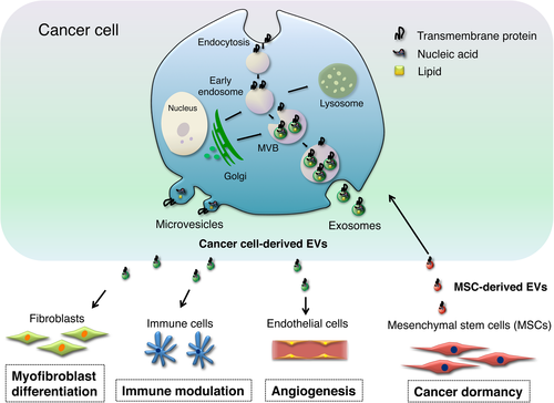
Exosomes are small EV, ranging between 50 and 150 nm in diameter, that originate from the fusion of multivesicular bodies (MVB) containing intraluminal vesicles.5, 6 The biogenesis of exosomes is mainly regulated by the machinery of endosomal sorting complexes required for transport (ESCRT) or lipid ceramide. The most common exosomal proteins are tetraspanins (CD9, CD63 and CD81), heat shock proteins (Hsp60, Hsp70 and Hsp90), membrane transporters and fusion proteins (Annexins, GTPases and flotillin), and MVB synthesis proteins (Alix and TSG101).7 Furthermore, exosomes contain MVB-associated proteins, as well as RNA encapsulated in a lipid bilayer with specific lipid components, such as sphingomyelin, cholesterol, ceramide and glycophospholipid.8 Microvesicles are large EV, ranging from 100 to 1000 nm in diameter, that are secreted by shedding or budding from the plasma membrane.1 Microvesicles have specific lipid components and are enriched in phosphatidylserine.9 The biogenesis of microvesicle shedding is stimulated by plasma membrane activation, such as intracellular calcium influx. However, there are only a few reports showing their function in cell-to-cell communication compared with exosomes. Finally, apoptotic bodies are heterogeneous EV that contain cytoplasm with condensed organelles and nuclear fragments. Apoptotic bodies are released into the extracellular space by apoptotic or dying cells. They also contain DNA fragments, RNA and cell organelles.10 For clarity, in this review we exclusively focus on exosomes as EV originating from MVE and microvesicles that are shed from the plasma membrane (Table 1 and Fig. 1).
| Vesicles | Exosomes | Microvesicles |
|---|---|---|
| Size | 50–150 nm | 100–1000 nm |
| Formation mechanisms | Multivesicular bodies fusion | Membrane shedding |
| Markers | CD63, CD81, CD9, Hsp60, Hsp70, Hsp90, Alix, TSG101, flotillin etc. | Annexin V and origin cell-specific markers |
| Components | Proteins, RNA, miRNA and lipids | Proteins, RNA, miRNA and lipids |
| Detection technology | Flow cytometry, ELISA, electron microscopy, western blot for exosome markers | Flow cytometry for microvesicles >300 nm, electron microscopy |

Previous studies have reported that the transfer of EV-contained miRNA and proteins are related to cancer development, such as initiation, invasion, metastasis and recurrence.11, 12 Among these phenotypes, we focused on EV participation in the key steps of cancer metastasis. This brief review identifies EV as an important messenger of cancer pathogenic components in cell-to-cell communication. EV have strong potential for use in liquid biopsy for disease diagnosis. Currently, there is some evidence for specific markers that distinguish cancer EV from normal EV.13-16 We will also briefly summarize reports about the potential use of EV for diagnostic purposes.
The Molecular Pathogenic Components of Extracellular Vesicles in Cancer
Extracellular vesicles have a vast array of content composed of proteins, mRNA, miRNA, DNA, lipids and transcriptional factors. The components are highly variable, functional, depend on cell origin, and exert powerful effects in recipient cells. Remarkably, cancer cell-derived EV contain functional oncogenic molecules, engendering a favorable microenvironment for cancer development. Among the pathogenic components, numerous studies have shown that the EV-contained miRNA and proteins modulate surrounding cells, such as endothelial cells,17 fibroblasts18 and immune cells (Fig. 1).19
In 2007, Valadi et al.20 showed that variable RNA, mRNA and miRNA can be transported between cells in EV. In 2010, we and two other groups independently reported that EV miRNA are transferred to recipient cells and translated into functional proteins.21-23 The vesicle-related RNA is called exosomal shuttle RNA (esRNA). Shuttling miRNA that may be oncomiR or tumor-suppressive is crucial for cancer malignancy. Notably, it has been shown that esRNA is poorly related to the RNA from EV-producing cells.24-26 This suggests that specific small RNA are actively loaded into the EV. Villarroya-Beltri et al.27 report that sumoylated heterogeneous nuclear ribonucleoprotein A2B1 (hnRNPA2B1) controls the sorting of miRNA into EV through the recognition of specific sequence motifs. Overall, the sort mechanisms are poorly understood and are likely to be associated with the mode of EV biogenesis. An increasing number of reports have shown that EV RNA contribute to cancer development, such as cell proliferation,28-30 drug resistance,31 angiogenesis,17 immune modulation19 and pre-metastatic niche formation.32
The lipid-bilayered EV can serve as suitable carriers for membrane proteins, allowing noncanonical cell–cell communication. The proteomic components of EV are specific, combining the common plasma membrane, cytosolic proteins and distinct cell-type specific proteins.33 EV membrane proteins can also affect cells via ligand-receptor binding and activation. However, EV proteins, including cell adhesion proteins and enzymes, have a pivotal role in promoting cancer metastasis. In cancer patients, cancer cell-derived EV represent elevated amounts of tumor-specific proteins in the blood.34 For instance, glioblastoma-derived EV promote the propagation of malignancy through the transfer of epidermal growth factor receptor variant III (EGFRvIII), an oncogenic form of EGFR.35 Furthermore, Luga et al.36 report that cancer associated fibroblasts secrete CD81-enriched EV that promote breast cancer cell protrusive activity and motility via Wnt-planar cell polarity, which results in metastasis and cell migration. Cancer cell-derived EV also transfer p-glycoproteins to induce drug resistance,37 transfer matrix metalloproteinases (MMP) to promote matrix degradation,38 and transfer natural killer group 2D (NKG2D) ligands to evade anti-cancer immune responses.39 These observations have been validated in numerous studies on the protein components of EV, and a few new initiatives are available online as free reference databases for EV investigators: Exocarta (http://www.exocarta.org),40 Vesiclepedia (microvesicles.org)41 and EVpedia (http://evpedia.info).42
The functional transfer of cancer pathogenic components in EV has been observed experimentally. Cancer cells deliver their EV pathogenic components not only to their surrounding cells but also to distant cells, and these EV can modulate to prepare niches that support metastasis. Therefore, understanding the profiles of EV pathogenic components may elucidate cancer malignancies.
Extracellular Vesicles as Key Regulators of Metastasis
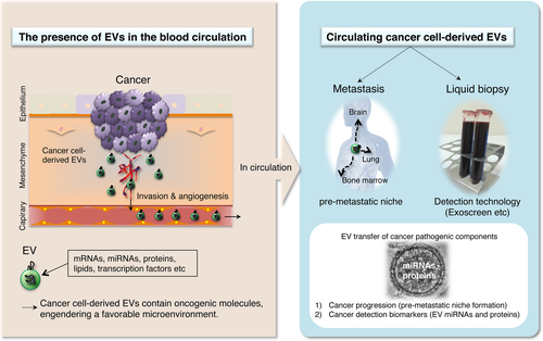
Metastasis is the leading cause of mortality in cancer patients. Therefore, there is an urgent need to develop predictive and early diagnostic markers for cancer metastasis and to clarify the molecular mechanisms of metastasis to develop effective treatment options. Several studies, including ours, have reported that EV mediate the formation of pre-metastatic niches for the metastasis of various organs. EV components promote metastasis and transfer metastatic potential. Furthermore, cancer cell-derived EV push selected host tissues toward a pre-metastatic phenotype.
Some reports show that EV mediate the formation of pre-metastatic niches in the lung. Jung et al.43 report that CD44v6-enriched EV from pancreatic cancer cells promote pre-metastatic niche formation in lymph nodes and lung tissues. Furthermore, they also found that these EV are rich in miR-494 and miR-542-3p, which target cadherin-17 and upregulate the MMP 2 and 9 transcription in pre-metastatic lung stroma cells.44 In renal cancer, Grange et al.45 report that EV from CD105-positive cancer stem cells stimulate angiogenesis and the formation of lung pre-metastatic niches. Peinado et al.46 report that melanoma-derived EV can determine the differentiation of bone marrow progenitor cells, resulting in the promotion of lung metastasis from melanoma cells. Using serum from melanoma patients, they found that melanoma-derived EV contain high TYRP2, VLA4, Hsp70, MET and Rab27a proteins. These EV travel to the bone marrow, lungs and lymph nodes during metastasis and are involved in the preparation of tumor niches by altering the extracellular matrix, including tissue inflammation, increasing vascular permeability and promoting other proangiogenic events.47 Finally, Le et al.48 report that miR-200-containing EV promote breast cancer cell lung metastasis. Transferring miR-200 can alter gene expression and promote the MET program for cancer cell colonization of distant tissues. In addition, they found that EV can promote metastasis and transform distal cells into metastatic cells. These data suggest that circulating EV from primary tumor developments and the formation of sites for metastasis may attract cancer cells to the niche, functioning as the first mediators of metastatic node formation.46, 49
Brain metastasis is also related to poor prognosis for cancer patients. One of the important events during brain metastasis is the destruction of the blood brain barrier (BBB), which consists of the endothelium and surrounding cells.50 Zhou et al.51 report that EV-mediated transfer of cancer-secreted miR-105 efficiently destroys tight junctions and the integrity of these natural barriers against metastasis. EV miR-105 targets the tight junction protein zona occludens protein 1 (ZO-1) and facilitates the migration of cancer cells to distant locations by disrupting tight junctions between endothelial cells, resulting in BBB breakdown. Furthermore, increased expression of circulating EV miR-105 can be detected during the pre-metastatic stage and correlated with the occurrence of metastasis in breast cancer patients. Thus, the present study showed that EV miR-105 destroys vascular endothelial barriers during early pre-metastatic niche formation and predicts metastasis in early-stage breast cancer patients. Similarly, we have shown a novel mechanism for brain metastasis mediated by EV that triggers the destruction of the BBB.52 EV miR-181c promotes breakdown of the BBB through abnormal localization of actin via the downregulation of its target 3-phosphoinostide-dependent protein kinase-1 (PDPK1). PDPK1 degradation by miR-181c leads to the downregulation of phosphorylated cofilin and the activated cofilin-induced modulation of actin dynamics. Moreover, serum levels of EV miR-181c were significantly higher in the serum from brain metastasis breast cancer patients compared with the serum from non-brain metastasis patients. In summary, these data suggest that EV influences the mechanisms of brain metastatic cancer.
Bone marrow metastasis can frequently occur following the development of metastatic cancer. Conversely, breast cancer patients often develop a recurrence years after resection of the primary tumor from cancer cells that have spread into metastatic niches, especially bone marrow. This was shown through the evidence that breast cancer cells survive for a long time in a dormant state. The mechanisms for cancer cells maintaining a dormant state before cancer recurrence are unknown. Recently, we revealed that EV promote cancer cell dormancy through their miRNA transfer in breast cancer.53 We found that the EV secreted by bone marrow mesenchymal stem cells (BM-MSC) regulate the cancer dormancy state of metastatic breast cancer stem cells. We also clarified that EV miR-23b from BM-MSC suppresses the expression of MARCKS, which encodes a protein that promotes cell cycling and motility in a bone marrow-metastatic human breast cancer cell line. Therefore, the bone marrow niche can release EV that deliver signals promoting metastatic cancer cell dormancy.
Both metastasis and recurrence are problematic issues delaying effective therapies for cancer treatments. Understanding the pathophysiological role of circulating EV in blood may be a good target for cancer treatment.12
Extracellular Vesicles as Biomarkers in Cancer Diagnostics
In general, EV reflect the physiological state of their original cells.3 Cancer cell-derived EV containing specific proteins and miRNA are secreted into tumor microenvironments and circulation.12, 54 EV have been isolated and characterized from different bodily fluids, such as serum, plasma, saliva, urine, ascites and breast milk.1, 12 Early detection of cancer is important to reduce mortality and improve survival. Liquid biopsies using circulating EV can clarify tumor characteristics without obtaining an invasive biopsy of the tumor. This has increased the interest in using EV RNA and proteins as cancer biomarkers. Currently, effort has been devoted to the utilization of EV as novel cancer diagnostic tools. EV miRNA are promising candidates as cancer biomarkers. Importantly, miRNA packaged in EV are intact in blood samples due to their resistance to RNases. This suggests the possibility of high reproducibility for this diagnostic approach.55 Table 2 shows representative studies reporting dysregulated miRNA in body fluid-derived EV from patients, suggesting its potential as a diagnostic tool to enhance early cancer detection.
| Primary tumor | Body fluids | Biomarkers | References |
|---|---|---|---|
| Lung cancer | Plasma | miR-151a-5p, miR-30a-3p,miR-200b-5p, miR-629, miR-100, miR-154-3p | 62 |
| Plasma | Let-7f, miR-30e-3p, miR-223, miR-301 | 63 | |
| Breast cancer | Serum | miR-200a, miR-200c, miR-205 | 64 |
| Serum | miR-21 | 65 | |
| Pancreatic cancer | Serum | miR-17-5p, miR-21 | 66 |
| Serum | miR-1246, miR-4644, miR-3976, miR-4306 | 67 | |
| Prostate cancer | Plasma, serum, urine | miR-107, miR-141, miR-375, miR-574-3p | 68 |
| Serum | miR-141 | 55 | |
| Glioblastoma | Serum | miR-320, miR-574-3p (and RNU6-1) | 69 |
| Cerebrospinal fluid | miR-21 | 70 | |
| Colorectal cancer | Serum | Let-7a, miR-1229, miR-1246, miR-150, miR-21, miR-223, miR-23a | 71 |
| Ovarian cancer | Serum | miR-21, miR-141, miR-200a, miR-200b, miR-200c, miR-203, miR-205, miR-214 | 72 |
| Cervical cancer | Cervicovaginal lavage | miR-21, miR-146a | 73 |
Cancer cell-derived EV also contain tumor-specific proteins. Recent proteomic studies have suggested that EV from cancer patients show specific protein profiles, thus indicating that the EV proteins might be useful for early detection of various cancers. Melo et al.13 report crucial evidence on using the circulating EV protein glypican-1 for diagnosis. Using mass spectrum analyses, they identified that glypican-1 was specifically enriched in breast cancer cell-derived EV. Glypican-1 positive circulating EV were detected in the serum of early pancreatic cancer patients with absolute specificity and sensitivity. The levels of glypican-1 positive circulating EV correlated with tumor burden and the survival of pre-surgical and post-surgical patients. The data indicated that circulating EV proteins may serve as a potential non-invasive diagnostic and screening tool to detect cancers. Currently, several promising cancer biomarkers have been found in patient body fluids and are summarized in Table 3.
| Primary tumor | Body fluid | EV protein markers | References |
|---|---|---|---|
| Melanoma | Plasma | TYRP2, VLA-4, Hsp70, Hsp90 | 46 |
| Serum | MRD-9, GFP78 | 74 | |
| Plasma | CD63, caveolin-1 | 75 | |
| Prostate cancer | Plasma | Survivin | 76 |
| Urine | δ-catenin | 77 | |
| Serum | PTEN | 78 | |
| Ovarian cancer | Serum | EpCam | 72 |
| Plasma | Claudin-4 | 73 | |
| Pancreatic cancer | Serum | Glypican-1 | 13 |
| Serum | CD44v6, Tspan8, EpCam, MET, CD104 | 67 | |
| Lung cancer | Urine | Leucine-rich α-2-glycoprotein (LRG1) | 15 |
| Serum | EGFR | 16 | |
| Serum | CD91 | 79 | |
| Colorectal cancer | Serum | CD147 | 14 |
| Nasopharyngeal carcinoma | Serum | Galactin-9 | 80 |
The identification and quantification of EV proteins in body fluids remain a major challenge compared with EV miRNA because EV proteins are highly heterogeneous and are difficult to collect and handle.56 However, there have been no superior techniques to isolate EV with a small amount of human body fluid. Furthermore, the ultracentrifugation normally used for EV isolation in basic research is unsuitable for most clinical settings. Consequently, we have to develop highly sensitive and specific detection methods for analyzing EV proteins.
Novel Extracellular Vesicles Detection Technologies in Cancer Diagnosis
Currently, there have been various reports using specific antibody-based technologies for analyzing EV proteins from human body fluids without isolation. These methods consist of flow cytometry,57 protein microarray (EV Array),58 diagnostic magnetic resonance59 and nanoplasmonic sensing technology.60 Despite these novel studies, the technological development of circulating EV detection is not yet mature. Hurdles that these technologies have to overcome include low sample throughput, time-consuming processes, and the variability and complexity of body fluids.61
In colon cancer, we have reported a sensitive and rapid analytical technology for profiling circulating EV proteins directly from colorectal cancer patient blood samples. Yoshioka et al.14 showed that circulating EV (CD147 and CD9 double-positive) can be used to detect colorectal cancer using “ExoScreen.” ExoScreen can simply detect disease-specific EV in the serum based on an amplified luminescent proximity homogeneous assay using photosensitizer-beads without an EV purification step. Remarkably, we can detect circulating cancer cell-derived EV by ExoScreen in only 5 μL serum from cancer patients. The receiver-operating characteristic curve indicated a diagnostic advent of CD147/CD9 double-positive EV compared with conventional tumor antigen markers. Therefore, ExoScreen using specific EV antigens provides a new liquid biopsy technique for sensitively detecting disease-specific EV.14
Conclusions
Currently, it is clear that the intercellular communication of EV constitutes an exciting field. Furthermore, many studies have strongly suggested great promises for EV transfer in cancer therapeutics and diagnostics. For tumor progression, cancer cell-derived EV modify tumor microenvironments and promote metastatic processes. EV pathogenic components, such as miRNA and proteins, clearly function to establish pre-metastatic niches. In addition, EV biomarkers from easily obtainable body fluids, such as blood and urine, are suitable for clinical application. This novel detection technology without isolation processes will have an outstanding impact on future precision medicine for cancer patients. Therefore, this is an attractive strategy for targeting cancer pathogenic components in EV with strong potential for cancer treatment (Fig. 2).

Acknowledgements
This work was supported in part by a Grant-in-Aid for the Japan Science and Technology Agency (JST) through the Center of Open Innovation Network for Smart Health (COINS) initiated by the Council for Science and a Grant-in-Aid for Basic Science and Platform Technology Program for Innovative Biological Medicine, and a Grant-in-aid for Project for Development of Innovative Research on Cancer Therapeutics (P-Direct).
Disclosure Statement
The authors have no conflict of interest to declare.


