Volume 203, Issue 2 p. 149
IMAGES IN HAEMATOLOGY
Open Access

Bone marrow pseudo-Gaucher cells demonstrating haemophagocytosis associated with extranodal NK/T cell lymphoma, nasal type

Anthony Jeffrey

Corresponding Author

Anthony Jeffrey

Department of Haematology, Royal North Shore Hospital, St Leonards, Australia

Northern Clinical School, Faculty of Medicine and Health, The University of Sydney, Sydney, Australia

Correspondence

Dr Anthony Jeffrey, Department of Haematology, Royal North Shore Hospital, St Leonards, Australia

Email: [email protected]

Search for more papers by this author
George Mason

George Mason

Department of Haematology, Royal North Shore Hospital, St Leonards, Australia

Northern Clinical School, Faculty of Medicine and Health, The University of Sydney, Sydney, Australia

Search for more papers by this author
Ian Kerridge

Ian Kerridge

Department of Haematology, Royal North Shore Hospital, St Leonards, Australia

Northern Clinical School, Faculty of Medicine and Health, The University of Sydney, Sydney, Australia

Northern Blood Research Centre, Kolling Institute, The University of Sydney, St Leonards, Australia

Search for more papers by this author
Poomahal Kumar

Poomahal Kumar

Department of Haematology, Royal North Shore Hospital, St Leonards, Australia

Northern Clinical School, Faculty of Medicine and Health, The University of Sydney, Sydney, Australia

Northern Blood Research Centre, Kolling Institute, The University of Sydney, St Leonards, Australia

Search for more papers by this author
First published: 31 July 2023

A 30 year old female presented with a 4 month history of an enlarging right posterior thigh mass. Biopsy of the lesion demonstrated an angioinvasive atypical lymphoid infiltrate with pleomorphic nuclei and prominent nucleoli. The immunohistochemical profile was consistent with a diagnosis of extranodal NK/T cell lymphoma: atypical lymphoid cells were positive for CD2, CD3, CD7, CD45RO, CD30, CD8 and cytotoxic molecules (Granzyme B, TIA1, Perforin). CD5 was lost in most cells. CD56 was expressed with weak intensity. TCR-beta was positive in a significant subset of the infiltrate. Ki-67% proliferation index was almost 100%. FDG-PET demonstrated FDG avid lymphadenopathy with extranodal involvement at the site of the leg lesion and in the left nasal cavity. Nasal biopsies demonstrated disease involvement, confirming a diagnosis of extranodal NK/T cell lymphoma, nasal type.

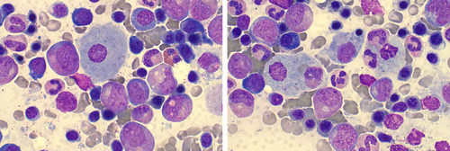
Bone marrow aspirate and trephine did not detect evidence of involvement with lymphoma by light microscopy, immunohistochemistry or flow cytometry. However, there was a diffuse increase in bone marrow macrophages with basophilic lamellar cytoplasm consistent with pseudo-Gaucher cells (left image, May-Grunwald Giemsa, ×100, oil immersion objective) and evidence of haemophagocytosis (right image). No organisms were seen by light microscopy or Ziehl-Neelson, Fite, Gram and Periodic acid-Schiff stains. The patient did not have any clinical features consistent with a lysosomal storage disorder and lysosomal enzymes including glucosylsphingosine were within normal ranges. There were no additional laboratory or clinical criteria to support a diagnosis of haemophagocytic lymphohistiocytosis (HLH). Pseudo-Gaucher cells have been reported in association with a variety of haematological malignancies, however this is the first report in extranodal NK/T cell lymphoma. Of note, the pseudo-Gaucher cells were identified in the absence of bone marrow involvement with lymphoma.

ACKNOWLEDGEMENTS

Open access publishing facilitated by The University of Sydney, as part of the Wiley - The University of Sydney agreement via the Council of Australian University Librarians.

      The full text of this article hosted at iucr.org is unavailable due to technical difficulties.