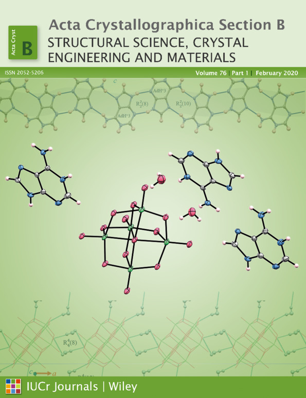
Crystal engineering of an adenine–decavanadate molecular device towards label-free chemical sensing and biological screening
Abstract
Due to the inherent geometrical interdependencies of nucleic acid structures, the ability to engineer biosensors that rely on the specific interactions of these compounds is of considerable importance. Additionally, sensing or screening in a label-free fashion is a capability of these structures that can be readily achieved by exploiting the fluorescent component. In this work, the [AdH]6[V10O28].4(H2O) (1) supramolecular structure is introduced using adenine and decavanadate moieties that allow probing of selectivity to specific nucleic acid binding events by optical changes. The structure of (1) is an alternating organic–inorganic hybrid architecture of cationic adeninium (AdH+) ribbons and anionic decavanadate (DV)–water sheets. The luminescent screening and anticancer activity of compound (1) on the two human mammary carcinoma cell lines MDA-MB-231 and MCF7 were investigated using fluorescent microscopy and MTT assays, respectively. It was found that compound (1) is cell permeable with no toxicity below 12.5 µM concentration and moderate cytotoxicity at concentrations as high as 200 µM in human breast cancer cell lines, making it a useful tool to study the cell nucleus in real time.


