New information on the Jurassic lepidosauromorph Marmoretta oxoniensis
Abstract
The earliest known crown-group lepidosaurs are known from the Middle Triassic; however, their stem group is poorly sampled, with only a few representative fossils found. This is partly due to the small size and delicate bones of early stem-lepidosaurs (= non-lepidosaurian lepidosauromorphs), which make both preservation in the fossil record and subsequent discovery less likely. The Middle Jurassic lepidosauromorph Marmoretta oxoniensis is re-examined using high-resolution micro-computed tomography to reveal parts of the skull anatomy that were previously unknown. These include a squamosal, postorbital, more complete parietal, pterygoids, and an articulated posterior section of the mandible. Some differences between this and other Marmoretta specimens were identified as a result, such as the arrangement of palatal teeth and the shape of the parabasisphenoid. The status of Marmoretta as a stem-lepidosaur or stem-squamate has been debated. To evaluate this, we tested the phylogenetic position of Marmoretta by including our new data in an adapted phylogenetic character matrix. We recover Marmoretta as a stem-lepidosaur and sister to Fraxinisaura rozynekae. Our findings support the hypothesis that both taxa belonged to a clade of non-lepidosaurian lepidosauromorphs that co-existed with lepidosaurs into the Middle Jurassic.
Lepidosaurs consist of more than 9000 extant species (Jones et al. 2013), including squamates (lizards, snakes and amphisbaenians) and Sphenodon, the only extant rhynchocephalian. The earliest fossils of crown-group lepidosaurs occur in the early Middle Triassic (c. 240 Ma; Jones et al. 2013) and their stem-lineage must extend back at least into the Permian, as indicated by the earliest occurrences of their extant sister taxon, Archosauromorpha (e.g. Ezcurra et al. 2014). However, the anatomy of stem-group lepidosaurs (i.e. non-lepidosaurian lepidosauromorphs) is not well known. Early stem-group lepidosaurs are currently represented by a few taxa primarily of Early–Middle Triassic age (Evans & Jones 2010), including the Early Triassic taxa Paliguana whitei (Carroll 1975) and Sophineta cracoviensis (Evans & Borsuk-Białynicka 2009), the Middle Triassic Fraxinisaura rozynekae (Schoch & Sues 2018), and, less certainly, the kuehneosaurs (specialized gliding reptiles with uncertain phylogenetic affinities, from the Early–Late Triassic; Evans & Jones 2010).
Marmoretta is a fossil lepidosauromorph from the Bathonian (166.1–168.3 Ma, Middle Jurassic; Gradstein et al. 2012) of the UK, known from several localities in southern England and the Isle of Skye, Scotland (Evans 1991; Waldman & Evans 1994). It is also known from the Upper Jurassic of Portugal (Evans 1991). Most studies have considered Marmoretta as a stem-group lepidosaur (Schoch & Sues 2018), in which case it might represent a relict lineage, being significantly younger than other stem-group lepidosaurs. However, a recent phylogenetic study found it as a stem-group squamate (Simões et al. 2018), which raises questions about its phylogenetic position. Nevertheless, Marmoretta has the potential to provide important anatomical data on deep lepidosaurian and lepidosauromorph divergences.
Most specimens of Marmoretta are fragmentary and disarticulated bones collected from screenwashing of bulk sediments (e.g. Evans 1991), however, specimens from the Isle of Skye include a semi-articulated partial skeleton (NMS G1992.47.1a–b; Waldman & Evans 1994). The original description of this specimen was carried out without removing the fossil material from the host matrix (a partially metamorphosed limestone, which was resistant to acid preparation). Only relatively superficial mechanical preparation was undertaken and only the bones on the surface of the blocks were described. Substantial further remains are enclosed within matrix and have not been studied until now.
Here, we provide a re-description and virtual reconstruction of the skull of Marmoretta based on synchrotron tomography of NMS G1992.47.1a–b and on micro-computed tomography (μCT) of the posterior portions of the mandibular rami from a different specimen, CAMSM X9991 (an incomplete specimen consisting of the posterior portion of the right lower jaw; Waldman & Evans 1994; Griffiths et al. 2021a). We use the new data from these scans in a phylogenetic analysis using Bayesian inference based on extensive revision of the matrix of Simões et al. (2018) (Griffiths et al. 2021b). We find that Marmoretta is a stem-group lepidosaur, and sister to Fraxinisaura.
Material and method
NMS G1992.47.1a–b consists of two blocks, one containing the skull and some postcranial material including 14 presacral vertebrae, partial ribs, an interclavicle and clavicles, and partial humerus, radius, ulna, femur and tibia (NMS G1992.47.1a) (Fig. 1), and a second, slightly smaller block, containing more postcranial material including a hand, seven presacral vertebrae with ribs, and the missing portions of humerus, radius, and ulna (split across both blocks) (NMS G1992.47.1b). We used high-resolution μCT to make three-dimensional (3D) visualizations of the specimen enclosed within the rock (Griffiths et al. 2021a). Here we focus on the skull description and phylogenetic implications. Synchrotron CT of the skull block (NMS G1992.47.1a) was carried out at The European Synchrotron Radiation Facility (ESRF) using propagation phase-contrast microtomography on the ID17 biomedical beamline. The images generated had an isotropic pixel size of 6.35 µm and were produced using a 90 keV monochromatic beam. Overall, 2499 images were produced from the combination of two radiographs with 0.1 s exposure times. The images were reconstructed with PyHST2 (Mirone et al. 2014) using the single-distance phase-retrieval approach (Paganin et al. 2002). The final images were then processed after production to change the bit depth from 32 to 16 bits, a weighted average was used for vertical and lateral stitching of the series of acquisition, a ring correction was applied (Lyckegaard et al. 2011), and finally volume cropping was done (V. Fernandez, pers. comm. 2019). The posterior portions of lower jaws (CAMSM X9991) were scanned at a resolution of 10.4 µm using a Nikon Metrology XT H 225 ST high-resolution CT scanner at the University of Bristol, School of Earth Sciences. The specimen was scanned using x-ray settings of 175 kV and 103 µA, with 3141 projections each captured for an exposure time of 0.5 s.
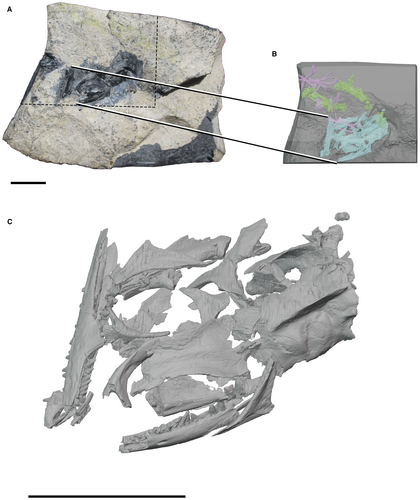
Image volumes were segmented using Mimics Research (http://biomedical.materialise.com/mimics), resulting in 3D models that were exported as .ply files and which were then imported to Blender (http://www.blender.org) for reconstruction and 2D rendering of the figures presented here (Griffiths et al. 2021a).
Institutional abbreviations
CAMSM, Sedgwick Museum of Earth Sciences, Cambridge, UK; NHMUK, Natural History Museum, London, UK; NMS, National Museums of Scotland, Edinburgh, UK.
Systematic palaeontology
DIAPSIDA Osborn, 1903
LEPIDOSAUROMORPHA Gauthier et al., 1988
Genus MARMORETTA Evans, 1991
Type and only species
Marmoretta oxoniensis Evans, 1991
Type specimen
NHMUK R12020, anterior portion of right maxilla from the Kirtlington Mammal Bed at the base of the Forest Marble, Old Cement Works Quarry, Kirtlington, Oxfordshire.
Referred specimens
NMS G1992.47.1a–b and CAMSM X9991 and other specimens (Panciroli et al. 2020) from the Isle of Skye, Scotland, and many isolated additional bones from Kirtlington Old Cement Works, England (Evans 1991, 1998), Leigh Delamere, England (Evans & Milner 1994), and Guimarota, Portugal (Evans 1991).
Diagnosis
(Revised from Evans 1991.) Small lepidosauromorph; large upper and lower temporal fenestrae; premaxillae paired, each with deep posterolateral maxillary facet; specialized maxillary/premaxillary overlap; small posteroventral process of the jugal; narrow fused frontals; fused parietal forming a broad parietal table, parietal foramen absent, large midline sagittal crest; dorsoventrally wide posterior (squamosal) process of the postorbital that overlaps on to a broad shallow facet on the squamosal; palatine with small teeth that decrease in size medially from a larger row along the medial choana margin to smaller scattered teeth on the ventral surface; pterygoids bear three rows of teeth which radiate anteriorly; long and slender dentary with subpleurodont teeth; coronoid with prominent coronoid process having a smooth concave posterior surface that emerges through the lower temporal fenestra.
Description
The skull is preserved and partially disarticulated in block NMS G1992.47.1a (Fig. 1). It includes mostly complete fused parietals, fused frontals, left and right prefrontals, almost complete right maxilla, partial right premaxilla, right postfrontal, right postorbital, left and right jugals, right squamosal, right quadrate and quadratojugal, partial left and right ectopterygoids, mostly complete left and right pterygoids, partial left and right palatines, parabasisphenoid, basioccipital, mostly complete right dentary, less complete left dentary, left and right coronoids, broken right prearticular, and a right articular. Post-depositional crushing has resulted in fragmentation and disarticulation of the lower jaws and cranial elements. Waldman & Evans (1994) reconstructed the skull based on the bones observable in the prepared specimen, which did not include new elements revealed by the μCT data, such as the squamosal and the full extent of the parietal crest. We present a new reconstruction of the skull of Marmoretta using information from NMS G1992.47.1a and CAMSM X9991 (Fig. 2), including the palatal region, which is poorly preserved.
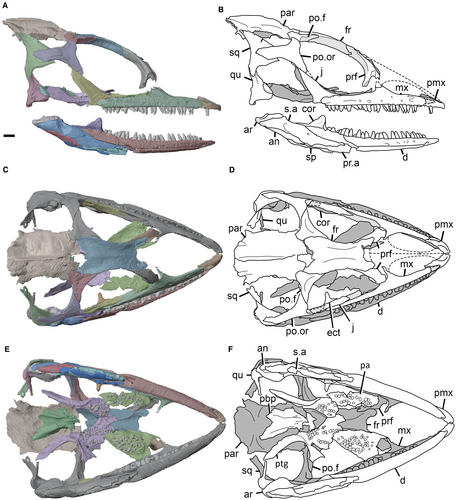
The dark grey portions of the articulated skull reconstruction are elements that have been preserved only on one side and have been duplicated and mirrored in Figure 2A, C, E. These include the right prefrontal (the right prefrontal is present although less complete than the left, therefore the left prefrontal has been mirrored in this reconstruction), and the entirety of the left mandibular ramus and skull except the jugal and prefrontal. The anteroventral process of the postorbital is missing, and is reconstructed based on the postorbital facet of the jugal, which is revealed in dorsal view. The anterior process of the maxilla is also missing, leaving the maxillary facet of the premaxilla exposed in lateral and dorsal view. Proposed positions for the nasals and dorsal processes of the premaxilla are also marked by dashed lines in Figure 2B, D.
The lack of a preserved squamosal–parietal contact renders the squamosal position provisional and also creates uncertainty with respect to the squamosal–quadrate articulation.
Cranium
Premaxilla
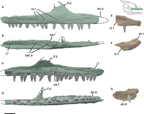
A partial right premaxilla is preserved, with the anterior and posterior portions missing. Its lateral surface is slightly convex. There are six alveoli, of which only one contains a tooth (Fig. 3E–G). It is likely that at least one more alveolus was present posteriorly, and another anteriorly, giving a minimum of eight marginal teeth in the premaxilla. A mediolaterally deep, V-shaped, maxillary facet is present on the posterolateral surface of the premaxilla. A subnarial ramus extends medially from the anteromedial surface. The ascending anterodorsal process is missing in NMS G1992.4.7.1a. However, specimens from Kirtlington (NHMUK R12022; Evans 1991) show that this process is long and tapers dorsally to separate the external nares across the midline anteriorly, thus dividing the external nares, unlike in Kuehneosaurus (Evans 2009).
Maxilla
Most of the right maxilla is preserved, but only a partial alveolar shelf of the left maxilla remains. The apex of the dorsal process of the right maxilla is broken, and the facets for the lacrimal and prefrontal are therefore not preserved (Fig. 3). The anterior portion of the right maxilla is also incomplete, although the length of the missing section is unknown. The maxilla is elongate and gracile anteroposteriorly, and the dorsal process appears to curve medially, possibly due to deformation. The preserved portion of the anterior process is relatively long, comprising 0.28 of the total anteroposterior length of the maxilla (Fig. 3). This is longer than in other stem-group lepidosaurs such as Sophineta, and most extant squamates, in which the anterior process (AP) is shorter relative to the total maxilla length (ML) (e.g. Sophineta, AP/ML = 0.13; Evans & Borsuk-Białynicka 2009; Iguana, 0.19; Japalura, 0.15; Hemidactylus, 0.11; Tropidophorus, 0.16; Cordylus, 0.21; Evans 2008). Rhynchocephalians also possess short anterior processes of the maxilla (Sphenodon, AP/ML = 0.13; Jones 2008), or even lack them entirely (e.g. Palaeopleurosaurus posidoniae and Pleurosaurus goldfussi; Jones 2008). The long anterior process of Marmoretta is similar to that of some squamates such as Lanthanotus borneensis (0.38) and varanids (e.g. Varanus salvator, 0.31; Evans 2008), but shorter than that of the Triassic stem-lepidosaur, Fraxinisaura (AP/ML = 0.51; Schoch & Sues 2018) and the extinct mosasaurians, in which the rostral part of the maxilla can form most of the bone.
A long shallow facet for the jugal is present posterodorsally on the medial surface of the maxilla. Two entrances for the superior alveolar canal are also visible on the dorsal surface of the alveolar shelf; the larger of the two is dorsal to the 16th alveolus, and the smaller is just anterior to this. The palatine facet is a horizontal groove on the alveolar shelf just posterior to the base of the dorsal process. A row of three neurovascular foramina open on the lateral surface of the maxilla, ventral and posterior to the dorsal process, and similar to those seen in Sophineta (Evans & Borsuk-Białynicka 2009).
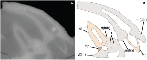
Twenty-three maxillary alveoli are present, 18 of which bear in situ teeth. This is slightly fewer than the estimated total of 25–30 maxillary teeth based on bulk-sample specimens from screenwashing at Kirtlington (Evans 1991). The difference is most likely due to incomplete preservation in NMS G1992.47.1a. The teeth are conical with a slight apicolingual curvature. Tooth implantation is pleurodont (sensu Bertin et al. 2018). There is a substantial difference in height between the labial and lingual walls of the maxilla, with the labial surface of the tooth root attached to the medial side of the labial wall (Fig. 4). This asymmetry of implantation is less evident in the dentary. However, here a basal plate supports the teeth lingually, a condition associated with ‘labial pleurodonty’ (Lessmann 1952; Zaher & Rieppel 1999; Bertin et al. 2018). With the exception of some smaller replacement teeth, the maxillary tooth row is approximately isodont, with tooth heights ranging from c. 0.8 to 0.9 mm.
Prefrontal
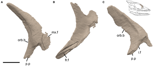
The prefrontals are crescentic in lateral view, and form the anterior margin of the orbit. Each prefrontal consists of an anteroposteriorly expanded ventral portion, which has a concave medial surface and convex lateral surface (Fig. 5). From this arises a tapering, rod-like dorsal process that bears a double facet for the frontal on its medial surface, divided by a narrow longitudinal ridge. Anteroventrally, the prefrontal bifurcates into a short anteromedial process and a longer posterolateral process that curves laterally at an acute angle to form the orbital margin. Specimens from Kirtlington show a broad and shallow facet in between the two prongs, probably for the reception of the lacrimals (Evans 1991), although these are not preserved in NMS G1992.47.1a.
Jugal
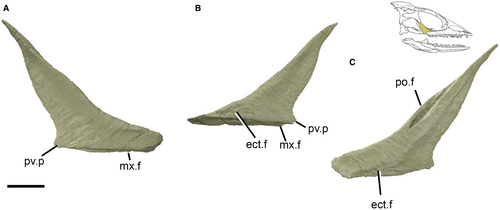
Both the left and right jugals are preserved. These are roughly triangular in lateral view, consisting of an anteroposteriorly broad ventral portion that articulates with the maxilla, and a tapering posterodorsal process that contacts the postorbital to form the ventral part of the postorbital bar (Fig. 6). The jugal facet extends further ventrally than the reconstructed ventral tip of the postorbital, and it appears that the ventral process of the postorbital is missing its distal part. The medial surface of the jugal bears a facet anteriorly, which most likely articulated with the ectopterygoid. The anterodorsal surface of the jugal forms the posteroventral rim of the orbit and is mediolaterally thickened compared with its posterior surface. A small posteroventral process is present, entering the anteroventral region of the temporal emargination. Although small, this process is more pronounced than in Sophineta (Evans & Borsuk-Białynicka 2009), but is smaller than that of Fraxinisaura, in which the posteroventral process of the jugal is dorsoventrally deep and extends further posteriorly (Schoch & Sues 2018). The absence of the lower temporal bar is a plesiomorphic feature in saurians, as well as being present in some non-saurian neodiapsids such as Acerosodontosaurus (Bickelmann et al. 2009) and Lanthanolania (Modesto & Reisz 2002).
Postorbital
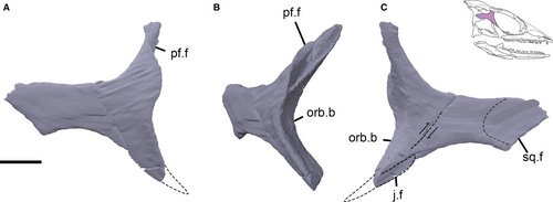
Only the right postorbital is preserved. It consists of three processes (Fig. 7). The ventral process forms the dorsal part of the postorbital bar and bears a facet for the jugal on its posterior surface. The dorsomedial process forms the anterior margin of the upper temporal fenestra and bears a facet for the postfrontal on its anterior surface. The posterior process forms the lateral margin of the upper temporal fenestra and bears a facet for the squamosal on its medial surface. The posterior process is broken and displaced dorsally and has been re-articulated to the anterior region of the postorbital in our reconstructions (Fig. 2A, B). The concave anterior surface of the dorsal and ventral processes forms a large part of the posterior orbital margin (Fig. 7). The right postorbital is dorsoventrally broad and mediolaterally thin, extending posteriorly to the posterior margin of the temporal region, where it articulates with the lateral surface of the squamosal in an overlapping contact (Fig. 7A). It is rhomboidal with a curved ventral border. The morphology of the posterior process differs from that seen in Kirtlington specimens (Evans 1991), in which the posterior process is narrower dorsoventrally than in NMS G1992.47.1a. The ventral process of the postorbital as reconstructed by Evans (1991) is also longer and more slender than in NMS G1992.47.1a, although this apparent difference is probably an artefact caused by the loss of the distal end of the ventral process in the Skye specimen, as indicated by the unoccupied lower half of the postorbital facet on the jugal.
Frontal
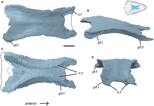
The frontals are fused into a median plate with a slightly raised area extending anteroposteriorly along the midline (Fig. 8A). The anteromedial and posterior portions of the bone are damaged and missing. The overall shape of the median frontal is approximately rectangular, transversely broader posteriorly than anteriorly, and narrowest at mid-orbit (c. 66% of the posterior transverse width). The ventral margins of the frontal bear distinct cristae cranii that follow the curve of the orbit and are somewhat shallower than in the early rhynchocephalian Diphydontosaurus (Whiteside 1986). The dorsal surface of the frontal is anteroposteriorly convex, as is most clearly evident in anterodorsal view (Fig. 8D). The lateral surface is embayed by the dorsal margin of the orbit, suggesting a juvenile or sub-adult ontogenetic stage (Evans 1991). Well-defined triangular facets for the postfrontals are evident in the posterolateral corners of the bone, tapering anteriorly. Shallow facets for the nasals are present on the preserved anterolateral surface of the frontal, with long prefrontal facets evident along the anterolateral margins.
Postfrontal
Only the right postfrontal is present in NMS G1992.47.1a (Fig. 9). The overall shape of the bone is triradiate, with a dorsal frontal process, posteromedial parietal process, and ventral postorbital process. The dorsal surface bears a facet for the frontal and the medial surface of the ventral process bears an elongate, triangular facet for the postorbital. This facet extends only for around one-third of the mediolateral width of the postfrontal, leaving a large posteromedial portion that participated in the anterior margin of the upper temporal opening. The posteromedial process is relatively short with a weak parietal facet on its medial surface.
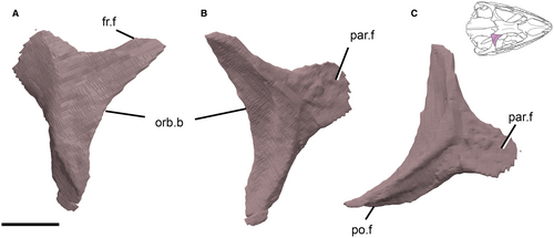
The postfrontal of Marmoretta is similar to that of Sophineta (Evans & Borsuk-Białynicka 2009), although in the latter taxon the anteromedial and dorsal processes are somewhat longer.
Parietal
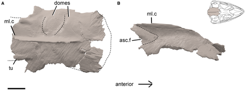
The parietal of Marmoretta is a single, fused element. The anterior portion of the parietal is broken on the right side, but is well-preserved on the left. This area is not embayed along the midline, and it is likely that a parietal foramen was absent, as noted by Evans (1991). Laterally, the parietal provides the dorsomedial margin of the upper temporal opening. This is best preserved on the left side, where the margin is slightly convex, rather than embayed. The dorsal surface of the parietal bears a prominent, mediolaterally narrow median (sagittal) crest. Either side of the crest, the dorsal surface is transversely convex. Two low, transversely orientated dome-like ridges form distinctive structures on the dorsal surface (Fig. 10). The first dome rises gradually from the frontoparietal suture, before diminishing sharply to form a transverse fossa approximately halfway along the length of the parietal. The second extends posteriorly from this fossa to form a slightly lower dome and shallow fossa. The posterior part of the parietal is inclined posterodorsally from this fossa, forming a short ascending flange at c. 45°, converging posteriorly to the level of the median crest (Fig. 10). Paired, anteroposteriorly oriented tubercles are present laterally at the base of the short ascending flange (Fig. 10). These tubercles have a hemispherical morphology and merge with the dorsal surface of the parietal anteriorly. The tubercles, and the posterior region of the parietal in general, are broken, but may have continued as lateral processes of the parietal, as in Huehuecuetzpalli (Reynoso 1998) and Dalinghosaurus (Evans & Wang 2005), or the short ascending flange may have extended posterodorsally, in a similar fashion to that seen in the Permian weigeltisaurid Coelurosauravus elivensis (Evans & Haubold 1987; Bulanov & Sennikov 2015).
The large parietal sagittal crest of Marmoretta is an unusual feature compared with other early lepidosauromorphs. Some Jurassic and Cretaceous rhynchocephalians (e.g. Palaeopleurosaurus, Kallimodon, Priosphenodon; Klein & Scheyer 2017) possess a short crest on a narrow parietal table, with distinctly ventrally orientated lateral flanges (Rieppel 1994). A midline crest on the parietal is also known in several early archosauromorphs (e.g. Protorosaurus, Macrocnemus, Trilophosaurus and the rhynchosaurs Mesosaurus and Howesia; Dilkes 1995; Heckert et al. 2006; Li et al. 2007; Gottmann-Quesada & Sander 2009; Pineiro et al. 2012). Simões et al. (2018, supp. info.) suggested that the sagittal crest occurs only in taxa with ventrally directed lateral margins of the parietal (i.e. with a narrow parietal table). Marmoretta is an exception in this case, in that the skull table is broad and the lateral margins are only moderately ventrolaterally inclined.
Squamosal
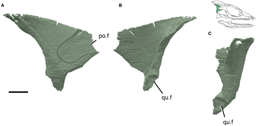
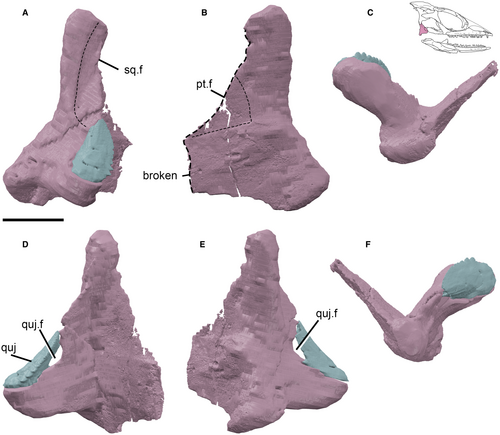
The right squamosal is preserved in NMS G1992.47.1a and is enclosed in matrix such that it was not described in previous studies (Evans 1991; Waldman & Evans 1994). As preserved, the squamosal is a large, triangular element. The lateral surface curves posteromedially to form a narrow contribution to the occipital region of the cranium (Fig. 11). It is a broadly plate-like bone, lacking clearly defined rami, unlike the tetraradiate squamosal in Sophineta or the triradiate squamosals of Pamelina, Huehuecuetzpalli and Megachirella (Reynoso 1998; Evans 2009; Evans & Borsuk-Białynicka 2009). There is a small posteroventral process, where the bone thickens, which bears a deep, wedge-shaped facet on the posteromedial surface for articulation with the dorsal (cephalic) condyle of the quadrate. The anteroventral process is broken distally, and most likely extended further ventrally, as implied by the presence of a facet on the anterolateral surface of the quadrate dorsal process. The morphology of that facet (Fig. 12) suggests that the ventral process of the squamosal terminated close to or in contact with the dorsal part of the quadratojugal (see Evans 1991). The squamosal lacks an emargination between the postorbital process and the anteroventral process. The lateral surface of the squamosal bears a broad, shallow facet anteroventrally for articulation with the postorbital (Fig. 11). This differs from the tongue and groove articulation of the postorbital and squamosal in Megachirella (Simões et al. 2018), but is somewhat similar to the same facet in the Lower Jurassic rhynchocephalian Gephyrosaurus bridensis (Evans 1980) and the overlapping contact of Sophineta, where a shallow postorbital facet is also present on the lateral face of the squamosal (Evans & Borsuk-Białynicka 2009). The squamosal tapers dorsally towards its contact with the parietal, although the contact itself is not preserved and cannot be determined. The posterior surface of the squamosal is distinctly concave in lateral view, and this may have supported the tympanic membrane, given that a tympanic crest or conch is absent from the quadrate and the retroarticular process is much reduced or absent (Fig. 11).
Quadrate
The right quadrate is preserved in NMS G1992.47.1a and is similar to the juvenile quadrate of Marmoretta (NHMUK R12040) described by Evans (1991) from Kirtlington Quarry. The quadrate consists of a mediolaterally expanded ventral portion that bears the articular condyles for the mandibles, a sheet-like anteromedial process, which extends to contact the quadrate ramus of the pterygoid, and a rod-like dorsal shaft that articulates with the squamosal dorsally via a convex condylar surface. The dorsal shaft also bears a large facet for articulation with the ventral process of the squamosal along its anterolateral surface. The medial surface of the quadrate shaft, which bears a low, horizontal ridge, may have received the columella of the stapes at the level of the dorsal margin of the quadratojugal.
In ventral view the anteromedial process of the quadrate forms a right angle with the axis of the lateral mandibular condyles. The medial condyle is mediolaterally narrow and anteroposteriorly longer than the lateral condyle, which is mediolaterally wide. The anteromedial process bears a broad, shallow facet for articulation with the pterygoid on its posteromedial surface, and is broken anteriorly (Fig. 12).
The quadrate conch is absent, as noted previously (Evans 1991). The presence of the quadrate conch was considered to be a synapomorphy of Lepidosauriformes (= total-group lepidosaurs excluding kuehneosaurs; equivalent to Lepidosauromorpha here) by Gauthier et al. (1988), who considered the conch to be present in Paliguana. The lack of a conch in Sphenodon represents a secondary loss (Gauthier et al. 1988), because the conch is present in basal rhynchocephalians such as Gephyrosaurus and Diphydontosaurus (Evans 1981; Whiteside 1986). Among early lepidosauromorphs, Sophineta also possesses a lateral conch, as does Megachirella (Evans & Borsuk-Białynicka 2009; Simões et al. 2018). In general, the quadrate morphology is similar to that of Sophineta, although Sophineta exhibits a larger depression between the lateral and medial condyles and a straighter dorsal process (Evans & Borsuk-Białynicka 2009).
Quadratojugal
The quadrate of NMS G1992.47.1a is articulated with a small, lenticular quadratojugal (Fig. 12). The quadratojugal lies ventral to the squamosal facet and may have contacted the squamosal. It articulates with the ventrolateral surface of the quadrate, enclosing a small quadrate–quadratojugal foramen laterally (Fig. 12).
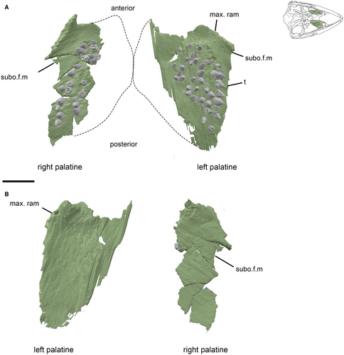
Palatine
Both palatines are both partially preserved in NMS G1992.47.1a. The thickened maxillary processes are present, but the medial and posterior portions that contact the pterygoids are missing, as are the anterior margins, which would contact the vomer. The palatines are thin, dorsally concave plates of bone that have roughly triangular outlines. A field of small teeth is present on the convex palatal surface (Fig. 13). The palatine thickens laterally as it approaches the maxillary process, but the margins of the choana and suborbital fenestra are not preserved. Palatine teeth are widespread among tetrapods, including stem-tetrapods (e.g. Ichthyostega), early amniotes (e.g. Petrolacosaurus), and many lepidosauromorphs (e.g. Sophineta, Sphenodon), but have been lost in many squamates (Matsumoto & Evans 2017). In Marmoretta the lateral row of palatal teeth is slightly enlarged (Fig. 13), differing from other early lepidosauromorphs except from rhynchocephalians such as Diphydontosaurus (Whiteside 1986). The condition in Marmoretta is weakly developed in comparison with rhynchocephalians, and we do not consider this to be a directly homologous character. The palatal teeth in NMS G1992.47.1a are less organized than those in the Kirtlington specimen, in which distinct tooth rows are apparent. This may be a case of interspecies difference or it may be due to the preservation of the Skye specimen, which has resulted in the teeth being disturbed and not preserved in their life position.
Pterygoid
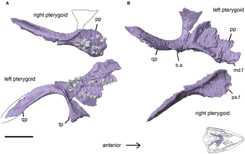
The pterygoids are anteroposteriorly long, each consisting of a large, sheet-like palatal process and a narrow quadrate process that extends posterolaterally from the posteromedial part of the palatal process. Both pterygoids are missing their anterior and lateral portions. The broad palatal process has a gently concave ventral surface, and is thickened on the medial edge, which forms the lateral margin of the interpterygoid vacuity (Fig. 14). The palatal surface bears three rows of teeth that radiate anterolaterally from a position just adjacent to the basal articulation. The transverse processes (pterygoid flanges) of both pterygoids are damaged, with only a remnant of the left process remaining. The left process consists of a roughly triangular extension that thickens along the posterior margin where it joins the main body of the pterygoid lateral to the basal articulation. Overall, the pterygoid is very similar to that of Fraxinisaura (Schoch & Sues 2018). There are no teeth present on the transverse process. The quadrate process of the pterygoid curves posterolaterally to meet the medial wing of the quadrate. There is no development of the pit (fossa columellae) on the dorsal surface of the pterygoid quadrate ramus that forms a mobile articulation with the base of the epipterygoid in squamates.
Ectopterygoid
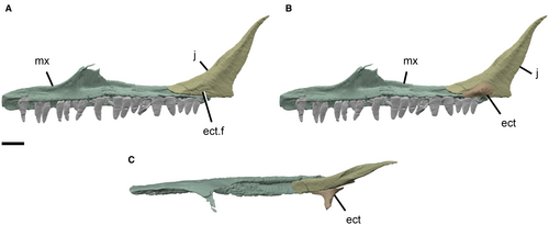
Both ectopterygoids are preserved, although the right bone is more complete than the left, and both are missing their medial portions, including the facet for articulation with the pterygoid. The ectopterygoids are small and consist of an expanded lateral plate for articulation with the maxilla and jugal (Fig. 15), from which a slender stem extends medially into the palate. The lateral articular surface is flat and dorsomedially deep, with a long, shallow ventral facet for the maxilla and a smaller posterodorsal facet for the jugal. The lateral flange of the ectopterygoid of Marmoretta is anteroposteriorly longer than that of Sophineta (Evans & Borsuk-Białynicka 2009) and Diphydontosaurus (Whiteside 1986). In Fraxinisaura the stem is thicker and not smoothly cylindrical (Schoch & Sues 2018).
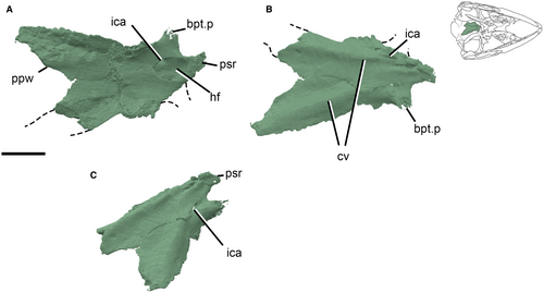
Parabasisphenoid
The parabasisphenoid is a midline bone that tapers anteriorly, resulting in an approximately triangular outline. It is embayed posteriorly between paired, posterolateral parasphenoid wings. The parasphenoid rostrum (cultriform process) extends anteriorly, but only its base is preserved. The basipterygoid processes extend anteroventrally, the right being broken and the left only partially preserved (Fig. 16). The posteroventral surface of the parabasisphenoid is concave, and the dorsal surface is also transversely concave and lacks the midline ridge seen in specimens referred to Marmoretta from Kirtlington Quarry (NHMUK R12055 and NHMUK R12057; Evans 1991). The internal carotid foramina perforate the ventral surface of the bone and enter the posterolateral part of the hypophysial fossa so that they are not visible in dorsal view. This also differs from the Kirtlington specimens NHMUK R12055 and NHMUK R12057 (Evans 1991) in which the foramina are located anteriorly within the fossa. It also differs from the parabasisphenoid in Fraxinisaura, which bears a patch of denticles on its ventral surface close to the base of the parabasisphenoid (Schoch & Sues 2018).
Basioccipital
The basioccipital forms an ovoid posteroventral occipital condyle (Fig. 17). The ventral surface of the bone bears a low transverse ridge, anterior to the occipital condyle. This becomes more prominent laterally on either side, forming two paired, ventrolaterally projecting basal tubera. These are relatively large and appear similar to inferred adult specimens referred to Marmoretta from Kirtlington (NHMUK R12058 (adult) compared with those of NHMUK R12059 (juvenile); Evans 1991). Facets for the exoccipitals are present dorsolaterally on the occipital condyle. The dorsal surface of the basioccipital bears a longitudinal median ridge that spans the posterior two-thirds of the bone; on either side of the ridge the bone is concave.

Mandible
Dentary
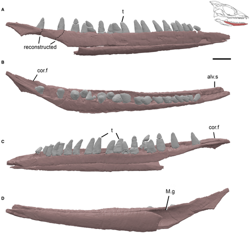
Both dentaries are incomplete but the right is the better preserved, although it is missing its anterior, posterior, and posteroventral sections. The dentary is long and slender, with the medial surface divided into dorsal and ventral parts by the Meckelian groove, which has been narrowed dorsoventrally by postmortem crushing (Fig. 18). As with the maxillary tooth row, the dentary teeth are implanted in the alveolar shelf, the labial wall of which is higher than the lingual wall, exposing most of the tooth bases lingually. The posterior portion of the right dentary had broken away from the main section of bone and has been repositioned accordingly for the reconstruction. This detached piece contains the posteriormost tooth and facets for the coronoid and surangular on its dorsomedial surface. The Meckelian groove is open medially in the anterior portion of the dentary, similar to NHMUK R12062 (Evans 1991).
Coronoid
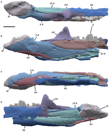
Both left and right coronoids are present in NMS G1992.47.1a and the left is present in CAMSM X9991. They are robust bones, consisting of a dorsoventrally broad, sheet-like anteromedial process, a narrow, tapering posterolateral process, and a prominent coronoid process (Fig. 19). The ventral surface of the coronoid bears a groove-like horizontal facet for articulation with the dorsal surface of the dentary. The anteromedial process extends ventral to this, covering a portion of the medial surface of the dentary. The lateral surface of the anteromedial flange bears a small posterior facet for the prearticular. The coronoid process curves medially to produce a smooth concave posterior surface that serves as an attachment site for the mandibular adductor (Evans 1991).
Splenial
The splenial is not preserved in NMS G1992.47.1a, however, it is present in articulation with the other bones of the posterior part of the mandible in CAMSM X9991. The splenial in CAMSM X9991 is incomplete, consisting of only the posteroventral and posterodorsal parts of the bone, which are broken and appear as separate fragments. These articulate with the dentary, coronoid and prearticular.
Prearticular
The right prearticular is present in both associated specimens of Marmoretta. In NMS G1992.47.1a it is broken in half dorsoventrally and is missing the anterior and posterior ends. In CAMSM X9991 the prearticular is preserved in articulation with the rest of the lower jaw bones, aiding the analysis of NMS G1992.47.1a (Fig. 19). On the medial surface of the bone there is a shallow impression bordered dorsally by a low ridge that runs anterodorsally–posteroventrally, ending about three-quarters of the way along the bone. This marks the dorsal extent of the splenial facet. On the lateral surface there is a long V-shaped facet for the dentary positioned anteriorly on the thickened dorsal margin. Posteriorly the prearticular tapers to a point at which the ventral surface is contacted by the angular, and the dorsal surface by the articular.
Surangular
The right surangular is present in both NMS G1992.47.1a and CAMSM X9991, although it is more complete in the latter. The bone is long, extending from the posteroventral surface of the dentary, adjacent to about the sixth from last tooth, to the ventral surface of the articular. On the anterolateral surface there is a long, broad and shallow facet for the posterior region of the dentary and, just ventral to the tip of the dentary, there is an anterior surangular foramen. Posteriorly the surangular expands into a broad cup-like facet for the articular. Ventrally the surangular contacts the prearticular anteroventrally and the angular posteroventrally (Fig. 19B, C).
Angular
The angular is not preserved in NMS G1992.47.1a, but the right bone is evident in CAMSM X9991. It is a small slender element that tapers at its anterior and posterior ends. The angular is positioned on the ventral surface of the lower jaw and contacts the surangular dorsolaterally, the prearticular and the articular dorsomedially (anterior–posterior), and the splenial ventrally.
Articular
The right articular is present in both associated specimens. It is a robust bone that comprises the posterior end of the lower jaw, with its dorsal surface articulating with the condyles of the quadrate. The ventral surface of the articular has a narrow but relatively deep medial facet for the prearticular. The medial surface of the bone continues dorsally from this facet and is mostly flat, expanding slightly at the dorsal surface. On the lateral side the articular is broad posteromedially and the ventrolateral surface narrows medially to form the lateral surface of the prearticular facet. The broad posteromedial portion of the bone is sheathed from below by the large surangular facet. Dorsally the articular slopes anteroposteriorly at an angle of c. 45°. The dorsal surface is divided by a central groove that is bordered by a tall projection medially, and a shorter, broader projection on the lateral side.
There is no development of a retroarticular process.
Discussion
Our high-resolution CT scans of referred specimens of Marmoretta (NMS G1992.47.1, CAMSM X9991) provide important new anatomical data. In particular it has clarified our understanding of the suspensorium and posterior region of the mandible, and demonstrated the extent of the parietal sagittal crest and the pleurodont nature of the marginal tooth implantation. Our reconstruction of the skull of Marmoretta retains much of the general form of previous studies (Evans 1991; Waldman & Evans 1994). However, the dorsoventral height of the postorbital region of the cranium and the posterior portion of the mandible suggest a distinctive, anteriorly tapering skull shape, augmented by the prominent sagittal crest.
The sagittal crest of Marmoretta differs from that of other reptiles in that it is combined with a transversely broad parietal table. The crest provides an attachment site for the external adductor muscle, which descends to attach to the medial surface of the coronoid eminence in the mandible. The coronoid eminence of Marmoretta bears a large concavity on the posteromedial surface for this adductor attachment, suggesting a strong closing force (King 1996). Although a comparatively powerful bite-force is postulated in small (<2.5 cm skull length) early Mesozoic diapsids, it is correlated with transversely narrow parietal tables and broad upper temporal openings in relation to the transverse width of the postorbital region (Pritchard et al. 2018). Marmoretta does not possess either of these features, although the adductor musculature in Marmoretta would have benefitted from extended dorsoventral length and may represent an ecomorphologically diverse approach to substantial bite-force in small diapsids.
The arrangement of the palatal teeth in NMS G1992.47.1a differs from that recorded by Evans (1991) based on specimens from Kirtlington Old Cement Quarry (NHMUK R12045, R12046, R12047). NMS G1992.47.1a possesses lateral palatine teeth that are slightly enlarged and are not positioned into distinct rows, unlike in the Kirtlington specimens. Also, the pterygoid of NMS G1992.47.1a bears three tooth rows as opposed to the two described in the Kirtlington specimens (Evans 1991; NHMUK R12052, R12054). However, this is likely to be due to the more complete preservation of the pterygoids in NMS G1992.47.1a compared with NHMUK R12052 and R12054.
Palatal teeth are considered an ancestral condition in amniotes and appear in one form or another in most major clades, although there is a general pattern of reduction in many lineages (Matsumoto & Evans 2017). Nevertheless, the morphology and inferred function of palatal teeth vary among taxa. The longitudinal rows of palatal teeth seen in Marmoretta suggest that they may have assisted with moving food towards the back of the mouth (Matsumoto & Evans 2015). In many extant lepidosaurs this function is carried out by a muscular tongue in conjunction with varying amounts of palatal dentition (Matsumoto & Evans 2017). The presence of anterior palatal teeth in Marmoretta (palatine and pterygoid, possibly vomer although this is unknown) and the lack of posterior palatal teeth (parabasisphenoid and transverse process) suggest that their main function was intraoral transport and that they were probably accompanied by a mobile tongue.
There are a few other differences between this specimen NMS G1992.47.1a and the Kirtlington specimens NHMUK R12037 (a juvenile postorbital) and NHMUK R12055 and NHMUK R12057 (parabasisphenoids) described by Evans (1991). These include the shape of the posterior process of the postorbital, which is dorsoventrally taller in NMS G1992.47, and the positioning of the internal carotid foramina within the hypophysial fossa, which are further posterior in this specimen. These may be examples of ontogenetic or intraspecies variation, or indicate that the assemblage from Kirtlington includes a different species to the specimens described here.
Phylogenetic analysis
Earlier studies have resulted in two hypotheses on the affinities of Marmoretta. Evans (1991) interpreted Marmoretta as a non-lepidosaurian lepidosauromorph, outside of the crown-group split between rhynchocephalians and squamates, based on material from Kirtlington, Oxfordshire. New data from specimens collected from the Isle of Skye (Waldman & Evans 1994) and subsequent analyses (Evans 2009; Evans & Borsuk-Białynicka 2009; Evans & Jones 2010; Jones et al. 2013) have generally reiterated this view. The recent phylogenetic analysis by Schoch & Sues (2018) also recovered Marmoretta as a stem-group lepidosaur, as sister to the Middle Triassic Fraxinisaura rozynekae. In contrast to this hypothesis, Simões et al. (2018) recovered Marmoretta, along with Megachirella from the Middle Triassic of Italy, as a stem-group squamate within Lepidosauria, using both parsimony analysis and Bayesian inference.
To evaluate the phylogenetic position of Marmoretta based on the new data, we used a modified version of the 347 characters in the morphological dataset of Simões et al. (2018). We added 32 new characters and removed two, making a total of 377 characters. These changes are based on an extensive review of their dataset and published comparative literature, and our modifications are described more completely in Griffiths et al. (2021b). Of the 32 new characters, two replaced existing characters and describe distinctive aspects of similarity among the squamosals of squamates that are absent outside the squamate crown-group (e.g. Evans 2008). Overall, our additions mostly reflect comparative observations that were framed by older literature, but which were not included in the original character list of Simões et al. (2018). These observations document variation among early crown-group reptiles and especially among early lepidosauromorphs, encoding character state variation that has been influential for existing phylogenetic hypotheses (e.g. Camp 1923; Parrington 1958). We also revised the scores of several taxa, focusing on those that have previously been considered as early lepidosauromorphs (e.g. Megachirella, Sophineta, Palaeagama, Gephyrosaurus and Diphydontosaurus) or potentially closely related taxa (e.g. Kuehneosaurus and Pamelina). We omitted some taxonomic units, and added others such as Fraxinisaura. A list of these modifications together with explanatory notes is included in Griffiths et al. (2021b).
We performed a non-time calibrated Bayesian analysis of the resulting data using the Mkv model with MrBayes v3.2.5 as described in Appendix S1 (see also Griffiths et al. 2021b), using a maximum clade credibility (MCC) tree to summarize the results of this analysis (Fig. 20).
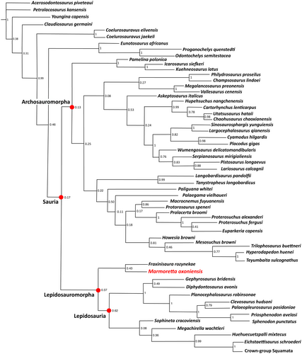
The MCC tree recovers Marmoretta as a stem-group lepidosaur (i.e. a non-lepidosaurian lepidosauromorph), in agreement with some previous studies (Evans 1991; Jones et al. 2013). We also find Marmoretta as a sister taxon of the Middle Triassic Fraxinisaura, within an early diverging and geologically long-lived clade of non-lepidosaurian lepidosauromorphs. This is consistent with the phylogenetic hypotheses posited by Schoch & Sues (2018), who noted the striking similarity of the maxillae of Marmoretta and Fraxinisaura, which both possess a low, triangular facial process and elongate anterior process. We find that this group (Marmoretta + Fraxinisaura) is supported by three unambiguous synapomorphies (the absence of the premaxillary process of the maxilla, char. 20.1; the absence of a parietal foramen, char. 73.1; and the absence of an infraorbital foramen on the palatine, char. 101.1). The clade containing Marmoretta + Fraxinisaura also possesses several lepidosauromorph synapomorphies, including a reduced lacrimal (under deltran char. 360.1), pleurodont implantation of maxillary dentition (under acctran char. 213.0), a quadratojugal foramen (unambiguous char. 42.1) and an hourglass-shaped frontal (under acctran char. 354.1).
Our phylogenetic findings therefore differ from those of Simões et al. (2018), who recovered Marmoretta as a stem-group squamate, nested within Lepidosauria (i.e. as a member of the crown-group). Consistent with our recovery of Marmoretta in the stem-group, we observe various features that are present in crown-group lepidosaurs but which are absent in Marmoretta. These features include subolfactory processes of the frontals (unambiguous char. 69.1) and the lateral conch of the quadrate (under deltran char. 121.1). The absence of a lateral conch of the quadrate in Marmoretta may be plesiomorphic for lepidosauromorphs, with the lateral conch probably appearing closer to the divergence of the crown-group in more derived stem-lepidosaurs. The quadrate conch is present in squamates and early rhynchocephalians (Evans 1980; Whiteside 1986; Simões et al. 2018) and, probably, convergently in kuehneosaurs (Evans 2009). Unfortunately, the condition in the quadrate is unknown in Fraxinisaura (Schoch & Sues 2018). Further, Marmoretta possesses several features that are not found in squamates (e.g. quadratojugal present, char. 38.0; absence of a notch for the squamosal on the cephalic head of the quadrate, char. 123.0; ventral exposure of the entry foramen for the internal carotid artery in the basisphenoid, char. 124.1), or in rhynchocephalians (e.g. absence of frontal tabs on the parietal, char. 78.1; presence of a splenial, char. 176.1; absence of a notochordal canal in adults, char. 229.1).
Megachirella from the Middle Triassic of Italy, like Marmoretta, was originally reported as a non-lepidosaurian lepidosauromorph (Renesto & Posenato 2003; Renesto & Bernardi 2014) but was subsequently recovered as a stem-squamate inside of the lepidosaurian crown-group by Simões et al. (2018). Our MCC tree recovers Megachirella as a stem-squamate, in accordance with Simões et al (2018). Megachirella shares several key features with lepidosaurs (e.g. a lateral quadrate conch, char. 121.1) and with squamates (e.g. the loss of the anteroventral process of the squamosal, char. 50), although both of these character states are also found in kuehneosaurs, which were not recovered as lepidosauromorphs in our analysis. We also recover Sophineta, which generally has been described as a non-lepidosaurian lepidosauromorph (Evans & Borsuk-Białynicka 2009, Jones et al. 2013), as an early-diverging stem squamate (in the MCC tree). However, it is notable that support for both Megachirella and Sophineta as stem squamates is poor in the MCC tree (posterior probability, 0.36 and 0.08, respectively), and both taxa are found in a trichotomy with squamates and rhynchocephalians in the 50% majority rule tree from our posterior sample (Fig. S1).
Our analysis also highlights substantial uncertainties regarding the phylogenetic positions of other taxa traditionally interpreted as basal lepidosauromorphs, with Paliguana recovered outside Lepidosauromorpha in both tree topologies (Fig. 20; Fig. S1). The anatomy, affinities and evolutionary implications of this taxon require further investigation.
Conclusion
New anatomical data on the skull of Marmoretta oxoniensis from the Middle Jurassic of the UK and the Late Jurassic of Portugal have significantly added to our knowledge of this taxon. Based on these new data, our phylogenetic analysis recovers Marmoretta as a member of the lepidosaurian stem lineage, and a sister taxon to the Middle Triassic Fraxinisaura. This differs from the hypothesis proposed by Simões et al. (2018), who recovered Marmoretta as a squamate, within the lepidosaurian crown-group. As a Middle Jurassic taxon, Marmoretta remains significantly younger than other stem-group lepidosaurs, including its closest known relative Fraxinisaura. Both taxa are members of a clade that co-existed with the crown-group for at least 80 myr, and probably became extinct before the end of the Mesozoic, leaving rhynchocephalians and squamates as the sole representatives of the lepidosaurian line.
Acknowledgements
The authors thank Dr Michael Waldman, who collected NMS G1992.47.1a–b, the National Museums Scotland for access to the specimen and permission to partially prepare it, and the John Muir Trust and Scottish Natural Heritage, who are responsible for permitting fieldwork on the protected fossil localities on the Elgol Coast Site of Special Scientific Interest. Thanks also to the Sedgwick Museum Cambridge for the loan of the CAMSM X9991 and permission to scan it, and to the scanning unit at the University of Bristol. The authors acknowledge the European Synchrotron Radiation Facility for the provision of synchrotron radiation facilities (proposal LS 2546), and thank Vincent Fernandez for assistance in using beamline ID17 to scan NMS G1992.47.1a, and for supplying methodological details. We acknowledge with thanks the comments and suggestions of Dr Hans-Dieter Sues and an anonymous referee on an earlier draft of this manuscript.
Author contributions
EFG carried out the investigation and formal analysis of the μCT data, prepared figures for publication based on scan data, and wrote the original manuscript draft. DPF carried out formal analysis of the phylogenetic data and wrote the phylogenetic discussion in the manuscript as well as the supplementary data. RBJB conceptualized, managed and supervised the project, and led the review and editing of the manuscript, assisted by all other authors. SEE assisted in the review and editing of the manuscript and the validation of character matrix scoring.
Open Research
Data archiving statement
Data for this study, including code, matrix and a list of MorphoSource DOIs, are available in the Dryad Digital Repository: https://doi.org/10.5061/dryad.ghx3ffbnm Scan data and 3D models are available on MorphoSource: http://www.morphosource.org/projects/000349957


