Utility of maximum perfusion intensity as an ultrasonographic marker of intraneural blood flow
Adeniyi A. Borire MBBS
Prince of Wales Clinical School, University of New South Wales, Sydney, New South Wales, Australia
Search for more papers by this authorLeo H. Visser MD, PhD
St. Elisabeth Ziekenhuis, Tilburg, The Netherlands
Search for more papers by this authorLuca Padua MD, PhD
Department of Geriatrics, Neurosciences, and Orthopedics, Università Cattolica del Sacro Cuore, Rome, Italy
Don Gnocchi Foundation, Milan, Italy
Search for more papers by this authorJames G. Colebatch DSc
Prince of Wales Clinical School, University of New South Wales, Sydney, New South Wales, Australia
Institute of Neurological Sciences, Prince of Wales Hospital, Sydney, New South Wales, Australia
Search for more papers by this authorWilliam Huynh MBBS, PhD
Institute of Neurological Sciences, Prince of Wales Hospital, Sydney, New South Wales, Australia
Brain and Mind Centre, University of Sydney, Sydney, New South Wales, Australia
Search for more papers by this authorNeil G. Simon MBBS, PhD
Brain and Mind Centre, University of Sydney, Sydney, New South Wales, Australia
St. Vincent's Clinical School, University of New South Wales, Sydney, New South Wales, Australia
Search for more papers by this authorMatthew C. Kiernan DSc
Brain and Mind Centre, University of Sydney, Sydney, New South Wales, Australia
Search for more papers by this authorCorresponding Author
Arun V. Krishnan MBBS, PhD
Prince of Wales Clinical School, University of New South Wales, Sydney, New South Wales, Australia
Institute of Neurological Sciences, Prince of Wales Hospital, Sydney, New South Wales, Australia
Correspondence to: A. Krishnan; e-mail: [email protected]Search for more papers by this authorAdeniyi A. Borire MBBS
Prince of Wales Clinical School, University of New South Wales, Sydney, New South Wales, Australia
Search for more papers by this authorLeo H. Visser MD, PhD
St. Elisabeth Ziekenhuis, Tilburg, The Netherlands
Search for more papers by this authorLuca Padua MD, PhD
Department of Geriatrics, Neurosciences, and Orthopedics, Università Cattolica del Sacro Cuore, Rome, Italy
Don Gnocchi Foundation, Milan, Italy
Search for more papers by this authorJames G. Colebatch DSc
Prince of Wales Clinical School, University of New South Wales, Sydney, New South Wales, Australia
Institute of Neurological Sciences, Prince of Wales Hospital, Sydney, New South Wales, Australia
Search for more papers by this authorWilliam Huynh MBBS, PhD
Institute of Neurological Sciences, Prince of Wales Hospital, Sydney, New South Wales, Australia
Brain and Mind Centre, University of Sydney, Sydney, New South Wales, Australia
Search for more papers by this authorNeil G. Simon MBBS, PhD
Brain and Mind Centre, University of Sydney, Sydney, New South Wales, Australia
St. Vincent's Clinical School, University of New South Wales, Sydney, New South Wales, Australia
Search for more papers by this authorMatthew C. Kiernan DSc
Brain and Mind Centre, University of Sydney, Sydney, New South Wales, Australia
Search for more papers by this authorCorresponding Author
Arun V. Krishnan MBBS, PhD
Prince of Wales Clinical School, University of New South Wales, Sydney, New South Wales, Australia
Institute of Neurological Sciences, Prince of Wales Hospital, Sydney, New South Wales, Australia
Correspondence to: A. Krishnan; e-mail: [email protected]Search for more papers by this authorABSTRACT
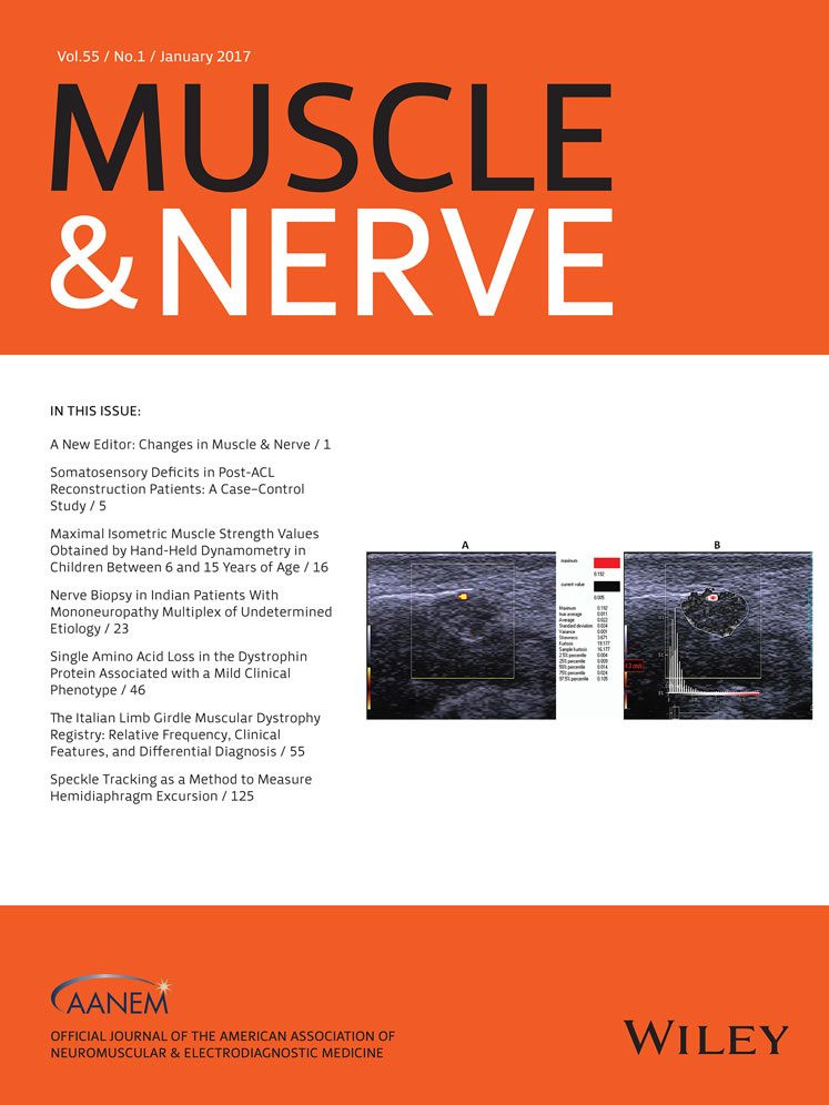
We quantified intraneural blood flow (INBF) using perfusion measurement software (PixelFlux), and compared it with the qualitative method of counting blood vessels (vessel score) in a cohort of carpal tunnel syndrome (CTS) patients. Methods: Forty-seven patients (67 wrists) with a clinical and electrophysiological diagnosis of CTS, and 20 healthy controls (40 wrists) were enrolled. Median nerve ultrasound (US) was performed at the carpal tunnel inlet to measure the cross-sectional area (CSA) and vessel score. Power Doppler sonograms from nerves with detectable INBF were processed with PixelFlux to obtain the maximum perfusion intensity (MPI). Results: Forty-nine percent of CTS patients had detectable INBF compared with none in the control group (P < 0.0001). MPI correlated significantly with vessel score (r = 0.945, P < 0.0001), CSA (r = 0.613, P < 0.0001), and electrophysiological severity (r = 0.440, P < 0.0001). MPI had higher intra- or interobserver reliability compared with vessel score (0.95 vs. 0.47). Conclusion: MPI is a better method for quantification of INBF. Muscle Nerve, 2016 Muscle Nerve 55: 77–83, 2017
REFERENCES
- 1 BeekmWan R, Visser LH. Sonography in the diagnosis of carpal tunnel syndrome: a critical review of the literature. Muscle Nerve 2003; 27: 26–33.
- 2 Simon NG, Ralph JW, Poncelet AN, Engstrom JW, Chin C, Kliot M. A comparison of ultrasonographic and electrophysiologic ‘inching’ in ulnar neuropathy at the elbow. Clin Neurophysiol 2015; 126: 391–398.
- 3 Cartwright MS, Hobson-Webb LD, Boon AJ, Alter KE, Hunt CH, Flores VH, et al.. Evidence-based guideline: neuromuscular ultrasound for the diagnosis of carpal tunnel syndrome. Muscle Nerve 2012; 46: 287–293.
- 4 Evans KD, Volz KR, Roll S, Hutmire C, Pargeon RL, Buford J, et al.. Establishing an imaging protocol for detection of vascularity within the median nerve using contrast-enhanced ultrasound. J Diagn Med Sonogr 2013; 29: 201–207.
- 5 Vanderschueren GA, Meys VE, Beekman R. Doppler sonography for the diagnosis of carpal tunnel syndrome: a critical review. Muscle Nerve 2014; 50: 159–163.
- 6 Ghasemi-Esfe AR, Khalilzadeh O, Mazloumi M, Vaziri-Bozorg MS, Ghaderi Niri S, Kahnouji H. Combination of high-resolution and color Doppler ultrasound in diagnosis of carpal tunnel syndrome Acta Radiol 2011; 52: 191–197.
- 7 Akcar N, Özkan S, Mehmetoglu Ö, Calisir C, Adapinar B. Value of power Doppler and gray-scale US in the diagnosis of carpal tunnel syndrome. Korean J Radiol 2010; 11: 632–639.
- 8 Dejacio C, Stradner M, Zauner D, Seel W, Simmet NE, Klammer A, et al.. Ultrasound for diagnosis of carpal tunnel syndrome: comparison of different methods to determine median nerve volume and value of power Doppler sonography. Ann Rheum Dis 2013; 72: 1934–1939.
- 9 Frijlink DW, Brekelmans GJF, Visser LH. Increased nerve vascularization detected by color Doppler sonography in patients with ulnar neuropathy at the elbow indicates axonal damage. Muscle Nerve 2013; 47: 188–193.
- 10 Goedee HS, Brekelmans GJ, van Asseldonk J, Beekman R, Mess WH, Visser LH. High resolution sonography in the evaluation of the peripheral nervous system in polyneuropathy—a review of the literature. Eur J Neurol 2013; 20: 1342–1351.
- 11 Jain S, Visser LH, Praveen TLN, Rao PN, Surekha T, Ellani R, et al.. High resolution sonography: a new technique to detect nerve damage in leprosy. PLoS Negl Trop Dis 2009; 3: 498–505.
- 12 Gallardo E, Noto Y, Simon NG. Ultrasound in the diagnosis of peripheral neuropathy: structure meets function in the neuromuscular clinic. J Neurol Neurosurg Psychiatry 2015; 86: 1066–1074.
- 13Practice parameter for electrodiagnostic studies in carpal tunnel syndrome: summary statement. Muscle Nerve 2002; 25: 918–922.
- 14
Stevens JC. AAEM minimonograph #26: The electrodiagnosis of carpal tunnel syndrome. Muscle Nerve 1997; 20: 1477–1486.
10.1002/(SICI)1097-4598(199712)20:12<1477::AID-MUS1>3.0.CO;2-5 CAS PubMed Web of Science® Google Scholar
- 15 Mondelli M, Filippou G, Gallo A, Frediani B. Diagnostic utility of ultrasonography versus nerve conduction studies in mild carpal tunnel syndrome. Arthritis Rheum 2008; 59: 357–366.
- 16 Hobson-Webb LD, Massey JM, Juel VC, Sanders DB. The ultrasonographic wrist-to-forearm median nerve area ratio in carpal tunnel syndrome. Clin Neurophysiol 2008; 119: 1353–1357.
- 17 Klauser A, Frauscher F, Schirmer M, Halpern E, Pallwein L, Herold M, et al.. The value of contrast-enhanced color Doppler ultrasound in the detection of vascularization of finger joints in patients with rheumatoid arthritis. Arthritis Rheum 2002; 46: 647–653.
- 18 Anderson T, McDicken WN. The difference between color Doppler velocity imaging and power Doppler imaging. Eur J Echocardiogr 2002; 3: 240–244.
- 19 Evans DH. Color flow and motion imaging. Proc Inst Mech Eng H 2010; 224: 241–253.
- 20 Shio K, Homma F, Kanno Y, Yamadera Y, Ohguchi Y, Nishimaki T, et al.. Doppler sonographic comparative study on usefulness of synovial vascularity between knee and metacarpophalangeal joints for evaluation of articular inflammation in patients with rheumatoid arthritis treated by infliximab. Mod Rheumatol 2006; 16: 220–225.
- 21 Scholbach T, Girelli E, Scholbach J. Tissue pulsatility index: a new parameter to evaluate renal transplant perfusion. Transplantation 2006; 81: 751–755.
- 22 Scholbach T, Girelli E, Scholbach J. Dynamic tissue perfusion measurement: a novel tool in follow-up of renal transplants. Transplantation 2005; 79: 1711–1716.
- 23
Scholbach T,
Dimos I,
Scholbach J. A new method of color Doppler perfusion measurement via dynamic sonographic signal quantification in renal parenchyma. Nephron Physiol 2004; 96: 99–104.
10.1159/000077380 Google Scholar
- 24 Wieczorek AP, Woz´niak MM, Stankiewicz A, Santoro GA, Bogusiewicz M, Rechberger T, et al.. Quantitative assessment of urethral vascularity in nulliparous females using high-frequency endovaginal ultrasonography. World J Urol 2011; 29: 625–632.
- 25 Evans KD, Volz KR, Pargeon RL, Fout LT, Buford J, Roll S. Use of contrast-enhanced sonography to investigate intraneural vascularity in a cohort of Macaca fascicularis with suspected median mononeuropathy. J Ultrasound Med 2014; 33: 103–111.
- 26 Lee D, van Holsbeeck MT, Janevski PK, Ganos DL, Ditmars DM, Darian VB. Diagnosis of carpal tunnel syndrome: ultrasound versus electromyography. Radiol Clin N Am 1999; 37: 859–872.
- 27 Sarria L, Cabada T, Cozcolluela R, Martinez-Berganza T, Garcia S. Carpal tunnel syndrome: usefulness of sonography. Eur Radiol 2000; 10: 1920–1925.
- 28 Katz JN, Simmons BP. Clinical practice. Carpal tunnel syndrome. N Engl J Med 2002; 346: 1807–1812.
- 29 Joy V, Therimadasamy AK, Chan YC, Wilder-Smith EP. Combined Doppler and B-mode sonography in carpal tunnel syndrome. J Neurol Sci 2011; 308: 16–20.
- 30 Evans KD, Volz KR, Hutmire C, Roll SC. Morphologic characterization of intraneural flow associated with median nerve pathology. J Diagn Med Sonogr 2012; 28: 11–19.
- 31
Hoskins P. Principles of Doppler ultrasound. P Hoskins, K Martin, A Thrush, editors. Diagnostic ultrasound: physics and equipment, 2nd ed. Cambridge, UK: Cambridge University Press; 2010. p 84–90.
10.1017/CBO9780511750885.009 Google Scholar
- 32 Huda W. Ultrasound. In: B Brown, R Shaw, B Martz, editors. Review of radiologic physics, 3rd ed. Baltimore: Lippincott Williams and Wilkins; 2010. p 170.
- 33
Visser LH,
Beekman R. Ultrasound of peripheral nerves. In: FO Walker, MS Cartwright, editors. Neuromuscular ultrasound. Philadelphia: Elsevier Saunders; 2011, p 30–31.
10.1016/B978-1-4377-1527-9.10002-6 Google Scholar
- 34 Mallouhi A, Pulzl P, Trieb T, Piza H, Bodner G. Predictors of carpal tunnel syndrome: accuracy of gray-scale and color Doppler sonography. AJR Am J Roentgenol 2006; 186: 1240–1245.
- 35 Tuncali D, Yuksel Barutcu A, Terzioglu A, Aslan G. Carpal tunnel syndrome: comparison of intra-operative structural changes with clinical and electrodiagnostic severity. Br J Plast Surg 2005; 58: 1136–1142.
- 36 Gupta RG, Gray M, Chao T, Bear D, Modafferi E, Mozaffar T. Schwann cells upregulate vascular endothelial growth factor secondary to chronic nerve compression injury. Muscle Nerve 2005; 31: 452–460.
- 37 Evans KD, Roll S, Volz KR, Freimer M. Relationship between intraneural vascular flow measured with sonography and carpal tunnel syndrome diagnosis based on electrodiagnostic testing. J Ultrasound Med 2012; 31: 729–736.
Citing Literature