Inborn errors of coenzyme A metabolism and neurodegeneration
Ivano Di Meo
Unit of Molecular Neurogenetics - Pierfranco and Luisa Mariani Centre for the Study of Mitochondrial Disorders in Children, Foundation IRCCS Neurological Institute C. Besta, Via Temolo 4, Milan 20126, Italy
Search for more papers by this authorMiryam Carecchio
Unit of Molecular Neurogenetics - Pierfranco and Luisa Mariani Centre for the Study of Mitochondrial Disorders in Children, Foundation IRCCS Neurological Institute C. Besta, Via Temolo 4, Milan 20126, Italy
Department of Child Neurology, Foundation IRCCS Neurological Institute C. Besta, Via Celoria 11, Milan 20133, Italy
Department of Medicine and Surgery, PhD Programme in Molecular and Translational Medicine, University of Milan Bicocca, Via Cadore 48, Monza 20900, Italy
Search for more papers by this authorCorresponding Author
Valeria Tiranti
Unit of Molecular Neurogenetics - Pierfranco and Luisa Mariani Centre for the Study of Mitochondrial Disorders in Children, Foundation IRCCS Neurological Institute C. Besta, Via Temolo 4, Milan 20126, Italy
Correspondence
Valeria Tiranti, Unit of Molecular Neurogenetics, Foundation Neurological Institute C. Besta, Via Temolo 4, 20126 Milan, Italy.
Email: [email protected]
Search for more papers by this authorIvano Di Meo
Unit of Molecular Neurogenetics - Pierfranco and Luisa Mariani Centre for the Study of Mitochondrial Disorders in Children, Foundation IRCCS Neurological Institute C. Besta, Via Temolo 4, Milan 20126, Italy
Search for more papers by this authorMiryam Carecchio
Unit of Molecular Neurogenetics - Pierfranco and Luisa Mariani Centre for the Study of Mitochondrial Disorders in Children, Foundation IRCCS Neurological Institute C. Besta, Via Temolo 4, Milan 20126, Italy
Department of Child Neurology, Foundation IRCCS Neurological Institute C. Besta, Via Celoria 11, Milan 20133, Italy
Department of Medicine and Surgery, PhD Programme in Molecular and Translational Medicine, University of Milan Bicocca, Via Cadore 48, Monza 20900, Italy
Search for more papers by this authorCorresponding Author
Valeria Tiranti
Unit of Molecular Neurogenetics - Pierfranco and Luisa Mariani Centre for the Study of Mitochondrial Disorders in Children, Foundation IRCCS Neurological Institute C. Besta, Via Temolo 4, Milan 20126, Italy
Correspondence
Valeria Tiranti, Unit of Molecular Neurogenetics, Foundation Neurological Institute C. Besta, Via Temolo 4, 20126 Milan, Italy.
Email: [email protected]
Search for more papers by this authorFunding information Mariani Foundation; Telethon GGP16234
Abstract
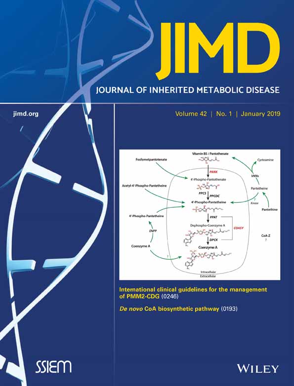
Two inborn errors of coenzyme A (CoA) metabolism are responsible for distinct forms of neurodegeneration with brain iron accumulation (NBIA), a heterogeneous group of neurodegenerative diseases having as a common denominator iron accumulation mainly in the inner portion of globus pallidus. Pantothenate kinase-associated neurodegeneration (PKAN), an autosomal recessive disorder with progressive impairment of movement, vision and cognition, is the most common form of NBIA and is caused by mutations in the pantothenate kinase 2 gene (PANK2), coding for a mitochondrial enzyme, which phosphorylates vitamin B5 in the first reaction of the CoA biosynthetic pathway. Another very rare but similar disorder, denominated CoPAN, is caused by mutations in coenzyme A synthase gene (COASY) coding for a bi-functional mitochondrial enzyme, which catalyzes the final steps of CoA biosynthesis. It still remains a mystery why dysfunctions in CoA synthesis lead to neurodegeneration and iron accumulation in specific brain regions, but it is now evident that CoA metabolism plays a crucial role in the normal functioning and metabolism of the nervous system.
REFERENCES
- 1Lipmann F, Kaplan NO. Coenzyme for acetylation, a pantothenic acid derivative. J Biol Chem. 1947; 167: 869.
- 2Srinivasan B, Sibon OCM. Coenzyme A, more than “just” a metabolic cofactor. Biochem Soc Trans. 2014; 42: 1075-1079. https://doi.org/10.1042/BST20140125.
- 3Leonardi R, Zhang Y-M, Rock CO, Jackowski S. Coenzyme A: back in action. Prog Lipid Res. 2005; 44: 125-153. https://doi.org/10.1016/j.plipres.2005.04.001.
- 4Baddiley J, Thain EM, Novelli GD, Lipmann F. Structure of coenzyme A. Nature. 1953; 171: 76. https://doi.org/10.1038/171076a0.
- 5Strauss E, Begley TP. The selectivity for cysteine over serine in coenzyme A biosynthesis. Chembiochem. 2005; 6: 284-286. https://doi.org/10.1002/cbic.200400340.
- 6Robishaw JD, Neely JR. Coenzyme A metabolism. Am J Phys. 1985; 248: E1-E9. https://doi.org/10.1152/ajpcell.1985.248.1.C1.
- 7Huang L, Khusnutdinova A, Nocek B, et al. A family of metal-dependent phosphatases implicated in metabolite damage-control. Nat Chem Biol. 2016; 12: 621-627. https://doi.org/10.1038/nchembio.2108.
- 8Sibon OC, Strauss E. Coenzyme A: to make it or uptake it? Nat Rev Mol Cell Biol. 2016; 17: 605-606. https://doi.org/10.1038/nrm.2016.110.
- 9Agrimi G, Russo A, Scarcia P, Palmieri F. The human gene SLC25A17 encodes a peroxisomal transporter of coenzyme A, FAD and NAD+. Biochem J. 2012; 443: 241-247. https://doi.org/10.1042/BJ20111420.
- 10Fiermonte G, Paradies E, Todisco S, Marobbio CMT, Palmieri F. A novel member of solute carrier family 25 (SLC25A42) is a transporter of coenzyme A and adenosine 3″,5-″diphosphate in human mitochondria. J Biol Chem. 2009; 284: 18152-18159. https://doi.org/10.1074/jbc.M109.0141182709381.
- 11Prohl C, Pelzer W, Diekert K, et al. The yeast mitochondrial carrier Leu5p and its human homologue Graves' disease protein are required for accumulation of coenzyme A in the matrix. Mol Cell Biol. 2001; 21: 1089-1097. https://doi.org/10.1128/MCB.21.4.1089-1097.200199563.
- 12Almannai M, Alsamri A, Alqasmi A, et al. Expanding the phenotype of SLC25A42-associated mitochondrial encephalomyopathy. Clin Genet. 2018; 93: 1097-1102. https://doi.org/10.1111/cge.13210.
- 13Srinivasan B, Baratashvili M, van der Zwaag M, et al. Extracellular 4′-phosphopantetheine is a source for intracellular coenzyme A synthesis. Nat Chem Biol. 2015; 11: 784-792. https://doi.org/10.1038/nchembio.1906.
- 14Gasmi L, McLennan AG. The mouse Nudt7 gene encodes a peroxisomal nudix hydrolase specific for coenzyme A and its derivatives. Biochem J. 2001; 357: 33-38. https://doi.org/10.1042/bj35700331221925.
- 15Ofman R, Speijer D, Leen R, Wanders RJA. Proteomic analysis of mouse kidney peroxisomes: identification of RP2p as a peroxisomal nudix hydrolase with acyl-CoA diphosphatase activity. Biochem J. 2006; 393: 537-543. https://doi.org/10.1042/BJ20050893.
- 16Zhou B, Westaway SK, Levinson B, Johnson MA, Gitschier J, Hayflick SJ. A novel pantothenate kinase gene (PANK2) is defective in Hallervorden-Spatz syndrome. Nat Genet. 2001; 28: 345-349. https://doi.org/10.1038/ng572.
- 17Gregory A, Hayflick S. Neurodegeneration with brain iron accumulation disorders overview. In: MP Adam, HH Ardinger, RA Pagon, et al., eds. GeneReviews. Seattle: University of Washington; 2014, 1993–2018. Available from: https://www.ncbi.nlm.nih.gov/books/NBK121988/.
- 18Hartig MB, Hörtnagel K, Garavaglia B, et al. Genotypic and phenotypic spectrum of PANK2 mutations in patients with neurodegeneration with brain iron accumulation. Ann Neurol. 2006; 59: 248-256. https://doi.org/10.1002/ana.20771.
- 19Schneider SA, Dusek P, Hardy J, et al. Genetics and pathophysiology of neurodegeneration with brain iron accumulation (NBIA). Curr Neuropharmacol. 2013; 11: 59-79. https://doi.org/10.2174/157015913804999469.
- 20Kotzbauer PT, Truax AC, Trojanowski JQ, Lee VM-Y. Altered neuronal mitochondrial coenzyme A synthesis in neurodegeneration with brain iron accumulation caused by abnormal processing, stability, and catalytic activity of mutant pantothenate kinase 2. J Neurosci. 2005; 25: 689-698. https://doi.org/10.1523/JNEUROSCI.4265-04.2005.
- 21Brunetti D, Dusi S, Morbin M, et al. Pantothenate kinase-associated neurodegeneration: altered mitochondria membrane potential and defective respiration in Pank2 knock-out mouse model. Hum Mol Genet. 2012; 21: 5294-5305. https://doi.org/10.1093/hmg/dds3803510755.
- 22Kruer MC, Hiken M, Gregory A, et al. Novel histopathologic findings in molecularly-confirmed pantothenate kinase-associated neurodegeneration. Brain. 2011; 134: 947-958. https://doi.org/10.1093/brain/awr0423105492.
- 23Leoni V, Strittmatter L, Zorzi G, et al. Metabolic consequences of mitochondrial coenzyme A deficiency in patients with PANK2 mutations. Mol Genet Metab. 2012; 105: 463-471.
- 24Aoun M, Corsetto PA, Nugue G, et al. Changes in red blood cell membrane lipid composition: a new perspective into the pathogenesis of PKAN. Mol Genet Metab. 2017; 121: 180-189. https://doi.org/10.1016/j.ymgme.2017.04.006.
- 25Di Meo I, Colombelli C, Srinivasan B, et al. Acetyl-4′-phosphopantetheine is stable in serum and prevents phenotypes induced by pantothenate kinase deficiency. Sci Rep. 2017; 7: 11260. https://doi.org/10.1038/s41598-017-11564-85595861.
- 26Siudeja K, Srinivasan B, Xu L, et al. Impaired coenzyme A metabolism affects histone and tubulin acetylation in Drosophila and human cell models of pantothenate kinase associated neurodegeneration. EMBO Mol Med. 2011; 3: 755-766. https://doi.org/10.1002/emmm.2011001803377114.
- 27Poli M, Derosas M, Luscieti S, et al. Pantothenate kinase-2 (Pank2) silencing causes cell growth reduction, cell-specific ferroportin upregulation and iron deregulation. Neurobiol Dis. 2010; 39: 204-210. https://doi.org/10.1016/j.nbd.2010.04.009.
- 28Santambrogio P, Dusi S, Guaraldo M, et al. Mitochondrial iron and energetic dysfunction distinguish fibroblasts and induced neurons from pantothenate kinase-associated neurodegeneration patients. Neurobiol Dis. 2015; 81: 144-153. https://doi.org/10.1016/j.nbd.2015.02.0304642744.
- 29Arber C, Angelova PR, Wiethoff S, et al. iPSC-derived neuronal models of PANK2-associated neurodegeneration reveal mitochondrial dysfunction contributing to early disease. PLoS One. 2017; 12: e0184104. https://doi.org/10.1371/journal.pone.01841045581181.
- 30Orellana DI, Santambrogio P, Rubio A, et al. Coenzyme A corrects pathological defects in human neurons of PANK2-associated neurodegeneration. EMBO Mol Med. 2016; 8: 1197-1211. https://doi.org/10.15252/emmm.2016063915048368.
- 31Kuo Y-M, Duncan JL, Westaway SK, et al. Deficiency of pantothenate kinase 2 (Pank2) in mice leads to retinal degeneration and azoospermia. Hum Mol Genet. 2005; 14: 49-57. https://doi.org/10.1093/hmg/ddi005.
- 32Garcia M, Leonardi R, Zhang Y-M, Rehg JE, Jackowski S. Germline deletion of pantothenate kinases 1 and 2 reveals the key roles for CoA in postnatal metabolism. PLoS One. 2012; 7: e40871. https://doi.org/10.1371/journal.pone.00408713398950.
- 33Zhang Y-M, Chohnan S, Virga KG, et al. Chemical knockout of pantothenate kinase reveals the metabolic and genetic program responsible for hepatic coenzyme A homeostasis. Chem Biol. 2007; 14: 291-302. https://doi.org/10.1016/j.chembiol.2007.01.0131892532.
- 34Brunetti D, Dusi S, Giordano C, et al. Pantethine treatment is effective in recovering the disease phenotype induced by ketogenic diet in a pantothenate kinase-associated neurodegeneration mouse model. Brain. 2014; 137: 57-68. https://doi.org/10.1093/brain/awt325.
- 35Bosveld F, Rana A, van der Wouden PE, et al. De novo CoA biosynthesis is required to maintain DNA integrity during development of the Drosophila nervous system. Hum Mol Genet. 2008; 17: 2058-2069. https://doi.org/10.1093/hmg/ddn105.
- 36Wu Z, Li C, Lv S, Zhou B. Pantothenate kinase-associated neurodegeneration: insights from a Drosophila model. Hum Mol Genet. 2009; 18: 3659-3672. https://doi.org/10.1093/hmg/ddp314.
- 37Zizioli D, Tiso N, Guglielmi A, et al. Knock-down of pantothenate kinase 2 severely affects the development of the nervous and vascular system in zebrafish, providing new insights into PKAN disease. Neurobiol Dis. 2016; 85: 35-48. https://doi.org/10.1016/j.nbd.2015.10.0104684146.
- 38Dusi S, Valletta L, Haack TB, et al. Exome sequence reveals mutations in CoA synthase as a cause of neurodegeneration with brain iron accumulation. Am J Hum Genet. 2014; 94: 11-22. https://doi.org/10.1016/j.ajhg.2013.11.0083882905.
- 39Rhee H-W, Zou P, Udeshi ND, et al. Proteomic mapping of mitochondria in living cells via spatially restricted enzymatic tagging. Science. 2013; 339: 1328-1331. https://doi.org/10.1126/science.12305933916822.
- 40Annesi G, Gagliardi M, Iannello G, Quattrone A, Iannello G, Quattrone A. Mutational analysis of COASY in an Italian patient with NBIA. Parkinsonism Relat Disord. 2016; 28: 150-151. https://doi.org/10.1016/j.parkreldis.2016.03.011.
- 41Evers C, Seitz A, Assmann B, et al. Diagnosis of CoPAN by whole exome sequencing: waking up a sleeping tiger's eye. Am J Med Genet A. 2017; 173: 1878-1886. https://doi.org/10.1002/ajmg.a.38252.
- 42Olzhausen J, Moritz T, Neetz T, Schüller H-J. Molecular characterization of the heteromeric coenzyme A-synthesizing protein complex (CoA-SPC) in the yeast Saccharomyces cerevisiae. FEMS Yeast Res. 2013; 13: 565-573. https://doi.org/10.1111/1567-1364.12058.
- 43Berti CC, Dallabona C, Lazzaretti M, et al. Modeling human coenzyme A synthase mutation in yeast reveals altered mitochondrial function, lipid content and iron metabolism. Microb Cell. 2015; 2: 126-135. https://doi.org/10.15698/mic2015.04.1965348974.
- 44Khatri D, Zizioli D, Tiso N, et al. Down-regulation of coasy, the gene associated with NBIA-VI, reduces bmp signaling, perturbs dorso-ventral patterning and alters neuronal development in zebrafish. Sci Rep. 2016; 6: 37660. https://doi.org/10.1038/srep376605124858.
- 45Zorzi G, Nardocci N. Therapeutic advances in neurodegeneration with brain iron accumulation. Int Rev Neurobiol. 2013; 110: 153-164. https://doi.org/10.1016/B978-0-12-410502-7.00008-9.
- 46Rana A, Seinen E, Siudeja K, et al. Pantethine rescues a Drosophila model for pantothenate kinase-associated neurodegeneration. Proc Natl Acad Sci U S A. 2010; 107: 6988-6993. https://doi.org/10.1073/pnas.09121051072872433.
- 47Schneider SA. Neurodegeneration with brain iron accumulation. Curr Neurol Neurosci Rep. 2016; 16: 9. https://doi.org/10.1007/s11910-015-0608-3.
- 48Christou Y-P, Tanteles GA, Kkolou E, et al. Open-label fosmetpantotenate, a phosphopantothenate replacement therapy in a single patient with atypical PKAN. Case Rep Neurol Med. 2017; 2017: 3247034. https://doi.org/10.1155/2017/3247034.
- 49Drecourt A, Babdor J, Dussiot M, et al. Impaired transferrin receptor palmitoylation and recycling in neurodegeneration with brain iron accumulation. Am J Hum Genet. 2018; 102: 266-277. https://doi.org/10.1016/j.ajhg.2018.01.0035985451.