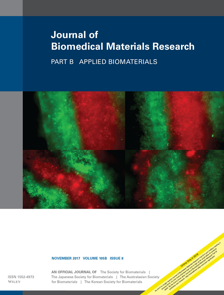
Novel silk protein barrier membranes for guided bone regeneration
Corresponding Author
Ralf Smeets
Department of Oral and Maxillofacial Surgery, University Medical Center Hamburg-Eppendorf, Hamburg, Germany
*These authors contributed equally to the study.
Correspondence to: R. Smeets; e-mail: [email protected]Search for more papers by this authorChristine Knabe
Department of Experimental Orofacial Medicine, Philipps University Marburg, Marburg, Germany
*These authors contributed equally to the study.
Search for more papers by this authorAndreas Kolk
Department of Oral- and Cranio-Maxillofacial Surgery, Klinikum rechts der Isar der Technischen Universität München, Munich, Germany
Search for more papers by this authorMichael Rheinnecker
Spintec Engineering GmbH, Kurbrunnenstraße 22, 52066 Aachen, Germany
Search for more papers by this authorAlexander Gröbe
Department of Oral and Maxillofacial Surgery, University Medical Center Hamburg-Eppendorf, Hamburg, Germany
Search for more papers by this authorMax Heiland
Department of Oral and Maxillofacial Surgery, University Medical Center Hamburg-Eppendorf, Hamburg, Germany
Search for more papers by this authorManuela Sachse
Department of Experimental Orofacial Medicine, Philipps University Marburg, Marburg, Germany
Search for more papers by this authorChristian Große-Siestrup
Spintec Engineering GmbH, Kurbrunnenstraße 22, 52066 Aachen, Germany
Search for more papers by this authorMichael Wöltje
Institute of Textile Machinery and High Performance Material Technology, Technische Universität Dresden, 01069 Dresden, Germany
*These authors contributed equally to the study.
Search for more papers by this authorHenning Hanken
Department of Oral and Maxillofacial Surgery, University Medical Center Hamburg-Eppendorf, Hamburg, Germany
*These authors contributed equally to the study.
Search for more papers by this authorCorresponding Author
Ralf Smeets
Department of Oral and Maxillofacial Surgery, University Medical Center Hamburg-Eppendorf, Hamburg, Germany
*These authors contributed equally to the study.
Correspondence to: R. Smeets; e-mail: [email protected]Search for more papers by this authorChristine Knabe
Department of Experimental Orofacial Medicine, Philipps University Marburg, Marburg, Germany
*These authors contributed equally to the study.
Search for more papers by this authorAndreas Kolk
Department of Oral- and Cranio-Maxillofacial Surgery, Klinikum rechts der Isar der Technischen Universität München, Munich, Germany
Search for more papers by this authorMichael Rheinnecker
Spintec Engineering GmbH, Kurbrunnenstraße 22, 52066 Aachen, Germany
Search for more papers by this authorAlexander Gröbe
Department of Oral and Maxillofacial Surgery, University Medical Center Hamburg-Eppendorf, Hamburg, Germany
Search for more papers by this authorMax Heiland
Department of Oral and Maxillofacial Surgery, University Medical Center Hamburg-Eppendorf, Hamburg, Germany
Search for more papers by this authorManuela Sachse
Department of Experimental Orofacial Medicine, Philipps University Marburg, Marburg, Germany
Search for more papers by this authorChristian Große-Siestrup
Spintec Engineering GmbH, Kurbrunnenstraße 22, 52066 Aachen, Germany
Search for more papers by this authorMichael Wöltje
Institute of Textile Machinery and High Performance Material Technology, Technische Universität Dresden, 01069 Dresden, Germany
*These authors contributed equally to the study.
Search for more papers by this authorHenning Hanken
Department of Oral and Maxillofacial Surgery, University Medical Center Hamburg-Eppendorf, Hamburg, Germany
*These authors contributed equally to the study.
Search for more papers by this authorAbstract
This study assesses the biocompatibility of novel silk protein membranes with and without modification, and evaluates their effect on facilitating bone formation and defect repair in guided bone regeneration. Two calvarian bone defects 12 mm in diameter were created in each of a total of 38 rabbits. Four different types of membranes, (silk-, hydroxyapatite-modified silk-, β-TCP-modified silk- and commonly clinically used collagen-membranes) were implanted to cover one of the two defects in each animal. Histologic analysis did not show any adverse tissue reactions in any of the defect sites indicating good biocompatibility of all silk protein membranes. Histomorphometric and histologic evaluation revealed that collagen and β-TCP modified silk membranes supported bone formation (collagen: bone area fraction p = 0.025; significant; β-TCP modified silk membranes bone area fraction: p = 0.24, not significant), guided bone regeneration and defect bridging. The bone, which had formed in defects covered by β-TCP modified silk membranes, displayed a more advanced stage of bone tissue maturation with restoration of the original calvarial bone microarchitecture when compared to the bone which had formed in defects, for which any of the other test membranes were used. Micro-CT analysis did not reveal any differences in the amount of bone formation between defects with and without membranes. In contrast to the collagen membranes, β-TCP modified silk membranes were visible in all cases and may therefore be advantageous for further supporting bone formation beyond 10 weeks and preventing soft tissue ingrowth from the periphery. © 2016 Wiley Periodicals, Inc. J Biomed Mater Res Part B: Appl Biomater, 105B: 2603–2611, 2017.
REFERENCES
- 1 Belser UC, Mericske-Stern R, Bernard JP, Taylor TD. Prosthetic management of the partially dentate patient with fixed implant restorations. Clin Oral Implants Res 2000; 11 Suppl 1: 126–145.
- 2 Bornstein MM, Lussi A, Schmid B, Belser UC, Buser D. Early loading of nonsubmerged titanium implants with a sandblasted and acid-etched (SLA) surface: 3-year results of a prospective study in partially edentulous patients. Int J Oral Maxillofac Implants 2003; 18(5): 659–666.
- 3 Wilson TG, Jr., Buser D. Advances in the use of guided tissue regeneration for localized ridge augmentation in combination with dental implants. Tex Dent J 1994; 111(7):5, 7–10.
- 4 Buser D, Dula K, Hess D, Hirt HP, Belser UC. Localized ridge augmentation with autografts and barrier membranes. Periodontol 2000 1999; 19: 151–163.
- 5 Buser D, Hoffmann B, Bernard JP, Lussi A, Mettler D, Schenk RK. Evaluation of filling materials in membrane–protected bone defects. A comparative histomorphometric study in the mandible of miniature pigs. Clin Oral Implants Res 1998; 9(3): 137–150.
- 6 Hammerle CH, Karring T. Guided bone regeneration at oral implant sites. Periodontol 2000 1998; 17: 151–175.
- 7 Kohal RJ, Trejo PM, Wirsching C, Hurzeler MB, Caffesse RG. Comparison of bioabsorbable and bioinert membranes for guided bone regeneration around non-submerged implants. An experimental study in the mongrel dog. Clin Oral Implants Res 1999; 10(3): 226–237.
- 8 Hardwick R, Hayes BK, Flynn C. Devices for dentoalveolar regeneration: an up-to-date literature review. J Periodontol 1995; 66(6): 495−505.
- 9 Kay SA, Wisner-Lynch L, Marxer M, Lynch SE. Guided bone regeneration: integration of a resorbable membrane and a bone graft material. Pract Periodont Aesthet Dent 1997; 9(2): 185−194; quiz 196.
- 10 von Arx T, Cochran DL, Schenk RK, Buser D. Evaluation of a prototype trilayer membrane (PTLM) for lateral ridge augmentation: An experimental study in the canine mandible. Int J Oral Maxillofac Surg 2002; 31(2): 190–199.
- 11
Knabe C,
Stiller M,
Ducheyne P. Dental graft materials, Oxford, UK. In: P Ducheyne, K Healy, DE Hutmacher, DW Grainger, CJ Kirkpatrick, editors. Comprehensive Biomaterials. Amsterdam: Elsevier Science; 2011. pp 305–324.
10.1016/B978-0-08-055294-1.00224-5 Google Scholar
- 12 Knabe C, Kraska B, Koch C, Gross U, Zreiqat H, Stiller M. A method for immunohistochemical detection of osteogenic markers in undecalcified bone sections. Biotech Histochem 2006; 81(1): 31–39.
- 13 Sodek J, Cheifitz S. Molecular regulation of osteogenesis. In: JE Davies, editor. Bone Engineering. Toronto, Canada: em squared Inc.; 2000. pp 31–43.
- 14 Aubin JE. Advances in the osteoblast lineage. Biochem Cell Biol 1998; 76(6): 899–910.
- 15 Aubin JE. Osteogenic cell differentiation. In: JE Davies, editor. Bone Engineering. Toronto, Canada: em squared Inc.; 2000. pp 19−30.
- 16 Farhadieh RD, Gianoutsos MP, Yu Y, Walsh WR. The role of bone morphogenetic proteins BMP-2 and BMP-4 and their related postreceptor signaling system (Smads) in distraction osteogenesis of the mandible. J Craniofac Surg 2004; 15(5): 714–718.
- 17 Knabe C, Koch C, Rack A, Stiller M. Effect of beta-tricalcium phosphate particles with varying porosity on osteogenesis after sinus floor augmentation in humans. Biomaterials 2008; 29(14): 2249–2258.
- 18 Knabe C, Nicklin S, Yu Y, Walsh WR, Radlanski RJ, Marks C, Hoffmeister B. Growth factor expression following clinical mandibular distraction osteogenesis in humans and its comparison with existing animal studies. J Craniomaxillofac Surg 2005; 33(6): 361–369.
- 19 Stiller M, Kluk E, Bohner M, Lopez-Heredia MA, Muller-Mai C, Knabe C. Performance of beta-tricalcium phosphate granules and putty, bone grafting materials after bilateral sinus floor augmentation in humans. Biomaterials 2014; 35(10): 3154–3163.
- 20 Tavakoli K, Yu Y, Shahidi S, Bonar F, Walsh WR, Poole MD. Expression of growth factors in the mandibular distraction zone: A sheep study. Br J Plast Surg 1999; 52(6): 434–439.
- 21 Stiller M, Rack A, Zabler S, Goebbels J, Dalugge O, Jonscher S, Knabe C. Quantification of bone tissue regeneration employing beta-tricalcium phosphate by three-dimensional non-invasive synchrotron micro-tomography—A comparative examination with histomorphometry. Bone 2009; 44(4): 619–628.
- 22 Dahlin C, Linde A, Gottlow J, Nyman S. Healing of bone defects by guided tissue regeneration. Plast Reconstr Surg 1988; 81(5): 672–676.
- 23 McAllister BS, Haghighat K. Bone augmentation techniques. J Periodontol 2007; 78(3): 377–396.
- 24 Schortinghuis J, Ruben JL, Raghoebar GM, Stegenga B. Therapeutic ultrasound to stimulate osteoconduction: A placebo controlled single blind study using e-PTFE membranes in rats. Arch Oral Biol 2004; 49(5): 413–420.
- 25 Schortinghuis J, Ruben JL, Raghoebar GM, Stegenga B, de Bont LG. Does ultrasound stimulate osteoconduction? A placebo-controlled single-blind study using collagen membranes in the rat mandible. Int J Oral Maxillofac Implants 2005; 20(2): 181–186.
- 26 Aaboe M, Pinholt EM, Schou S, Hjorting-Hansen E. Incomplete bone regeneration of rabbit calvarial defects using different membranes. Clin Oral Implants Res 1998; 9(5): 313–320.
- 27 Wiltfang J, Merten HA, Peters JH. Comparative study of guided bone regeneration using absorbable and permanent barrier membranes: A histologic report. Int J Oral Maxillofac Implants 1998; 13(3): 416–421.
- 28 Mueller AA, Rahn BA, Gogolewski S, Leiggener CS. Early dural reaction to polylactide in cranial defects in rabbits. Pediatr Neurosurg 2005; 41(6): 285–291.
- 29 He H, Huang J, Chen G, Dong Y. Application of a new bioresorbable film to guided bone regeneration in tibia defect model of the rabbits. J Biomed Mater Res A 2007; 82(1): 256–262.
- 30 Bergsma JE, de Bruijn WC, Rozema FR, Bos RR, Boering G. Late degradation tissue response to poly(l-lactide) bone plates and screws. Biomaterials 1995; 16(1): 25–31.
- 31 Luttikhuizen DT, van Amerongen MJ, de Feijter PC, Petersen AH, Harmsen MC, van Luyn MJ. The correlation between difference in foreign body reaction between implant locations and cytokine and MMP expression. Biomaterials 2006; 27(34): 5763–5770.
- 32 Stavropoulos F, Dahlin C, Ruskin JD, Johansson C. A comparative study of barrier membranes as graft protectors in the treatment of localized bone defects. An experimental study in a canine model. Clin Oral Implants Res 2004; 15(4): 435–442.
- 33 Kolk A, Handschel J, Drescher W, Rothamel D, Kloss F, Blessmann M, Heiland M, Wolff KD, Smeets R. Current trends and future perspectives of bone substitute materials—From space holders to innovative biomaterials. J Craniomaxillofac Surg 2012; 40(8): 706–718.
- 34 von Arx T, Broggini N, Jensen SS, Bornstein MM, Schenk RK, Buser D. Membrane durability and tissue response of different bioresorbable barrier membranes: A histologic study in the rabbit calvarium. Int J Oral Maxillofac Implants 2005; 20(6): 843–853.
- 35 Rothamel D, Schwarz F, Sager M, Herten M, Sculean A, Becker J. Biodegradation of differently cross-linked collagen membranes: An experimental study in the rat. Clin Oral Implants Res 2005; 16(3): 369–378.
- 36 Schwarz F, Rothamel D, Herten M, Sager M, Becker J. Angiogenesis pattern of native and cross-linked collagen membranes: An immunohistochemical study in the rat. Clin Oral Implants Res 2006; 17(4): 403–409.


