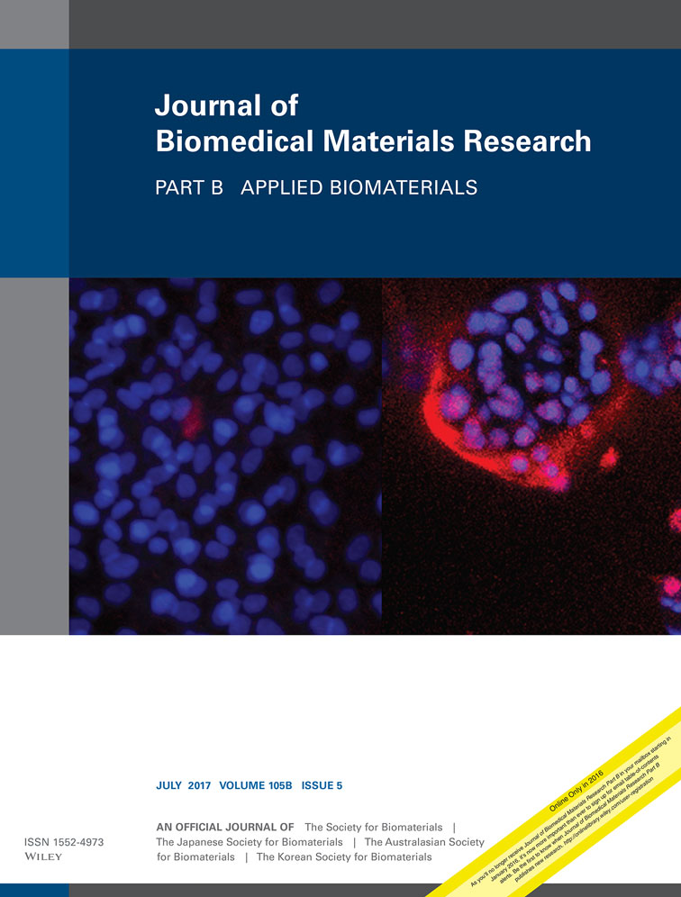
PCL/PHBV blended three dimensional scaffolds fabricated by fused deposition modeling and responses of chondrocytes to the scaffolds
Wasana Kosorn
Biomedical Engineering Research Unit, National Metal and Materials Technology Center, Klong Luang, Pathumthani, 12120 Thailand
Search for more papers by this authorMorakot Sakulsumbat
Biomedical Engineering Research Unit, National Metal and Materials Technology Center, Klong Luang, Pathumthani, 12120 Thailand
Search for more papers by this authorPaweena Uppanan
Biomedical Engineering Research Unit, National Metal and Materials Technology Center, Klong Luang, Pathumthani, 12120 Thailand
Search for more papers by this authorPakkanun Kaewkong
Biomedical Engineering Research Unit, National Metal and Materials Technology Center, Klong Luang, Pathumthani, 12120 Thailand
Search for more papers by this authorSurapol Chantaweroad
Biomedical Engineering Research Unit, National Metal and Materials Technology Center, Klong Luang, Pathumthani, 12120 Thailand
Search for more papers by this authorJaturong Jitsaard
Biomedical Engineering Research Unit, National Metal and Materials Technology Center, Klong Luang, Pathumthani, 12120 Thailand
Search for more papers by this authorKriskrai Sitthiseripratip
Biomedical Engineering Research Unit, National Metal and Materials Technology Center, Klong Luang, Pathumthani, 12120 Thailand
Search for more papers by this authorCorresponding Author
Wanida Janvikul
Biomedical Engineering Research Unit, National Metal and Materials Technology Center, Klong Luang, Pathumthani, 12120 Thailand
Correspondence to: W. Janvikul; e-mail: [email protected]Search for more papers by this authorWasana Kosorn
Biomedical Engineering Research Unit, National Metal and Materials Technology Center, Klong Luang, Pathumthani, 12120 Thailand
Search for more papers by this authorMorakot Sakulsumbat
Biomedical Engineering Research Unit, National Metal and Materials Technology Center, Klong Luang, Pathumthani, 12120 Thailand
Search for more papers by this authorPaweena Uppanan
Biomedical Engineering Research Unit, National Metal and Materials Technology Center, Klong Luang, Pathumthani, 12120 Thailand
Search for more papers by this authorPakkanun Kaewkong
Biomedical Engineering Research Unit, National Metal and Materials Technology Center, Klong Luang, Pathumthani, 12120 Thailand
Search for more papers by this authorSurapol Chantaweroad
Biomedical Engineering Research Unit, National Metal and Materials Technology Center, Klong Luang, Pathumthani, 12120 Thailand
Search for more papers by this authorJaturong Jitsaard
Biomedical Engineering Research Unit, National Metal and Materials Technology Center, Klong Luang, Pathumthani, 12120 Thailand
Search for more papers by this authorKriskrai Sitthiseripratip
Biomedical Engineering Research Unit, National Metal and Materials Technology Center, Klong Luang, Pathumthani, 12120 Thailand
Search for more papers by this authorCorresponding Author
Wanida Janvikul
Biomedical Engineering Research Unit, National Metal and Materials Technology Center, Klong Luang, Pathumthani, 12120 Thailand
Correspondence to: W. Janvikul; e-mail: [email protected]Search for more papers by this authorAbstract
In this study, poly(ε-caprolactone)/poly(3-hydroxybutyrate-co−3-hydroxyvalerate) (PCL/PHBV) blended porous scaffolds were fabricated by fused deposition modeling (FDM). PCL/PHBV filaments, initially prepared at different weight ratios, that is, 100/0, 75/25, 50/50, and 25/75, were fabricated by the lay-down pattern of 0/90/45/135° to obtain scaffolds with dimension of 6.0 × 6.0 × 2.5 mm3 and average filament diameters and channel sizes in the ranges of 370–390 µm and 190–210 µm, respectively. To enhance the surface hydrophilicity of the materials, the scaffolds were subsequently subjected to a low pressure oxygen plasma treatment. The untreated and plasma-treated scaffolds were comparatively characterized, in terms of surface properties, mechanical strength, and biological properties. From SEM, AFM, water contact angle, and XPS results, the surface roughness, wettability, and hydrophilicity of the blended scaffolds were found to be enhanced after plasma treatment, while the compressive strength of the scaffolds was scarcely changed. It was, however, found to increase with an increasing content of PHBV incorporated. The porcine chondrocytes exhibited higher proliferative capacity and chondrogenic potential when being cultured on the scaffolds with greater PHBV contents, especially when they were plasma-treated. The PCL/PHBV scaffolds were proven to possess good physical, mechanical, and biological properties that could be appropriately used in articular cartilage regeneration. © 2016 Wiley Periodicals, Inc. J Biomed Mater Res Part B: Appl Biomater, 105B: 1141–1150, 2017.
REFERENCES
- 1 Liu C, Xia Z, Czernuszka JT. Design and development of three-dimensional scaffolds for tissue engineering. Chem Eng Res Des 2007; 85: 1051–1064.
- 2 Puppi D, Chiellini F, Piras AM, Chiellini E. Polymeric materials for bone and cartilage repair. Prog Polym Sci 2010; 35: 403–440.
- 3 Ergun A, Yu X, Valdevit A, Ritter A, Kalyon DM. In vitro analysis and mechanical properties of twin screw extruded single-layered and coextruded multilayered poly(caprolactone) scaffolds seeded with human fetal osteoblasts for bone tissue engineering. J Biomed Mater Res A 2011; 99A: 354–366.
- 4 Reignier J, Huneault MA. Preparation of interconnected poly(3-caprolactone) porous scaffolds by a combination of polymer and salt particulate leaching. Polymer 2006; 47: 4703–4717.
- 5 Lebourg M, Anton JS, Gomez Ribelles JL. Porous membranes of PLLA-PCL blend for tissue engineering applications. Eur Polym J 2008; 44: 2207–2218.
- 6 Du Y, Chen X, Koh Y-H, Lei B. Facilely fabricating PCL nano fibrous scaffolds with hierarchicalpore structure for tissue engineering. Mater Lett 2014; 122: 62–65.
- 7 Annabi N, Fathi A, Mithieux SM, Weiss AS, Dehghani F. Fabrication of porous PCL/elastin composite scaffolds for tissue engineering applications. J Supercrit Fluids 2011; 59: 157–167.
- 8 Salerno A, Di Maioc E, Iannaceb S, Nettia PA. Solid-state supercritical CO2 foaming of PCL and PCL-HA nano-composite: Effect of composition, thermal history and foaming process on foam pore structure. J Supercrit Fluids 2011; 58: 158–167.
- 9
Parthasarathy J,
Starly B,
Raman S. A design for the additive manufacture of functionally graded porous structures with tailored mechanical properties for biomedical applications. J Manuf Processes 2011; 13: 160–170.
10.1016/j.jmapro.2011.01.004 Google Scholar
- 10 Fang Z, Starly B, Sun W. Computer-aided characterization for effective mechanical properties of porous tissue scaffolds. Comput-Aided Des 2005; 37: 65–72.
- 11
Sun W,
Starly B,
Nam J,
Darling A. Bio-CAD modeling and its applications in computer-aided tissue engineering. Comput-Aided Des 2005; 37: 1097–1114.
10.1016/j.cad.2005.02.002 Google Scholar
- 12 Woodfield TBF, Malda J, de Wijn J, Péters F, Riesle J, van Blitterswijk CA. Design of porous scaffolds for cartilage tissue engineering using a three-dimensional fiber-deposition technique. Biomaterials 2004; 25: 4149–4161.
- 13 Zein I, Hutmacher D, Tan KC, Teoh SH. Fused deposition modeling of novel scaffold architectures for tissue engineering applications. Biomaterials 2002; 23: 1169–1185.
- 14 Kim JY, Cho D-W. Blended PCL/PLGA scaffold fabrication using multi-head deposition system. Microelectron Eng 2009; 86: 1447–1450.
- 15 Chiono V, Ciardelli G, Vozzi G, Sotgiu MG, Vinci B, Domenici C, Giusti P. Poly(3-hydroxybutyrate-co−3-hydroxyvalerate)/poly(ε-caprolactone) blends for tissue engineering applications in the form of hollow fibers. J Biomed Mater Res A 2008; 85A: 938–953.
- 16 Gardel LS, Correia-Gomes C, Serra LA, Gomes ME, Reis RL. A novel bidirectional continuous perfusion bioreactor for the culture of large-sized bone tissue-engineered constructs. J Biomed Mater Res B Appl Biomater 2013; 101: 1377–1386.
- 17 Link DP, Gardel LS, Correlo VM, Gomes ME, Reis RL. Osteogenic properties of starch poly(ε-caprolactone) (SPCL) fiber meshes loaded with osteoblast-like cells in a rat critical-sized cranial defect. J Biomed Mater Res A. 2013; 101: 3059–3065.
- 18
Yoshinari M,
Matsuzaka K,
Inoue T. Surface modification by cold-plasma technique for dental implants-Bio-functionalization with binding pharmaceuticals. Jpn Dent Sci Rev 2011; 47: 89–101.
10.1016/j.jdsr.2011.03.001 Google Scholar
- 19 Chu PK, Chen JY, Wang LP, Plasma-surface modification of biomaterials. Mater Sci Eng R Rep 2002; 36: 143–206.
- 20 Ma Z, Mao Z, Gao C. Surface modification and property analysis of biomedical polymers used for tissue engineering. Colloids Surf B 2007; 60: 137–157.
- 21 Jacobs T, Geyter ND, Morent R, Desmet T, Dubruel P, Leys C. Plasma treatment of polycaprolactone at medium pressure. Surf Coat Int 2011; 205: S543–547.
- 22 Yildirim ED, Pappas D, Güçeri S, Sun W. Enhanced cellular functions on polycaprolactone tissue scaffolds by O2 plasma surface modification. Plasma Processes Polym 2011; 8: 256–267.
- 23 Domingos M, Intranuovo F, Gloria A, Gristina R, Ambrosio L, Bártolo PJ, Favia P. Improved osteoblast cell affinity on plasma-modified 3-D extruded PCL. Acta Biomater 2013; 9: 5997–6005.
- 24 Lee H-U, Jeong Y-S, Jeong S-Y, Park S-Y, Bae J-S, Kim H-G, Cho C-R. Role of reactive gas in atmospheric plasma for cell attachment and proliferation on biocompatible poly ε-caprolactone film. Appl Surf Sci 2008; 254: 5700–5705.
- 25 Lee H-U, Kang Y-H, Jeong S-Y, Koh K, Kim J-P, Bae J-S, Cho C-R. Long-term aging characteristics of atmospheric-plasma-treated poly(ε-caprolactone) films and fibers. Polym Degrad Stab 2011; 96: 1204–1209.
- 26 Ferreira BMP, Pinheiro LMP, Nascente PAP, Ferreira MJ, Duek EAR. Plasma surface treatments of poly(L-lactic acid) (PLLA) and poly(hydroxybutyrate-co-hydroxyvalerate) (PHBV). Mater Sci Eng C 2009; 29: 806–813.
- 27 Xiang HX, Chen SH, Cheng, Zhou Z, Zhu MF. Structural characteristics and enhanced mechanical and thermal properties of full biodegradable tea polyphenol/poly(3-hydroxybutyrate-co-3-hydroxyvalerate) composite films. Express Polym Lett 2013; 7: 778–786.
- 28 Shepherd DE, Seedhom BB. Thickness of human articular cartilage in joints of the lower limb. Ann Rheum Dis 1999; 58: 27–34.
- 29 Fu X, Sammons RL, BertÓti I, Jenkins MJ, Dong H. Active screen plasma surface modification of polycaprolactone to improve cell attachment. J Biomed Mater Res B 2012: 314–320.
- 30 Wan Y, Tu C, Yang J, Bei J, Wang S. Influences of ammonia plasma treatment on modifying depth and degradation of poly(L-lactide) scaffolds. Biomaterials 2006; 27: 2699–2704.
- 31 Gogolewski S, Jovanovic M, Perren SM, Dillon JG, Hughes MK. Tissue response and in vivo degradation of selected polyhydroxyacids: polylactides (PLA), poly(3-hydroxybutyrate) (PHB), and poly(3-hydroxybutyrate)-co-(3-hydroxyvalerate) (PHB/VA). J Biomed Mater Res A 1993; 27: 1135–1148.
- 32 Kösea GT, Korkusuzb F, Özkulc A, Soysald Y, Özdemire T, Yildize C, Hasircif V. Tissue engineered cartilage on collagen and PHBV matrices. Biomaterials 2005; 26: 5187–5197.
- 33 Kösea GT, Korkusuzb F, Korkusuzc P, Puralid N, Özkule A, Hasırcı V. Bone generation on PHBV matrices: An in vitro study. Biomaterials 2003; 24: 4999–5007.
- 34 GlowackiJ, Trepman E, Folkman J. Cell shape and phenotypic expression in chondrocytes. Proc Soc Exp Biol Med 1983; 172: 93–98.
- 35 Brodkin KR, García AJ, Levenston ME, Chondrocyte phenotypes on different extracellular matrix monolayers. Biomaterials 2004; 25: 5929–5938.
- 36 Matsumoto E, Furumatsu T, Kanazawa T, Tamura M, Ozaki T. ROCK inhibitor prevents the dedifferentiation of human articular chondrocytes. Biochem Biophys Res Commun 2012; 420: 124–129.


