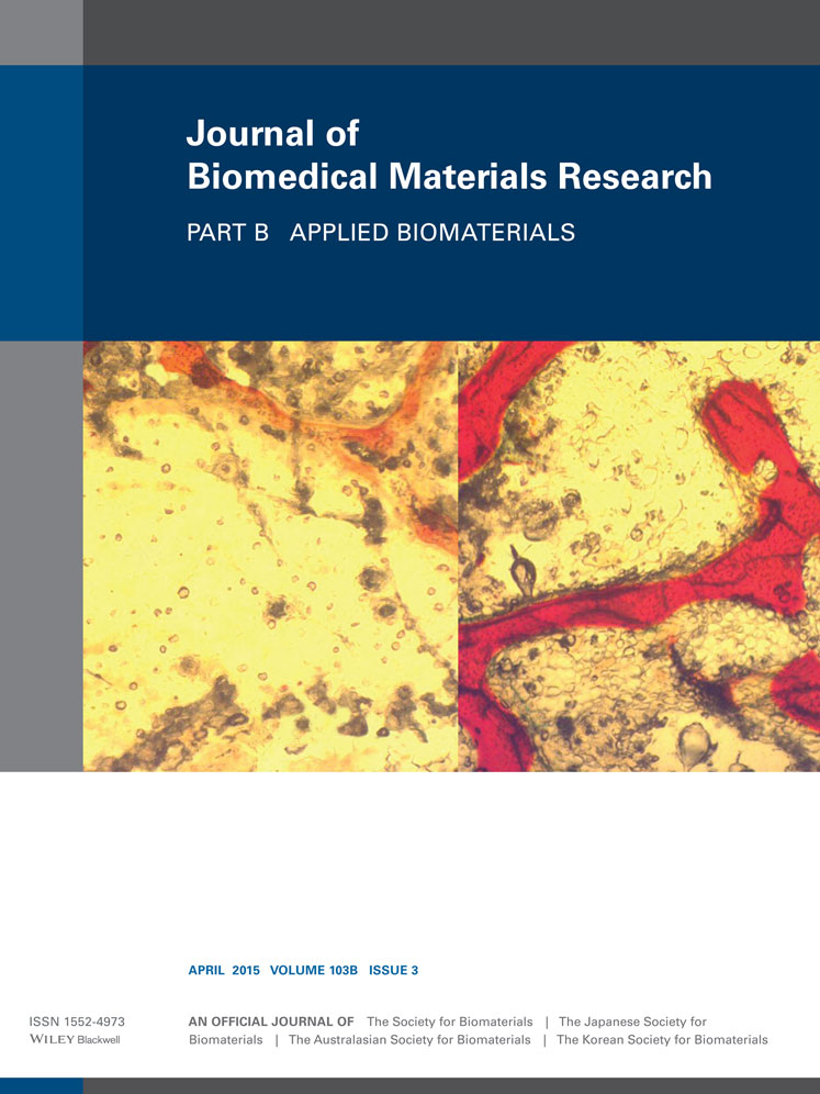
SrO- and MgO-doped microwave sintered 3D printed tricalcium phosphate scaffolds: Mechanical properties and in vivo osteogenesis in a rabbit model
Solaiman Tarafder
W.M. Keck Biomedical Materials Research Laboratory, School of Mechanical and Materials Engineering, Washington State University, Pullman, Washington, 99164
Search for more papers by this authorWilliam S. Dernell
Department of Veterinary Clinical Sciences, College of Veterinary Medicine, Washington State University, Pullman, Washington, 99164
Search for more papers by this authorAmit Bandyopadhyay
W.M. Keck Biomedical Materials Research Laboratory, School of Mechanical and Materials Engineering, Washington State University, Pullman, Washington, 99164
Search for more papers by this authorCorresponding Author
Susmita Bose
W.M. Keck Biomedical Materials Research Laboratory, School of Mechanical and Materials Engineering, Washington State University, Pullman, Washington, 99164
Correspondence to: S. Bose (e-mail: [email protected])Search for more papers by this authorSolaiman Tarafder
W.M. Keck Biomedical Materials Research Laboratory, School of Mechanical and Materials Engineering, Washington State University, Pullman, Washington, 99164
Search for more papers by this authorWilliam S. Dernell
Department of Veterinary Clinical Sciences, College of Veterinary Medicine, Washington State University, Pullman, Washington, 99164
Search for more papers by this authorAmit Bandyopadhyay
W.M. Keck Biomedical Materials Research Laboratory, School of Mechanical and Materials Engineering, Washington State University, Pullman, Washington, 99164
Search for more papers by this authorCorresponding Author
Susmita Bose
W.M. Keck Biomedical Materials Research Laboratory, School of Mechanical and Materials Engineering, Washington State University, Pullman, Washington, 99164
Correspondence to: S. Bose (e-mail: [email protected])Search for more papers by this authorAbstract
The presence of interconnected macro pores allows guided tissue regeneration in tissue engineering scaffolds. However, highly porous scaffolds suffer from having poor mechanical strength. Previously, we showed that microwave sintering could successfully be used to improve mechanical strength of macro porous tricalcium phosphate (TCP) scaffolds. This study reports the presence of SrO and MgO as dopants in TCP scaffolds improves mechanical and in vivo biological performance. We have used direct three dimensional printing (3DP) technology for scaffold fabrication. These 3DP scaffolds possessed multiscale porosity, that is, 3D interconnected designed macro pores along with intrinsic micro pores. A significant increase in mechanical strength, between 37 and 41%, was achieved due to SrO and MgO doping in TCP as compared with pure TCP. Maximum compressive strengths of 9.38 ± 1.86 MPa and 12.01 ± 1.56 MPa were achieved by conventional and microwave sintering, respectively, for SrO-MgO-doped 3DP scaffolds with 500 μm designed pores. Histomorphological and histomorphometric analysis revealed a significantly higher osteoid, bone and haversian canal formation induced by the presence of SrO and MgO dopants in 3DP TCP as compared with pure TCP scaffolds when tested in rabbit femoral condyle defect model. Increased osteon and thus enhanced network of blood vessel formation, and osteocalcin expression were observed in the doped TCP scaffolds. Our results show that these 3DP SrO-MgO-doped TCP scaffolds have the potential for early wound healing through accelerated osteogenesis and vasculogenesis. © 2014 Wiley Periodicals, Inc. J Biomed Mater Res Part B: Appl Biomater, 103B: 679–690, 2015.
REFERENCES
- 1Kneser U, Schaefer DJ, Polykandriotis E, Horch RE. Tissue engineering of bone: The reconstructive surgeon's point of view. J Cell Mol Med 2006; 10: 7–19.
- 2Becker ST, Warnke PH, Behrens E, Wiltfang J. Morbidity after iliac crest bone graft harvesting over an anterior versus posterior approach. J Oral Maxillofacial Sur 2011; 69: 48–53.
- 3Silber JS, Anderson DG, Daffner SD, Brislin BT, Leland JM, Hilibrand AS, Vaccaro AR, Albert TJ. Donor site morbidity after anterior iliac crest bone harvest for single-level anterior cervical discectomy and fusion. Spine 2003; 28: 134–139.
- 4Zimmermann G, Moghaddam A. Allograft bone matrix versus synthetic bone graft substitutes. Injury 2011; 42 (S2): S16–S21.
- 5Gross RH. The use of bone grafts and bone graft substitutes in pediatric orthopaedics. J Pediatric Orthopaedics 2012; 32: 100–105.
- 6Kolk A, Handschel J, Drescher W, Rothamel D, Kloss F, Blessmann M, et al. Current trends and future perspectives of bone substitute materials—From space holders to innovative biomaterials. J Cranio-Maxillofacial Surg 2012; 40: 706–718.
- 7Tarafder S, Bodhak S, Bandyopadhyay A, Bose S. Effect of Electrical Polarization and Composition of Biphasic Calcium Phosphates on Early Stage Osteoblast Interactions. J Biomed Mater Res Part B: Appl Biomater 2011; 97B: 306–314.
- 8Tarafder S, Balla VK, Davies NM, Bandyopadhyay A, Bose S. Microwave-sintered 3D printed tricalcium phosphate scaffolds for bone tissue engineering. J Tissue Eng Regen Med 2013; 7: 631–641.
- 9LeGeros RZ. Calcium Phosphate-Based Osteoinductive Materials. Chem Rev 2008; 108: 4742–4753.
- 10Bose S, Tarafder S. Calcium phosphate ceramic systems in growth factor and drug delivery for bone tissue engineering: A review. Acta Biomater. 2012; 8: 1401–1421.
- 11Rey C. Calcium phosphate biomaterials and bone mineral. Differences in composition, structures and properties. Biomaterials 1990; 11: 13–15.
- 12Bandyopadhyay A, Bernard S, Xue W, Bose S. Calcium Phosphate-Based Resorbable Ceramics: Influence of MgO, ZnO, and SiO2 Dopants. J Am Ceram Soc 2006; 89: 2675–2688.
- 13Banerjee SS, Tarafder S, Davies NM, Bandyopadhyay A, Bose S. Understanding the influence of MgO and SrO binary doping on the mechanical and biological properties of b-TCP ceramics. Acta Biomater 2010; 6: 4167–4174.
- 14Seyednejad H, Gawlitta D, Kuiper RV, de Bruin A, van Nostrum CF, Vermonden T, et al. In vivo biocompatibility and biodegradation of 3D-printed porous scaffolds based on a hydroxyl-functionalized poly(ε-caprolactone). Biomaterials 2012; 33: 4309–4318.
- 15Jones AC, Arns CH, Hutmacher DW, Milthorpe BK, Sheppard AP, Knackstedt MA. The correlation of pore morphology, interconnectivity and physical properties of 3D ceramic scaffolds with bone ingrowth. Biomaterials 2009; 30: 1440–1451.
- 16Karageorgiou V, Kaplan D. Porosity of 3D biomaterial scaffolds and osteogenesis. Biomaterials 2005; 26: 5474–5491.
- 17Ramay HRR, Zhang M. Biphasic calcium phosphate nanocomposite porous scaffolds for load-bearing bone tissue engineering. Biomaterials 2004; 25: 5171–5180.
- 18Sicchieri LG, Crippa GE, de Oliveira PT, Beloti MM, Rosa AL. Pore size regulates cell and tissue interactions with PLGA-CaP scaffolds used for bone engineering. J Tissue Eng Regen Med 2012; 6: 155–162.
- 19Yang S, Leong K-F, Du Z, Chua C-K. The Design of Scaffolds for Use in Tissue Engineering. Part II. Rapid Prototyping Techniques. Tissue Eng 2002; 8: 1–11.
- 20Qi QQ, Chen JD, Gao SZ, Bu J, Qiu ZP. Preparation Cell Scaffolds with Well Defined Pore Structure Through Elastic Porogen/Pressure Filtration. Adv Mater Res 2011; 236-238: 1897–1901.
- 21Sachlos E, Czernuszka J. Making tissue engineering scaffolds work. Eur Cells Mater 2003; 5: 29–40.
- 22Tarafder S, Davies NM, Bandyopadhyay A, Bose S. 3D Printed Tricalcium Phosphate Scaffolds: Effect of SrO and MgO Doping on in vivo osteogenesis in rat distal femoral defect model. Biomater Sci 2013; 1: 1250–1259.
- 23Butscher A, Bohner M, Roth C, Ernstberger A, Heuberger R, Doebelin N, et al. Printability of calcium phosphate powders for three-dimensional printing of tissue engineering scaffolds. Acta Biomater 2012; 8: 373–385.
- 24Fielding GA, Bandyopadhyay A, Bose S. Effects of silica and zinc oxide doping on mechanical and biological properties of 3D printed tricalcium phosphate tissue engineering scaffolds. Dent Mater 2012; 28: 113–122.
- 25Khalyfa A, Vogt S, Weisser J, Grimm G, Rechtenbach A, Meyer W, et al. Development of a new calcium phosphate powder-binder system for the 3D printing of patient specific implants. J Mater Sci: Mater Med 2007; 18: 909–916.
- 26Kalita SJ, Ferguson M. Fabrication of 3-D Porous Mg/Zn doped Tricalcium Phosphate Bone-Scaffolds via the Fused Deposition Modelling. Am J Biochem Biotechnol 2006; 2: 57–60.
- 27Yadoji P, Peelamedu R, Agrawal D, Roy R. Microwave sintering of Ni-Zn ferrites: comparison with conventional sintering. Mater Sci Eng B 2003; 98: 269–278.
- 28Bose S, Dasgupta S, Tarafder S, Bandyopadhyay A. Microwave-processed nanocrystalline hydroxyapatite: Simultaneous enhancement of mechanical and biological properties. Acta Biomater 2010; 6: 3782–3790.
- 29Chanda A, Dasgupta S, Bose S, Bandyopadhyay A. Microwave sintering of calcium phosphate ceramics. Mater Sci Eng: C 2009; 29: 1144–1149.
- 30Nightingale SA, Worner HK, Dunne DP. Microstructural Development during the Microwave Sintering of Yttria-Zirconia Ceramics. J Am Ceram Soc 2005; 80: 394–400.
10.1111/j.1151-2916.1997.tb02843.x Google Scholar
- 31Seeley Z, Bandyopadhyay A, Bose S. Tricalcium phosphate based resorbable ceramics: Influence of NaF and CaO addition. Mater Sci Eng C 2008; 28: 11–17.
- 32Seeley Z, Bandyopadhyay A, Bose S. Influence of TiO2 and Ag2O addition on tricalcium phosphate ceramics. J Biomed Mater Res A 2007; 82: 113–121.
- 33Bose S, Tarafder S, Banerjee SS, Davies NM, Bandyopadhyay A. Understanding in vivo response and mechanical property variation in MgO, SrO and SiO2 doped β-TCP. Bone 2011; 48: 1282–1290.
- 34Pors Nielsen S. The biological role of strontium. Bone 2004; 35: 583–588.
- 35Lakhkar NJ, Lee I-H, Kim H-W, Salih V, Wall IB, Knowles JC. Bone formation controlled by biologically relevant inorganic ions: Role and controlled delivery from phosphate-based glasses. Adv Drug Deliv Rev 2013; 65: 405–420.
- 36Bose S, Fielding G, Tarafder S, Bandyopadhyay A. Understanding of dopant-induced osteogenesis and angiogenesis in calcium phosphate ceramics. Trends Biotechnol 2013: 31: 594–605.
- 37Staiger MP, Pietak AM, Huadmai J, Dias G. Magnesium and its alloys as orthopedic biomaterials: A review. Biomaterials 2006; 27: 1728–1734.
- 38Locke M. Structure of long bones in mammals. J Morphol 2004; 262: 546–565.
- 39Robling AG, Castillo AB, Turner CH. Biomechanical and Molecular Regulation of Bone Remodeling. Annu Rev Biomedl Eng 2006; 8: 455–498.
- 40Lan Levengood SK, Polak SJ, Wheeler MB, Maki AJ, Clark SG, Jamison RD, et al. Multiscale osteointegration as a new paradigm for the design of calcium phosphate scaffolds for bone regeneration. Biomaterials 2010; 31: 3552–3563.
- 41Habibovic P, Yuan H, van der Valk CM, Meijer G, van Blitterswijk CA, de Groot K. 3D microenvironment as essential element for osteoinduction by biomaterials. Biomaterials 2005; 26: 3565–3575.
- 42Hing KA, Annaz B, Saeed S, Revell PA, Buckland T. Microporosity enhances bioactivity of synthetic bone graft substitutes. J Mater Sci: Mater Med 2005; 16: 467–475.
- 43Perera FH, Martínez-Vázquez FJ, Miranda P, Ortiz AL, Pajares A. Clarifying the effect of sintering conditions on the microstructure and mechanical properties of [beta]-tricalcium phosphate. Ceram Int 2010; 36: 1929–1935.
- 44Marchia J, Dantasa ACS, Greila P, Bressianib JC, Bressianib AHA, Müller FA. Influence of Mg-substitution on the physicochemical properties of calcium phosphate powders. Mater Res Bull 2007; 42: 1040–1050.
- 45Yin X, Calderin L, Stott MJ, Sayer M. Density functional study of structural, electronic and vibrational properties of Mg- and Zn-doped tricalcium phosphate biomaterials. Biomaterials 2002; 23: 4155–4163.
- 46Yang F, Yang D, Tu J, Zheng Q, Cai L, Wang L. Strontium Enhances Osteogenic Differentiation of Mesenchymal Stem Cells and In Vivo Bone Formation by Activating Wnt/Catenin Signaling. Stem Cells 2011; 29: 981–991.
- 47Pazzaglia UE, Congiu T, Marchese M, Spagnuolo F, Quacci D. Morphometry and Patterns of Lamellar Bone in Human Haversian Systems. The Anat Rec 2012; 295: 1421–1429.
- 48Swaminathan R. Biochemical markers of bone turnover. Clinica Chimica Acta 2001; 313: 95–105.
- 49Glover SJ, Eastell R, McCloskey EV, Rogers A, Garnero P, Lowery J, et al. Rapid and robust response of biochemical markers of bone formation to teriparatide therapy. Bone 2009; 45: 1053–1058.


