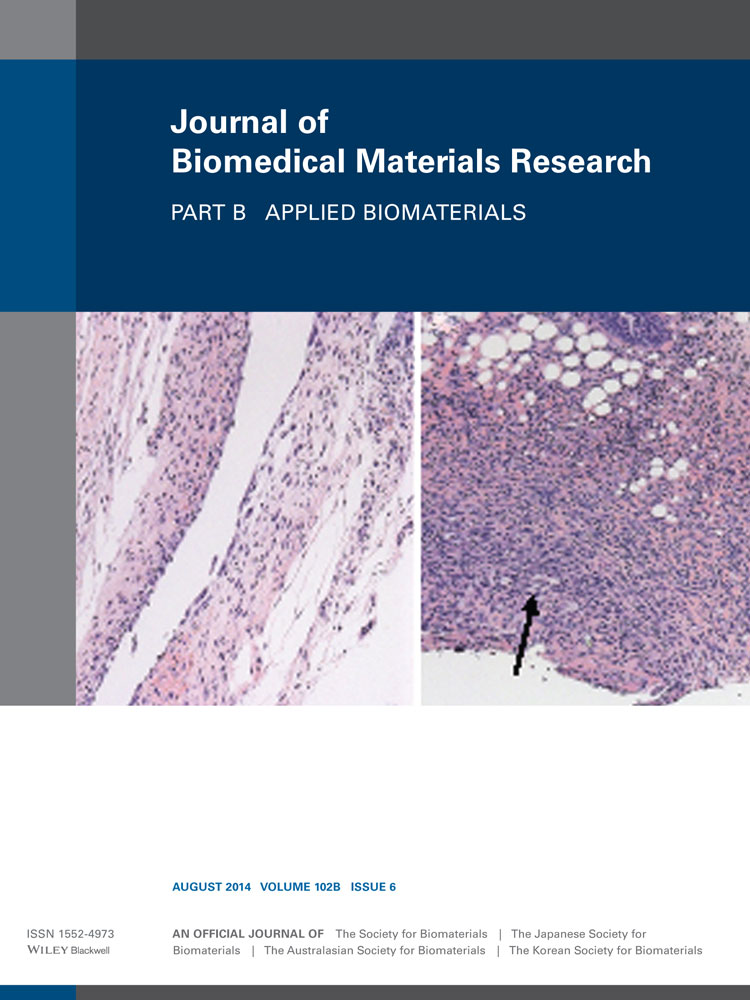
Chitosan–cellulose composite for wound dressing material. Part 2. Antimicrobial activity, blood absorption ability, and biocompatibility
April L. Harkins
Department of Chemistry, Marquette University, P. O. Box 1881, Milwaukee, Wisconsin, 53201
Search for more papers by this authorSimon Duri
Department of Chemistry, Marquette University, P. O. Box 1881, Milwaukee, Wisconsin, 53201
Search for more papers by this authorLuther C. Kloth
Department of Chemistry, Marquette University, P. O. Box 1881, Milwaukee, Wisconsin, 53201
Search for more papers by this authorCorresponding Author
Chieu D. Tran
Department of Chemistry, Marquette University, P. O. Box 1881, Milwaukee, Wisconsin, 53201
Correspondence to: C.D. Tran (e-mail: [email protected])Search for more papers by this authorApril L. Harkins
Department of Chemistry, Marquette University, P. O. Box 1881, Milwaukee, Wisconsin, 53201
Search for more papers by this authorSimon Duri
Department of Chemistry, Marquette University, P. O. Box 1881, Milwaukee, Wisconsin, 53201
Search for more papers by this authorLuther C. Kloth
Department of Chemistry, Marquette University, P. O. Box 1881, Milwaukee, Wisconsin, 53201
Search for more papers by this authorCorresponding Author
Chieu D. Tran
Department of Chemistry, Marquette University, P. O. Box 1881, Milwaukee, Wisconsin, 53201
Correspondence to: C.D. Tran (e-mail: [email protected])Search for more papers by this authorAbstract
Chitosan (CS), a polysaccharide derived from chitin, the second most abundant polysaccharide, is widely used in the medical world because of its natural and nontoxic properties and its innate ability for antibacterial and hemostasis effects. In this study, the novel composites containing CS and cellulose (CEL) (i.e., [CEL + CS]), which we have previously synthesized using a green and totally recyclable method, were investigated for their antimicrobial activity, absorption of anticoagulated whole blood, anti-inflammatory activity through the reduction of tumor necrosis factor-α (TNF-α) and interleukin-6 (IL-6), and the biocompatibility with human fibroblasts. The [CEL + CS] composites were found to inhibit the growth of both Gram positive and negative micro-organisms. For examples, the regenerated 100% lyophilized chitosan material was found to reduce growth of Escherichia coli (ATCC 8739 and vancomycin resistant Enterococcus faecalis (ATCC 51299) by 78, 36, and 64%, respectively. The composites are nontoxic to fibroblasts; that is, fibroblasts, which are critical to the formation of connective tissue matrix were found to grow and proliferate in the presence of the composites. They effectively absorb blood, and at the same rate and volume as commercially available wound dressings. The composites, in both air-dried and lyophilized forms, significantly inhibit the production of TNF-α and IL-6 by stimulated macrophages. These results clearly indicate that the biodegradable, biocompatible and nontoxic [CEL + CS] composites, particularly those dried by lyophilizing, can be effectively used as a material in wound dressings. © 2014 Wiley Periodicals, Inc. J Biomed Mater Res Part B: Appl Biomater, 102B: 1199–1206, 2014.
REFERENCES
- 1Dai T, Tegos GP, Barkatovskaya M, Castano AP, Hamblin MR. Chitosan acetate bandage as a topical antimicrobial dressing for infected burns. Antimicrob Agents Chemother 2009; 53: 393–400.
- 2Bordenave N, Grelier S, Coma V. Hydrophobization and antimicrobial activity of chitosan and paper-based packaging materials. Biomacromolecules 2010; 11: 88–96.
- 3Rabea EI, Badawy M, Stevens CV, Smagghe G, Steurbaut W. Chitosan as antimicrobial agent: Applications and mode of action. Biomacromolecules 2003; 4: 1457–1465.
- 4Altiok D, Altiok E, Tihminlioglu F. Physical, antibacterial and antioxidant properties of chitosan films incorporated with thyme oil for potential wound healing applications. J Mater Sci Mater Med 2010; 21: 2227–2236.
- 5Burkatovskaya M, Tegos GP, Swietlik E, Demidova TN, Castano AP, Hamblin MR. Use of chitosan bandage to prevent fatal infections developing from highly contaminated wounds in mice. Biomaterials 2006; 27: 4157–4164.
- 6Kiyozumi T, Kanatani Y, Ishihara M, Saitoh D, Shimizu J, Yura H, Suzuki S, Okada Y, Kikuchi M. Medium (DMEM/F12)-containing chitosan hydrogel as adhesive and dressing in autologous skin grafts and accelerator in the healing process. J Biomed Mater Res B 2006; 79B: 129–136.
- 7Jain D, Banerjee R. Comparison of ciprofloxacin hydrochloride-loaded protein, lipid, and chitosan nanoparticles for drug delivery. J Biomed Mater Res B 2008; 86: 105–112.
- 8Varshosaz J, Tabbakhian M, Salmani Z. Designing of a thermosensitive chitosan/poloxamer in situ gel for ocular delivery of ciprofloxacin. Open Drug Delivery J 2008; 2: 61–70.
- 9Naficy S, Razal JM, Spinks GM, Wallace GG. Modulated release of dexamethasone from chitosan-carbon nanotube films. Sens Actuators A 2009; 155: 120–124.
- 10Finkenstadt VL, Millane RP. Crystal structure of Valonia cellulose 1β. Macromolecules 1998; 31: 7776–7783.
- 11Augustine AV, Hudson SM, Cuculo JA. Cellulose sources and exploitation. In: JF Kennedy, GO Philipps, PA Williams, editors. Aspects of Cellulose Structure. New York: E. Horwood; 1990. p 59.
- 12Dawsey TR. Cellulosic polymers, blends and composites. In: RD Gilbert, editor. Water Soluble Cellulose Derivative and Their Commercial Use. New York: Carl Hanser Verlag; 1994. p 157.
- 13Cai J, Liu Y, Zhang L. Dilute solution properties of cellulose in LiOH/urea aqueous system. J Polym Sci B Pol Phys 2006; 44: 3093–30105.
- 14Fink HP, Weigel P, Purz HJ, Ganster J. Structure formation of regenerated cellulose materials from NMMO- solutions. Prog Polym Sci 2001; 26: 1473–1524.
- 15Miao J, Zhang F, Takieddin M, Mousa S, Linhardt RJ. Adsorption of doxorubicin on poly(methyl methacrylate)-chitosan-heparin-coated activated carbon beads. Langmuir 2012; 28: 4396–4403.
- 16McCarthy SJ, Gregory KW, Morgan JW. Tissue dressing assemblies systems, and methods formed from hydrophilic polymer sponge structures such as chitosan. WO Patent No. 062896, 2005.
- 17El-Mekawy A, Hudson S, El-Baz A, Hamza H, El-Halafawy K. Preparation of chitosan films mixed with superabsorbent polymer and evaluation of its haemostatic and antibacterial activities. J Appl Polym Sci 2010; 116: 3489–3496.
- 18Sandoval M, Albornoz C, Munoz S, Fica M, Garcia-Huidobro I, Mertens R, et al. Addition of chitosan may improve the treatment efficacy of triple bandage and compression in the treatment of venous leg ulcers. J Drugs Dermatol 2010; 10: 75–80.
- 19Jayakumar R, Prabaharan M, Sudheesh PT, Nair SV, Tamura H. Biomaterials based on chitin and chitosan in wound dressing applications. Biotech Adv 2011; 29: 322–337.
- 20Tran CD, Duri S, Harkins A. Recycle synthesis, characterization and properties of polysaccharide ecocomposite materials. J Biomed Mater Res A 2013; 101: 2248–2257.
- 21Pinto RJB, Fernandes SCM, Freire CSR, Sadocco P, Causio J, eto CP, Trindade T. Antibacterial activity of optically transparent nanocomposite films based on chitosan or its derivatives and silver nanoparticles. Carbohydr Res 2012; 348: 77–83.
- 22Terrill P, Sussman G, Bailey M. Absorption of blood by moist wound healing dressings. Prim Intention 2003; 1: 7–10.
- 23Kloth LC, Berman JE, Laatsch LJ, Kirchner PA. Bactericidal and cytotoxic effects of chloramine-T on wound pathogens and human fibroblasts in vitro. Adv Skin Wound Care 2007; 20: 331–345.
- 24Rabea EI, Badawy MET, Stevens CV, Smagghe G, Steurbaut W. Chitosan as antimicrobial agent: Applications and mode of action. Biomacromolecules 2003; 4: 1457–1465.
- 25Kong M, Chen XG, Xing K, Park HJ. Antimicrobial properties of chitosan and mode of action: A state of the art review. Int J Food Microbiol 2010; 144: 51–63.
- 26Jayakumar R, Prabaharan M, Sudheesh PT, Nair SV, Tamura H. Biomaterials based on chitin and chitosan in wound dressing applications. Biotech Adv 2011; 29: 322–337.
- 27Burkatovskaya M, Tegos GP, Swietlik E, Demidova TN, Castano AP, Hamblin MR. Use of chitosan bandage to prevent fatal infections developing from highly contaminated wounds in mice. Biomaterials 2006; 27: 4157–4164.
- 28Burkatovskaya M, Castano AP, Demidova-Rice TN, Tegos GP, Hamblin MR. Effect of chitosan acetate bandage on wound healing in infected and noninfected wounds in mice. Wound Repair Regen 2008; 16: 425–431.
- 29Dai T, Tegos GP, Burkatovskaya M, Castano AP, Hamblin MR. Chitosan acetate bandage as a topical antimicrobial dressing for infected burns. Antimicrob Agents Chemother 2009; 53: 393–400.
- 30Ignatova M, Starbova K, Markova N, Naolova N, Rashkov I. Electrospun nano-fibre mats with antibacterial properties from quarternized chitosan and poly(vinyl alcohol). Carbohydr Res 2006; 341: 2098–2107.
- 31Ignatova M, Manolova N, Rashkov I. Novel antibacterial fibers of quarternized chitosan and poly(vinyl pyrrolidine) prepared by electrospinning. Eur Polym J 2007; 43: 1112–1122.
- 32Cai ZX, Mo XM, Zhang KH, Fan LP, Yin AL, He CL, Wang HS. Fabrication of chitosan/silk fibroin composite nanofibers for wound-dressing applications. Int J Mol Sci 2010; 11: 3529–3539.
- 33Aziz MA, Cabral JD, Brooks HJ, Moratti SC, Hanton LR. Antimicrobial properties of a chitosan dextran-based hydrogel for surgical use. Antimicrob Agents Chemother 2012; 56: 280–287.
- 34Wu YB, Yu SH, Mi FL, Wu CW, Shyu SS, Peng CK, Chao AC. Preparation and characterization on mechanical and antibacterial properties of chitosan/cellulose blends. Carbohydr Polym 2004; 57: 435–440.
- 35Spoering AL, Lewis K. Biofilms and planktonic cells of Pseudomonas aeruginosa have similar resistance to killing by antimicrobials. J Bacteriol 2001; 183: 6746–6751.
- 36Costerton JW, Ellis B, Kan L, Johnson F, Khoury AE. Mechanism of electrical enhancement of efficacy of antibiotics in killing biofilm bacteria. Antimicrob Agents Chemother 1994; 38: 2803–2809.
- 37Fulton JA, Blasiole KN, Cottingham T, Tornero M, Graves M, Smith LG, Mirza S, Mostow EN. Wound dressing absorption: A comparative study. Adv Skin Wound Care 2012; 25: 315–320.
- 38Chatelet C, Damour O, Domard A. Influence of the degree of acetylation on some biological properties of chitosan films. Biomaterials 2001; 22: 261–268.
- 39Silva SS, Santos MI, Coutinho OP, Mano JF, Reis RL. Physical properties and biocompatibility of chitosan/soy blended membranes. J Mater Sci Mater Med 2005; 16: 575–579.
- 40Silva SS, Luna SM, Gomes ME, Benesch J, Pashkuleva I, Mano JF, Reis RL. Plasma surface modifications of chitosan membranes: Characterization and preliminary cell response studies. Macromol Biosci 2008; 8: 568–576.
- 41Fakhry A, Schneider GB, Zaharias R, Senel S. Chitosan supports the initial attachment and spreading of osteoblasts preferentially over fibroblasts. Biomaterials 2004; 25: 2075–2079.
- 42Mori T, Okumura M, Matsuura M, Ueno K, Tokura S, Okamoto Y, Minami S, Fujinaga T. Effects of chitin and its derivatives on the proliferation and cytokine production of fibroblasts in vitro. Biomaterials 1997; 18: 947–951.
- 43Mori R, Kondo T, Ohshima T, Ishida Y, Mukaida N. Accelerated wound healing in tumor necrosis factor receptor p55-deficient mice with reduced leukocyte infiltration. FASEB J 2002; 16: 963–974.
- 44Werner S, Grose R. Regulation of wound healing by growth factors and cytokines. Physiol Rev 2003; 83: 835–870.
- 45Anderson JM. Biological responses to materials. Ann Rev Mater Res 2001; 31: 81–110.
- 46Anderson JM, Rodriguez A, Chang DT. Foreign body reaction to biomaterials. Semin Immunol 2008; 20: 86–100.
- 47Xia Z, Triffitt JT. A review on macrophage responses to biomaterials. Biomed Mater 2006; 1: R1–R9.
- 48Lee HS, Stachelek SJ, Tomczyk N, Finley MJ, Composto RJ, Eckmann DM. Correlating macrophage morphology and cytokine production resulting from biomaterial contact. Soc Biomater 2012; doi: 10.1002/jbm.a.34309.
10.1002/jbm.a.34309 Google Scholar
- 49Oliveira MI, Santos SG, Oliveira MJ, Torres AL, Barbosa MA. Chitosan drives anti- inflammatory macrophage polarisation and pro-inflammatory dendritic cell stimulation. Eur Cells Mater 2012; 24: 136–153.


