Biphasic Synovial Sarcoma in the Abdominal Cavity
Funding: This work was supported by the National Natural Science Foundation of China (Project No. 82160348), the Yunnan Province Major Special Plan (No. 202302AA310018-D-8), the Youth Talent Project of Yunnan Province's “Xingdian Talent Support Program” (No. XDYC-QNRC-2022-0608), and the 2024 Senior Health Technology and Medical Discipline Leader of Yunnan Provincial Health Commission (No. D-2024056).
Abbreviations
-
- CT
-
- computed tomography
-
- SS
-
- synovial sarcoma
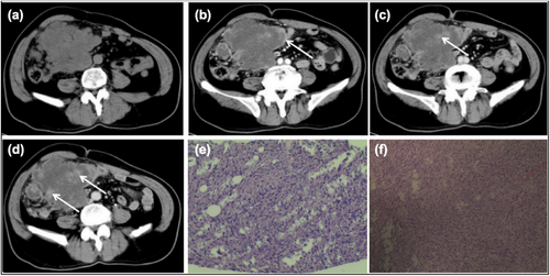
A 63-year-old man was admitted to the hospital with a > 1-year history of repeated acid reflux and belching and a 1-month history of an abdominal mass. On admission, the patient was in good condition, and his vital signs were stable. Laboratory examinations revealed no significant abnormalities. Abdominal computed tomography (CT) revealed a soft tissue mass with uneven density, measuring approximately 13.3 cm × 9.0 cm and extending from the right abdomen into the pelvic cavity. Enhanced CT showed mild uneven enhancement of the mass during the arterial phase, with significant wall enhancement and visible septa. Significant enhancement of the solid components within the mass was observed during the venous and delayed phases with a CT value of approximately 50.3 HU, indicating “progressive enhancement.” Curved blood vessels were visible around the mass, and the surrounding structures were compressed and displaced (Figure 1). During the operation, a tumor measuring approximately 15 cm × 10 cm was observed in the lower right retroperitoneum. It was tightly adhered to the small intestine, ascending colon, and greater omentum, and showed invasive growth. No metastasis was found in the liver, gallbladder, or stomach during the surgical exploration. The pathological diagnosis was synovial sarcoma (SS) with extensive necrosis. Immunohistochemistry revealed epithelial–mesenchymal biphasic differentiation of the tumor cells.

Imaging and pathological findings of our patient. (a) Axial plain CT image. (b) Enhanced abdominal CT image showing significant enhancement of the tumor wall during the arterial phase (arrow). (c and d) Venous and delayed phase contrast-enhanced CT images illustrating the “progressive enhancement” characteristic (arrow). (e and f) Pathological analysis showing the tumor tissue with a nodular appearance and an outer capsule. Tumor cells are arranged in a glandular epithelioid pattern, with enlarged, atypical nuclei, visible nucleoli, and mitotic figures. CT, computed tomography.
SS is a rare malignant soft tissue tumor originating from primitive mesenchymal cells. It most commonly affects the limb joints, particularly those of the lower limbs. Cases of SS arising from the abdominal cavity, especially involving the ascending colon and small intestine, are rare and prone to preoperative misdiagnosis. This case exhibited unique imaging features, including significant arterial phase enhancement of the tumor wall, consistent with progressive enhancement. Magnetic resonance imaging further revealed characteristic signs, such as the “triple signal sign,” “liquid–liquid plane,” and “grape bowl sign,” which can help differentiate SS from other abdominal tumors.
Author Contributions
Yubo Wang: writing–original draft (lead), resources (equal). Jiageng Li: writing–original draft (equal). Yang Fu: writing–original draft (equal). Bin Yang: resources (equal), writing–review and editing (lead).
Acknowledgments
The authors have nothing to report.
Ethics Statement
The authors have nothing to report.
Consent
The patient provided written informed consent at the time of entering this study.
Conflicts of Interest
The authors declare no conflicts of interest.
Open Research
Data Availability Statement
The data supporting the findings of this study are available on request from the corresponding author. The data are not publicly available due to privacy or ethical restrictions.


