Patterning of the hepato-pancreatobiliary boundary by BMP reveals heterogeneity within the murine liver bud
Potential conflict of interest: Nothing to report.
Supported by National Institutes of Health grant R01DK087753 (to K.D.T.).
Abstract
During development, the endoderm initiates organ-restricted gene expression patterns in a spatiotemporally controlled manner. This process, termed induction, requires signals from adjacent mesodermal derivatives. Fibroblast growth factor (FGF) and bone morphogenetic protein (BMP) emanating from the cardiac mesoderm and the septum transversum mesenchyme (STM), respectively, are believed to be simultaneously and uniformly required to directly induce hepatic gene expression from the murine endoderm. Using small molecule inhibitors of BMP signals during liver bud induction in the developing mouse embryo, we found that BMP signaling was not uniformly required to induce hepatic gene expression. Although BMP inhibition caused an overall reduction in the number of induced hepatoblasts, the STM-bounded posterior liver bud demonstrated the most severe loss of the essential hepatic transcription factor, hepatocyte nuclear factor 4-α (HNF4α), whereas the sinus venosus–lined anterior liver bud was less affected. We found that the posterior liver bud progenitors were anteriorly displaced and aberrantly activated pancreatobiliary markers, including sex-determining region Y-box 9 (SOX9). Additionally, we found that ectopically expressed SOX9 inhibited HNF4α and that BMP was indirectly required for hepatoblast induction. Finally, because previous studies have demonstrated that FGF signals are essential for anterior but not posterior liver bud induction, we examined synchronous BMP and FGF inhibition and found this led to a nearly complete loss of hepatoblasts. Conclusion: BMP signaling is required to maintain the hepato-pancreatobiliary boundary, at least in part, by indirectly repressing SOX9 in the hepatic endoderm. BMP and FGF signals are each required for the induction of spatially complementary subsets of hepatoblasts. These results underscore the importance of studying early inductive processes in the whole embryo. (Hepatology 2018;68:274-288).
Abbreviations
-
- Afp
-
- α-fetoprotein
-
- Alk8
-
- activin receptor-like kinase 8
-
- BMP
-
- bone morphogenetic protein
-
- CAS3
-
- cleaved caspase-3
-
- CDX2
-
- caudal type homeobox 2
-
- DMH1
-
- dorsomorphin homolog 1, 4-[6-[4-(1-Methylethoxy) phenyl]pyrazolo[1,5-a]pyrimidin-3-yl]-quinoline
-
- DMSO
-
- dimethyl sulfoxide
-
- dn
-
- dominant-negative
-
- FGF
-
- fibroblast growth factor
-
- FOXA1
-
- forkhead box A1
-
- FOXA2
-
- forkhead box A2
-
- E
-
- embryonic day
-
- HNF4α
-
- hepatocyte nuclear factor 4-α
-
- PDX1
-
- pancreatic and duodenal homeobox 1
-
- pH3
-
- phosphohistone H3
-
- PROX1
-
- prospero homeobox protein 1
-
- pSMAD
-
- phosphorylated SMAD
-
- S
-
- somite
-
- SOX9
-
- sex-determining region Y-box 9
-
- SOX17
-
- sex-determining region Y-box 17
-
- STM
-
- septum transversum mesenchyme
-
- SV
-
- sinus venosus
-
- VMEL
-
- ventral midline of the endoderm lip
The liver bud emerges from the endoderm as a simple thickened epithelium wholly composed of the embryonic liver precursor, termed the hepatoblast.1 Hepatoblasts are induced at 7-8 somites (S) proximal to the developing heart. We previously identified two distinct populations of endoderm that give rise to the liver bud.2 One population resides at the ventral midline of the endoderm lip (VMEL); the other is present at two bilaterally symmetrical positions lateral to the endodermal lip. Although lateral and VMEL progenitors converge at the endoderm lip to produce the nascent liver between 10S and 11S, fate mapping and ablation studies have shown that the lateral progenitors contribute to much of the middle and posterior liver bud, whereas the VMEL progenitors contribute most pronouncedly to the anterior liver bud.2-4 By embryonic day (E) 9.0, a liver bud that expresses liver-specific genes such as α-fetoprotein (Afp) and hepatocyte nuclear factor 4-α (Hnf4α) has formed. Explant experiments have suggested a role for secreted signals, including fibroblast growth factors (FGFs), and bone morphogenetic proteins (BMPs) in the induction of liver specific gene expression.5, 6
In the canonical BMP pathway, BMP ligands bind the type I and type II BMP receptors to form a complex that results in receptor phosphorylation and activation. The activated receptor complex in turn phosphorylates receptor regulated SMAD-1, -5, or -8. Phosphorylated SMAD-1, -5, or -8 (pSMAD1/5/8) can bind to SMAD4, and the heteromeric complex translocates into the nucleus to regulate target gene expression. Because SMAD1/5/8 activation is unique to the BMP pathway, pSMAD1/5/8 can be used as a readout for active BMP signaling.7, 8
Although a role for BMP signals for murine liver development has mainly been established in the context of pre-hepatic/mesoderm explants, a complete understanding is impeded by the redundancy of the pathway components and the requirement for this pathway in multiple earlier developmental processes.1 For example, a subset of BMP4 null embryos are delayed in liver budding6; however, it is also clear that BMP4 is required to appropriately establish mesoderm derivatives that are involved in liver bud induction such as the cardiovascular system and septum transversum mesenchyme (STM).9, 10 Thus, the hepatic delay observed in BMP4 null embryos may be secondary to or exacerbated by its earlier requirement in mesoderm formation. Regardless of the tissue that utilizes BMP, a requirement for BMP in liver induction is evolutionarily conserved and has been established in zebrafish using BMP receptor I (activin receptor-like kinase 8 [Alk8]) mutants and a dominant-negative BMP receptor11 and its role in liver induction supported by chick explant and ex vivo cell/bead implantation experiments.12
In the present study, we used intact developing mouse embryos to gain a more complete understanding of BMP's role in mammalian hepatic induction. Specifically, we used a whole embryo culture approach that allows for the isolation of embryos immediately before liver bud induction and for their normal development through liver budding. We used small molecule inhibitors that act as antagonists for BMP type I receptors, preventing SMAD1/5/8 phosphorylation.13 This approach dampens BMP signals throughout the embryo in a temporally specific manner, bypassing earlier requirements for BMP signals. Using this technique, we found that the posterior liver bud is more affected by reduced BMP signals than the anterior liver bud and uncover a previously undescribed role for BMP in demarcating the hepato-pancreatobiliary boundary. Together, these experiments highlight the need to study the role of inductive signals in the context of the developing embryo.
Materials and Methods
EMBRYO CULTURE AND SMALL MOLECULE ADMINISTRATION
All animal studies were approved by the Institutional Animal Care and Use Committee, University of Massachusetts, Amherst (protocol #2015-0042). Embryo culture was performed as described.3 CD-1 animals (Charles River) were used to generate embryos. The small molecules used include 5 μM dorsomorphin (Tocris), 12.5 μM dorsomorphin homolog 1, 4-[6-[4-(1-Methylethoxy) phenyl]pyrazolo[1,5-a]pyrimidin-3-yl]-quinoline (DMH1; Calbiochem), and 40 μM SU5402 (Tocris). DMH1 is capable of inhibiting signaling from ALK2 and ALK313 whereas dorsomorphin can inhibit signaling from ALK2, ALK3, and ALK6.14 Each drug was dissolved in dimethyl sulfoxide (DMSO) and control embryos of similar somite stages were provided with an equal volume of DMSO.
HISTOLOGY AND IMMUNOHISTOCHEMISTRY
Embryos were fixed overnight in 4% paraformaldehyde, dehydrated, cleared in xylene, embedded in paraffin (Paraplast Plus), and serially sectioned (7.5 μm). All subsequent analyses were performed after dewaxing and rehydration. Immunofluorescence protocols and antibodies are described in the Supporting Information. For analysis, sagittally sectioned liver buds were divided into anterior (liver bud bounded by the sinus venosus [SV]) and posterior (liver bud surrounded by the STM). Middle sections comprised those that lacked a “dorsal” component of the liver; lateral sections comprised the remaining sections.
SECTION IN SITU HYBRIDIZATION
Section in situ hybridization was performed as described previously15 (see also the Supporting Information) using antisense probes for Afp16 and sex-determining region Y-box 17 (Sox17) (IMAGE clone: 1529001).
ELECTROPORATION
Embryos at 7-9S were used for electroporation as described previously,17 except as described hereafter. The electroporation chamber was produced with 0.007-inch-thick platinum electrodes placed 0.5 cm apart and the chamber created using RTV615 silicone rubber (Momentive) cubes affixed to a glass slide with Epoxy (MG Chemicals). Embryos were soaked in 3- to 4-μg/μL plasmid for 5 minutes before transfer to a chamber filled with Hank's balanced salt solution. Embryos were oriented so that the hepatic endoderm was facing the anode. After electroporation, embryos were allowed to recover in equilibrated culture medium (75% rat serum, 25% Dulbecco's modified Eagle's medium) in a 37°C, 5% O2, and 5% CO2 incubator for 2 hours and then transferred to rolling culture for approximately 30 hours.
DiI LABELING
Embryos were dissected at 5-8S and individual embryos labeled with DiI (CM-DiI, Molecular Probes) as described previously.3 Bright field and fluorescent images were produced using an ANDOR Zyla camera and NIS Elements BR software to record the position of the labeled cells and cultured as documented above.
DATA ANALYSIS
All statistical analysis (P value generation) on samples was performed using the two-tailed Student t test unless otherwise noted.
Results
INHIBITION OF BMP SIGNALING LEADS TO LOSS OF HNF4Α IN THE POSTERIOR LIVER BUD
Because of the stringent early developmental requirements for components of the BMP pathway and the genetic redundancy within the pathway, we used small molecule inhibitors in the context of the whole embryo to examine the role of BMP signaling in liver development. This method allows for strict temporal control, limits any secondary defects, and promotes unbiased down-regulation of the canonical signaling pathway. Embryos were cultured from pre-induction (4-7S; E8.25) through liver bud stages (E9.25 or 19-21S; E9.75 or 26-27S) in either the BMP inhibitor DMH113 or an equal volume of vehicle (DMSO). The DMH1 concentration used (12.5 μM) was the maximum dose compatible with grossly normal embryonic development. To measure the efficacy of this dose, 7-8S embryos were cultured for 4, 8, or 12 hours and pSMAD1/5 levels were measured by way of western blotting. Compared with controls, DMH1 reduced pSMAD1/5 up to 80% (Supporting Fig. S1A). Section immunofluorescence of treated embryos with an anti-pSMAD1/5/8 antibody demonstrates a uniform overall reduction in pSMAD1/5/8 compared with controls (Supporting Fig. S1B).
Despite looking grossly normal after culture (Supporting Fig. S1C), careful histological analysis of sagittal sections revealed that DMH1 treatment alters liver bud and STM morphology (Supporting Fig. S3A). The orientation of sections in relation to the embryonic axis and to the remainder of the embryo is described in more detail in Supporting Figure S2A,B and Figure 1A. Consistent with published results,6 BMP inhibition is accompanied by an overall reduction in the number of HNF4α positive (+) hepatoblasts compared with controls (Fig. 1B-C). Intriguingly, HNF4α, an early and essential hepatoblast-specific transcription factor,18-20 is most dramatically reduced in the portion of the liver bud lined by the STM (Fig. 1B, blue dashed line on the liver bud), which is hereafter termed the posterior liver bud. The portion of the liver bud lined by the SV (Fig. 1B, yellow dashed line on the liver bud), henceforth termed the anterior liver bud, is less affected. These data are quantified in Figure 1C,D. Importantly, these results are recapitulated using the less specific BMP inhibitor, dorsomorphin13 (Fig. 1E).
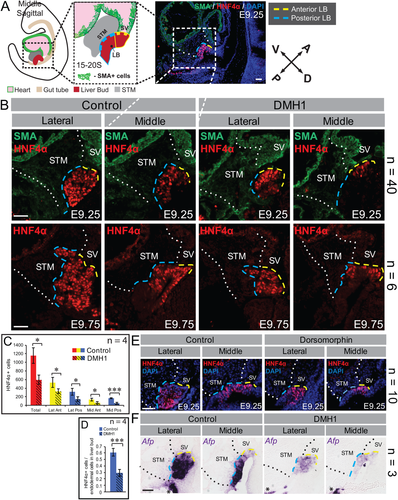
To further probe the abnormal distribution of hepatoblasts in DMH1 treated embryos, we examined the early nonessential liver marker Afp, which at this stage, is highly expressed in the posterior liver bud of controls (Fig. 1F). BMP inhibition significantly hampers Afp in the presumptive posterior liver bud while the weaker expression in the anterior liver bud is not affected compared with controls (Fig. 1F). Expression of the critical pan-endoderm pioneer factors forkhead box A1 (FOXA1) and forkhead box A2 (FOXA2; Supporting Fig. S3B,C) and the hepato-pancreatobiliary domain marker prospero homeobox protein 1 (PROX1)21 (Supporting Fig. S3D) were also examined. Other than putative liver bud size, no alterations between drug-treated and control embryos were found. Taken together, these data suggest that BMP inhibition does not affect the prepatterning of the hepato-pancreatobiliary domain but is specifically required for appropriate hepatic emergence.
To tease apart the role for BMP signaling during hepatic induction from its role in hepatic growth, we treated progressively older embryos with DMH1 for at least 12 hours (Supporting Fig. S4). We observed a consistent loss of posterior specification when the drug was administered to embryos 12S and younger (Supporting Fig. S4A). When embryos were subjected to BMP inhibition at 13-15S or 16-18S, a less severe posterior-restricted loss was observed (Supporting Fig. S4B,C). Similar to that observed with younger embryos, loss of BMP signals prior to 18S results in overall smaller liver buds. By 19-21S, BMP abrogation no longer restricts posterior induction nor overall growth (Supporting Fig. S4D). These data suggest that between 4-12S, BMP signals are vital for the induction of HNF4α+ hepatoblasts in the posterior liver bud and that BMP signals simultaneously suport hepatic maintenance or growth. Interestingly, these results are consistent with the role of BMP signaling on hepatic specification and growth in zebrafish.11
BMP SIGNALING IS NOT REQUIRED FOR SURVIVAL OR PROLIFERATION OF POSTERIOR HEPATOBLASTS
To assess whether the reduced number of HNF4α+ hepatoblasts in the liver bud was due to increased cell death or reduced cell proliferation, cleaved caspase-3 (CAS3) or phospho-histone H3 (pH3), respectively, were examined using immunofluorescence. DMH1 treatment does not affect apoptosis by E9.25 (Supporting Fig. S5A). We surmised that perhaps the treated cells died earlier, and examined apoptosis levels after only 12 hours of culture but did not observe any increased cell death in FOXA2+ cells in the nascent liver bud region (Supporting Fig. S5B). Similarly, we do not observe statistically significant differences in the rate of cell proliferation of the HNF4α+ hepatoblasts in the drug treated versus the control embryos (Supporting Fig. S5C,D). These results demonstrate that abrogating BMP signals affects neither hepatoblast proliferation nor cell death.
INHIBITION OF BMP SIGNALING CAUSES AN ANTERIOR SHIFT OF THE POSTERIOR LIVER BUD
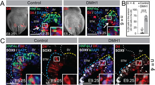
Because our results suggest that upon BMP inhibition endoderm normally fated to contribute to the hepatoblast population is present but not directed towards the hepatic lineage, we next sought to examine the fate of these cells. We demonstrated previously that the VMEL progenitors contribute mainly to the anterior liver bud, whereas the lateral hepatic progenitors mainly contribute hepatoblasts that populate the middle and posterior liver bud.2-4 Hence, to assess the fate of the posterior liver bud, we labeled a subset of the lateral hepatic progenitors with the vital fluorescent dye, DiI, and cultured DMH1-treated and control embryos through the liver bud stage. As expected, all control embryos contain DiI-labeled HNF4α+ cells in the posterior liver bud (Fig. 2A). All BMP-inhibited embryos contain DiI-labeled lateral progenitors that aberrantly localize to the anterior bud (Fig. 2A). To quantify these differences, we counted the number of DiI-labeled cells in the anterior liver bud of control and BMP-inhibited embryos. After BMP inhibition, approximately 75% of the DiI-labeled lateral progenitors are localized to the anterior, compared with only 3%-4% in controls (Fig. 2B). Furthermore, most DiI-labeled cells in the hepatic domain of drug-treated embryos fail to express HNF4α but instead express the early pancreatobiliary marker, sex-determining region Y-box 9 (SOX9; Fig. 2C). These results support the observation that reduced BMP signaling did not result in cell death (Supporting Fig. S5A,B), but rather causes a subset of lateral hepatic progenitors to be anteriorly displaced. To assess whether this anterior shift was specific to the lateral liver progenitors, we labeled the VMEL progenitors and found that DMH1 did not affect the localization of this liver progenitor population compared with controls (Supporting Fig. S6). Taken together, these data highlight the progenitor-specific effects of BMP inhibition.
BMP PATTERNS THE HEPATO-PANCREATOBILIARY BOUNDARY
Numerous studies in zebrafish and in mouse support the hypothesis that the liver and pancreas arise from a common precursor pool that may be directed to one lineage or the other based on extrinsic cues.6, 22-24 Similarly, the data presented above (Fig. 2) suggests the adoption of a pancreatobiliary fate by the presumptive posterior hepatoblasts upon BMP inhibition. To determine whether reduced hepatogenic signals initiate a reciprocal increase in pancreatic fate, we examined markers of pancreas and gall bladder after BMP inhibition.
The early “ventral pancreas bud”, discernable as early as E9.0, contains both ventral pancreas and gall bladder progenitors.24 By E9.5, cells within the more aptly named pancreatobiliary bud begin sorting into an anteriorly situated pancreatic and duodenal homeobox 1 (PDX1)-low/SOX17-high gall bladder primordium and the posteriorly located PDX1-high/SOX17-low ventral pancreas bud. By E10.5, sorting is complete, and the gall bladder and ventral pancreas are marked by SOX17 and PDX1, respectively.25 Similar to SOX17, SOX9 is expressed throughout the early pancreatobiliary domain, but later becomes restricted to the ventral (and dorsal) pancreas bud.26 In E9.25 control embryos, PDX1 is expressed posterior to the hepatic domain, within the pancreatobiliary domain (Fig. 3A, pink line). After drug treatment, PDX1+ cells are located more anteriorly than controls and are found in the posterior liver bud domain (Fig. 3A). Extending culture for another 12 hours yields an increased number of PDX1+ cells in the posterior hepatic domain of treated embryos (Fig. 3B). These results are complimented by the expansion of SOX9 throughout the liver bud of drug-treated embryos over developmental time. In controls, SOX9 is most highly expressed in PDX1+ portion of the pancreatobiliary domain (Fig. 3A,B).
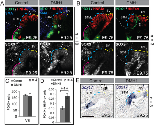
Unlike the liver, the ultimate size of the pancreas is governed by the number of pancreatic progenitors.27 Thus, we opted to count the number of PDX1+ cells in the ventral foregut of drug treated and control embryos and found that despite the spatial expansion, DMH1 treated embryos had no significant increase in the number of PDX1+ cells (Fig. 3C). However, due to the decrease in hepatoblasts, the ratio of pancreatic/hepatic cells was altered significantly (Fig. 3D). Finally, the early pancreatobiliary marker Sox17 displays aberrant expression in the posterior liver bud and in a portion of the anterior liver bud after DMH1 treatment (Fig. 3E). Taken together, these data demonstrate that after BMP inhibition, the pancreatobiliary domain expands anteriorly into the region usually occupied by the posterior liver bud, suggesting that a role of BMP is to delineate the hepato-pancreatobiliary boundary.
SOX9 NEGATIVELY REGULATES HNF4α
The anterior expansion of SOX9 after BMP inhibition suggests that a function of BMP is to exclude SOX9 from the hepatic domain. SOX9 negatively regulates the intestinal transcription factor caudal type homeobox 2 (CDX2) at the pancreato-duodenal boundary, restricting expansion of the duodenum into the pancreatic domain.28 Hence, we hypothesized that SOX9 may perform a similar function at the hepato-pancreatobiliary boundary by repressing HNF4α.
To test this hypothesis, we sought to overexpress SOX9 in the hepatic domain of wild-type embryos. A SOX9 expression plasmid coexpressing GFP was constructed (Sox9-hrGFP; Supporting Fig. S7A-C). Sox9-hrGFP or hrGFP was electroporated into the foregut of E8.5 embryos and cultured through E9.5. Detection of Sox9-hrGFP expressing cells was verified using the SOX9 and GFP antibodies (Fig. 4A). After electroporation with hrGFP (control), approximately 85% of the GFP+ cells in the thickened hepatic domain also express HNF4α (Fig. 4B,C). However, after electroporation with Sox9-hrGFP, only ∼10% of the GFP+ cells in the thickened hepatic endoderm coexpress HNF4α (Fig. 4B,C). These data support the hypothesis that SOX9 is sufficient to repress HNF4α in the hepatic domain.
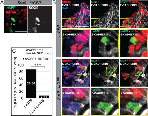
To test whether SOX9 is sufficient to direct endoderm in the hepatic region to a pancreatic or biliary fate, we examined the expression of PDX1, an essential pancreatic gene, and SOX17, an essential early bile duct marker. Ectopic SOX9 failed to promote either PDX1 or SOX17 expression in the hepatic domain (data not shown), suggesting that SOX9 alone is not sufficient to promote pancreatic or biliary fate in these cells. Taken together, these results are similar to those of Shih et al.27 demonstrating that ectopic expression of SOX9 suppressed CDX2 in the duodenum but did not promote a pancreatic bud formation.
BMP SIGNALING IS NOT DIRECTLY REQUIRED FOR HEPATOBLAST SPECIFICATION
To address which components of the BMP pathway are involved in hepatic induction, we examined the expression of BMP ligands in and around the prehepatic/hepatic endoderm domain. Based on their ability to induce hepatic gene expression in vitro and because they are expressed in a manner consistent with a role in hepatic induction, we examined the expression of Bmp2, Bmp4, and Bmp7 using section in situ hybridization.6, 29, 30 The BMP type I receptors Alk3 (Bmpr1a) and Alk2 (Acvr1), as well as the type II receptor Bmpr2, were also examined given that null mutants of each display early embryonic lethality and may play a role in hepatic induction.31-33 The embryonic stages examined include 3-6S, representing prehepatic endoderm; 8-9S, coinciding with the onset of hepatic gene expression/induction; 11-12S, representing the onset of a morphologically thickened hepatic endoderm; and 14-15S, when the nascent liver bud can be readily identified. Bmp2 is expressed in the mesenchyme surrounding the anterior hepatic endoderm at 8-9S and 11-12S, but is absent from the hepatic endoderm at these stages (Supporting Fig. S8A). Surprisingly, Bmp4 is not expressed either in the mesoderm derivatives surrounding the hepatic endoderm nor in the endoderm itself before (3-6S) or at the liver induction stage (8-9S; Supporting Fig. S8B). Bmp2 and Bmp4 are expressed most strongly in the anterior STM between 14S and 15S. Bmp7, on the other hand, is expressed strongly in the hepatic endoderm and proximal mesenchyme during induction stages (8-9S; Supporting Fig. S8C). However, it is interesting to note that Bmp7 mutants do not have a liver phenotype.34, 35 Alk3 is expressed strongly in the liver bud at E8.75 (14-15S); however, weak expression is observed at the liver induction stages (8-9S; Supporting Fig. S8D). Alk2 is expressed strongly in the hepatic endoderm at induction (8-9S) but is expressed weakly in the nascent liver bud (11-12S, 14-15S; Supporting Fig. S8E). Bmpr2 is expressed weakly in the hepatic endoderm at 11-12S and 14-15S (Supporting Fig. S8F).
Because the above expression analysis did not implicate specific pathway components in the direct transmission of BMP signals to the (pre)hepatic endoderm immediately before or during hepatic induction, we next examined pSMAD1/5/8 levels. Interestingly, despite the widespread embryonic expression of Smad-1, 5, and 8 36 we observe only weak levels of pSMAD1/5/8 in the hepatic endoderm at 4-6S and 8-9S stages using immunofluorescence (Fig. 5A). However, a progressive increase in pSMAD1/5/8 intensity in the specified liver bud observed between 11S and 14S (Fig. 5A). Contrary to our expectations, these data suggest that BMP signals play an indirect role in the endoderm to liver bud transition (induction) while supporting a direct role for BMP in post-inductive liver bud growth.

To further assess the requirement for BMP signals in hepatoblast induction, the prehepatic endoderm was targeted by electroporation with either a dominant-negative (dn) Alk3-hrGFP,37 a dominant-negative Alk2-hrGFP,38 or an hrGFP plasmid and the embryos cultured through E9.5 (dnAlk3-hrGFP and dnAlk2-hrGFP validation; Supporting Fig. S9A-E). If BMP signals are directly required for hepatic specification, then electroporation of the dominant-negative constructs into prehepatic endoderm would preclude induction of HNF4α. Instead, similar to the hrGFP controls, approximately 80% of dnAlk3-hrGFP and approximately 90% of dnAlk2-hrGFP electroporated cells (GFP+) in the presumptive liver bud are also HNF4α+ (Fig. 5B,C). These results further support the hypothesis that BMP signals are not directly required in the naïve murine endoderm for hepatoblast induction.
INHIBITION OF BOTH BMP AND FGF SIGNALING LEADS TO A DRAMATIC LOSS OF HEPATIC INDUCTION
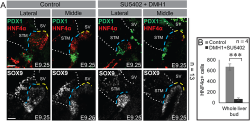
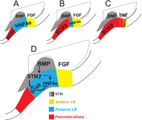
We previously demonstrated that small molecule–mediated inhibition of FGF signaling leads to a selective loss of HNF4α in the anterior but not posterior liver bud of cultured embryos.16 Whereas E9.25 controls displayed pronouncedly thickened liver buds,39 loss of FGF signaling produced increased cell death throughout the bud and an abnormal thinning and loss of HNF4α in the anterior liver bud.16 Indeed, after simultaneous exposure to the BMP and FGF inhibitors DMH1 and SU5402, respectively, HNF4α expression is almost completely lost, much of the presumptive liver bud is abnormally thin, and SOX9 ectopically expressed throughout the hepatic domain (Fig. 6A,B). These results support the hypothesis that BMP and FGF signaling act on complementary subsets of prehepatic endoderm. This view of hepatic induction is summarized in Figure 7. Finally, the small anterior population of HNF4α+ cells that remains after dual drug treatment (Fig. 6A) is reproducible and may indicate the involvement of a third signaling pathway in liver bud induction.
Discussion
The most persuasive studies implicating a role for BMP signals in murine hepatic induction include explant experiments that demonstrate a requirement for BMP in initiating the hepatic markers Albumin and Afp.6, 28 Herein, we expand on these studies and investigate the role BMP plays in hepatic induction in the context of the whole embryo. Given the radical tissue remodeling and distinct migrations that occur in each tissue layer during early postgastrulation,40-42 including the prehepatic endoderm and adjacent mesoderm (JRA and KDT; unpublished data), static explant studies may yield results that differ from those in the dynamic environment of the developing embryo. Indeed, a global reduction of BMP signaling in the embryo during the period of hepatic induction results in an overall reduction in liver bud size that is most severe in the posterior liver bud. In agreement with the conclusions made by the explant experiments, our data support a role for BMP signaling in liver specification; however, when performed in the context of an embryo, an important role for BMP signals in defining the hepato-pancreatobiliary boundary is also apparent (Fig. 7).
Inhibition of the BMP pathway profoundly disturbs the posterior liver bud. There are at least two possibilities to explain these results. The first is that the hepatic endoderm induction occurs differentially along the anterior/posterior axis. Posterior hepatoblast induction requires BMP signals, whereas anterior induction requires FGF signals.16 In agreement with this idea, we have demonstrated that the anterior and posterior hepatic endoderm is adjacent to distinct and potentially inductive mesoderm derivatives. Furthermore, simultaneous inhibition of the signals required for anterior and posterior induction result in a profound overall loss of hepatoblasts (Fig. 6). An alternative possibility is that BMP is uniformly required for hepatic induction, but DMH1 fails to inhibit anterior signals as efficiently as the posterior signals because the BMP ligands are distributed in an anterior–posterior gradient. In support of this hypothesis, Bmp2 and Bmp4 are expressed more highly in the anterior STM (Supporting Fig. S8). To further examine this possibility, we cultured embryos in different concentrations of DMH1. If the gradient hypothesis were correct, it would predict that the boundary of HNF4α+ cells in the posterior liver bud would shift with the dose. However, at lower doses of DMH1 (6 μM), no effects on the liver bud were observed, and at a higher dose (24 μM), where there are more obvious changes in overall embryo morphology, a similar hepatic phenotype was found (data not shown). Taken together, our data favor the idea that BMP signals above a particular threshold are required to induce the posterior liver bud. Whether this heterogeneity and the ability to respond to signaling pathways is intrinsic or guided by the availability of extrinsic factors is a topic for future investigations.
The discovery of distinct inductive requirements for subsets of hepatoblasts during normal development has implications for protocols designed to differentiate stem cells into hepatocytes. Generally, such protocols utilize transforming growth factor-β members such as activin-A and BMP4 to produce definitive endoderm from embryonic stem cells, followed by a mixture of FGFs and BMPs to produce hepatoblast-like cells and then by hepatocyte growth factor to produce hepatocyte-like cells.43-45 Thus far, only hepatocyte-like cells have been produced, indicating that the current regime requires refinement. Our data suggest that up to three populations of induced hepatoblasts are present in the liver bud—one that responds to FGF signaling, a second that requires factors downstream of BMP signaling, and potentially a third, represented by those induced upon BMP and FGF inhibition, that responds to a yet unknown signaling pathway. A 2010 study used a cocktail of FGF10, retinoic acid, and an inhibitor of Activin/Nodal receptor to obtain hepatic progenitors that were able to mature into hepatocytes,46 suggesting that retinoic acid might be the missing third signal that induces the small population of HNF4α+ cells that remain after DMH1 and SU5402 treatment.
BMP SIGNALING IS INDIRECTLY REQUIRED FOR HEPATOBLAST INDUCTION
Surprisingly, our data suggest that BMP signals indirectly support hepatoblast specification. Although a requirement for active BMP signals by the prehepatic endoderm has been assumed, little direct evidence supports this hypothesis. In murine explants, the BMP inhibitor, Noggin, reduces expression of albumin and Afp.6 Because these explants contain both endoderm and mesoderm, the tissue directly responding to BMP is unclear. Furthermore, although pSMAD1/5/8 immunofluorescence is detected in the ventral midline of 3-9S embryos in whole mount,29 our section immunofluorescence indicates that pSMAD1/5/8 activity is most pronounced in the mesoderm adjacent to the VMEL and lateral liver progenitors (Fig. 5A, Supporting Fig. S1B, and data not shown). Furthermore, our in situ data suggests that the adjacent mesoderm rather than the hepatic endoderm is equipped to respond to BMP signals before and during hepatic induction (3-10S). Finally, we demonstrate that ectopic expression of dnAlk2 or dnAlk3 construct does not inhibit induction of HNF4α in the posterior liver bud (Fig. 5). Instead, we propose that the STM or the endothelial cells associated with the hepatic endoderm are the tissues that respond to BMP signals. Indeed, endothelial cells not only support expansion of the liver bud47 but also play a role in hepatic induction from mouse embryonic stem cells.48 These data suggest that an STM or endothelial-derived secondary signal may be necessary for proper specification and maintenance of liver developmental program.
We propose that one way that BMP regulates liver specification is by indirectly suppressing SOX9 in the posterior liver bud. Furthermore, we suggest that SOX9 expression in the liver bud precludes activation of liver-specific genes such as HNF4α. In support of this hypothesis, BMP inhibition during hepatic induction stages produces ectopic SOX9 expression (Fig. 3A,B), and we have demonstrated that ectopic SOX9 alone is sufficient to prevent HNF4α expression throughout the liver bud, although its mechanism remains unclear (Fig. 4). It is interesting to note that some SOX9+ cells in the anterior of the liver bud in both control and DMH1 treated embryos are also HNF4α+ (Fig. 2B). In these cells, SOX9 expression is weaker compared with surrounding SOX9+ cells, suggesting that any coexpressing cell may be in transition and in the process of turning off SOX9 or that low levels of SOX9 is not necessarily sufficient to suppress HNF4α in the early liver bud.
In conclusion, we show that in the context of the developing embryo, BMP signals are required for appropriately inducing the liver bud and that these requirements are complementary to those of FGF signals. Furthermore, we highlight a requirement for BMP signals in establishing the posterior hepatic boundary. Together, these experiments further underscore the importance of assessing complex embryonic processes in their native environment.
Acknowledgment
We thank Peter Chien and Kamal Joshi for help with western blotting and Kamal Joshi and members of the Tremblay and Mager laboratories for their insight and support. We also thank the Alfandari, Peyton, Jerry, and Farkas laboratories from University of Massachusetts, Amherst, for supplying cells or complementary DNA.
REFERENCES
Author names in bold designate shared co-first authorship.


