IL-Y, a synthetic member of the IL-12 cytokine family, suppresses the development of type 1 diabetes in NOD mice
Abstract
The IL-12 family of heterodimeric cytokines, consisting of IL-12, IL-23, IL-27, and IL-35, has important roles in regulating the immune response. IL-12 family members are comprised of a heterodimer consisting of α and β chains: IL-12 (p40 and p35), IL-23 (p40 and p19), IL-27 (Ebi3 and p28), and IL-35 (Ebi3 and p35). Given the combinatorial nature of the IL-12 family, we generated adenoviral vectors expressing two putative IL-12 family members not yet found naturally, termed IL-X (Ebi3 and p19) and IL-Y (p40 and p28), as single-chain molecules. Single chain IL-Y (scIL-Y), but not scIL-X, was able to stimulate significantly a unique cytokine/chemokine expression profile as well as activate STAT3 in mice, in part, through a pathway involving IL-27Rα in splenocytes. Adenoviral-mediated, intratumoral delivery of scIL-Y increased tumor growth in contrast to the anti-tumor effects of scIL-12 and scIL-23. Similarly, treatment of prediabetic NOD mice by intravenous injection of Ad.scIL-Y prevented the onset of hyperglycemia. Analysis of cells from Ad.scIL-Y-treated NOD mice demonstrated that scIL-Y reduced expression of inflammatory mediators such as IFN-γ. Our data demonstrate that a novel, synthetic member of the IL-12 family, termed IL-Y, confers unique immunosuppressive effects in two different disease models and thus could have therapeutic applications.
Introduction
The IL-12 family of heterodimeric cytokines currently has four members, termed IL-12, IL-23, IL-27, and IL-35, consisting of one of two α (p40, Ebi3) and one of three β chains (p19, p28, p35) 1. IL-12 is comprised of a p40 and p35 heterodimer, whereas IL-23, IL-27, and IL-35 are comprised of heterodimers p40 and p19, Ebi3 and p28, Ebi3 and p35, respectively. The α chains are structurally homologous to the type I cytokine IL-6, whereas the β chains are homologous to the soluble form of the IL-6 receptor. There are notable differences in the expression of the subunits for these cytokines with IL-12, IL-23, and IL-27 being expressed and secreted by APC such as DCs and macrophages (MΦ). Activation of innate receptors, like TLRs, induces the expression of these cytokines. However, T-cell help in the form of IFN-γ or CD40L also can enhance their expression 2-4. IL-35 is expressed primarily by FoxP3+ Treg cells, but its subunits also are expressed by γδ T cells, CD8+ T cells, and placental trophoblasts 5.
Members of the IL-12 family of cytokines also exhibit different functions. Both IL-12 and IL-23 are proinflammatory cytokines with the former being able to induce Th1 cells while the latter plays a major role in the induction of Th17 cells. IL-27 exhibits a pleotropic functional phenotype capable of augmenting both pro and anti-inflammatory responses 6, 7. For example, IL-27 suppresses expression of certain anti-inflammatory cytokines, which skews the adaptive immune response toward proinflammatory 8, 9. In contrast, IL-27 also upregulates the expression of IL-10, leading to the induction of type I regulatory T (Tr-1) cells 10-12. IL-35 is a suppressive cytokine inhibiting effector T cells responses 1, 5 and suppressing the development of Th17 cells as well the proliferation of effector T cells 13. The suppression of T cells mediated by IL-35 is IL-10 dependent 14, 15.
Previously, we and others generated adenoviral-based vectors expressing either IL-12 or IL-23 and demonstrated their ability to induce anti-tumor immunity following intratumor injection 16-18. Both IL-12 and IL-23 can induce potent anti-tumor immune responses, but at different kinetics. IL-12 is effective in the early stages of tumor formation whereas IL-23 is more effective during the later stages of tumor growth. IL-23 also is more effective in inducing anti-tumor immunity. The anti-tumor activities of IL-12 and IL-23 also were significantly enhanced when their two subunits were expressed as a single chain using a 15 amino acid linker 17. The enhanced function of the linked cytokines is reminiscent of previous studies with IL-12 (p35 and p40), G-CSF (GM-CSF fusion), and GM-CSF (IL-3 fusion) 19-21.
Given the promiscuous use of receptor and ligand subunits between IL-12, IL-23, IL-27, and IL-35, additional pairings between these components or interactions with other partners are possible. Thus, we generated adenoviral vectors expressing two additional hypothetical IL-12 family heterodimers not yet found naturally, termed IL-Y (p40 and p28) and IL-X (Ebi3 and p19), as single chain molecules. Here, we demonstrate that single chain IL-Y (scIL-Y) in particular is able to induce a specific subset of chemokines/cytokines from primary splenocytes, in part, through an IL-27R pathway. Moreover, we demonstrate that scIL-Y is immunosuppressive in vivo, blocking anti-tumor responses and preventing onset of hyperglycemia in the NOD mice, a model for type 1 diabetes (T1D). Although scIL-Y induced IFN-γ expression in cultured naïve splencoytes, treatment of NOD mice with Ad.scIL-Y decreased expression of IFN-γ in T cells. Interestingly, scIL-Y decreased the frequency of FoxP3+Helios+ Treg cells while having little effect on FoxP3 single-positive Treg cells. Overall, the data suggest that a novel heterodimeric IL-12 family member, scIL-Y, comprised of p40 and p28, is able to antagonize Th1-driven immune responses. Thus scIL-Y could have therapeutic applications for treating autoimmune diseases.
Results
Novel IL-12 family heterodimer, scIL-Y, exhibits biological activity
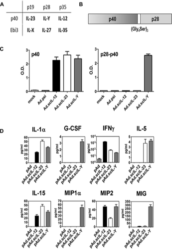
We generated adenoviral vectors expressing the two hypothetical IL-12 family members, termed IL-Y and IL-X, comprised of p40 and p28 and Ebi3 and p19, respectively (Fig. 1A and B). These novel IL-12 family members were generated as single chain molecules using a 15 amino acid (Gly4Ser)3 spacer as described for scIL-23 17. We verified that scIL-Y and scIL-X were secreted following infection of A549 cells by ELISA as described in the Materials and method section. We initially tested the biological activity of Ad.scIL-Y and Ad.scIL-X by treating naïve splenocytes with conditioned media (CM) from Ad.scIL-Y and Ad.scIL-X infected MCA205 cells. As shown in Fig. 1D, we analyzed these supernatants using a limited multiplex Luminex© assay for the expression of a subset of cytokines/chemokines. We chose a subset of cytokine/chemokines representative of these diverse immune responses. In contrast, Ad.scIL-X had a minimal effect on cytokine/chemokine expression (data not shown).
scIL-Y induces phosphorylation of STAT3
Members of the IL-12 cytokine family signal through cell surface receptors consisting of heterodimers 1. Each monomer binds individually to its corresponding receptor subunit. For example, IL-12p35 binds to the IL-12Rβ2 subunit and IL-12p40 to IL-12Rß1 subunit of the IL-12R. The potential receptor for scIL-Y would consist of IL-27Rα, binding p28, and IL-12Rβ1, binding p40. Immediately following activation, the IL-12 family member receptors activate the JAK/STAT pathway. To determine if scIL-Y stimulation leads to phosphorylation of either STAT3 or STAT4, which would be activated by the potential scIL-Y receptor, we stimulated C57BL/6 splenocytes with CM for 48 h and cells prepared for intracellular staining to detect phosphorylated STATs (pSTAT). As shown in Fig. 2A, scIL-12 CM induced pSTAT4, consistent with previous observations 22, whereas none of the other CMs tested (scIL-23, scIL-27, scIL-35, scIL-Y) induced pSTAT4 indicating the specificity of the CMs. In contrast, phosphorylation of STAT3 was induced by scIL-23, scIL-27, and scIL-Y in wild-type splenocytes. Quantitative analysis demonstrated that pSTAT3 was significantly induced by scIL-Y in comparison to control splenocytes (Fig. 2B). We verified activation of STAT3 by Western blot analysis in which phosphorylation of STAT3 was induced 3 h following stimulation with scIL-Y CM (Supporting Information Fig. 1). Interestingly, the analysis revealed that activation of STAT3 was slower than with the other IL-12 family members as very little phosphorylation of STAT3 was detected at 10 min or 1 h following stimulation with CM (Supporting Information Fig. 1). This could be due to a reduced affinity of scIL-Y for its receptor compared to scIL-23 and scIL-27. Importantly, pSTAT3 was significantly reduced in IL-27Rα KO splenocytes treated with either scIL-Y or scIL-27 CM, suggesting that IL-27Rα is part of the scIL-Y receptor. There also was a reduction of pSTAT3 in scIL-23 and scIL-35 CM-treated splenocytes, but not to the extent as seen with scIL-Y or scIL-27. No significant differences were observed with pSTAT4 in IL-27Rα KO splenocytes.
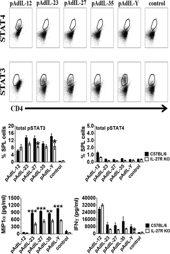
We next examined the effect of scIL-Y on cytokine/chemokine production by activated splenocytes. Analysis of the splenocyte supernatants by ELISA revealed that the loss of IL-27Rα reduced the level MIP1α stimulated by scIL-23, scIL-27, and scIL-Y CM, but not scIL-12 (Fig. 2C). A similar trend was observed for IFN-γ. However, the reduction of IFN-γ was only significant for scIL-Y stimulated IL-27Rα KO splenocytes.
Activation of APCs by scIL-Y
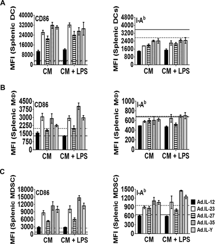
To determine the effect of scIL-Y on APCs, splenocytes were cultured in the presence of CM ± LPS. After 48 h, the cells were collected and assessed for activation by measuring the upregulation of CD86 and MHC II. No difference in the activation of DCs (CD11b+CD11c+) was observed with any CM tested (Fig. 3A). A similar pattern for MΦ (CD11b+F4/80+) was observed, with the exception of a greater upregulation of CD86 by scIL-35 CM + LPS (Fig. 3B). In addition, both CD86 and MHC II were significantly upregulated by scIL-35 and scIL-Y CM (± LPS) on myeloid derived-suppressor cells (MDSCs; Gr-1+CD11b+) (Fig. 3C).
scIL-Y suppresses the anti-tumor immune response
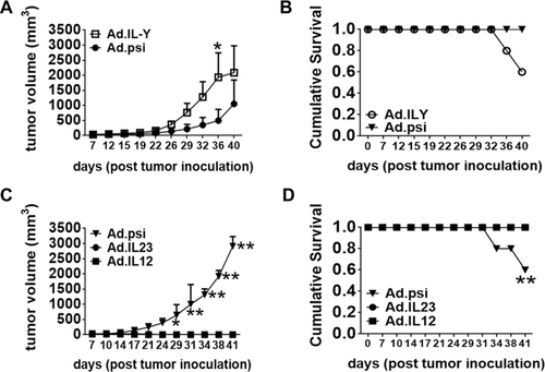
To determine if scIL-Y had any immune activity in vivo, the effect of Ad.scIL-Y on murine tumor growth was evaluated, given that we previously demonstrated significant anti-tumor effects following intra-tumor injection of Ad.IL-12 and Ad.IL-23. MCA205 fibrosarcoma cells were implanted subcutaneously in the flank of C57BL/6 mice and either Ad.scIL-Y or Ad.psi5 injected intratumorally on days 7, 9, and 11. Interestingly, mice treated with Ad.scIL-Y exhibited enhanced tumor growth, compared to control mice (Fig. 4A and B). In contrast, tumor-bearing mice treated with either Ad.scIL-12 or Ad.scIL-23 (Fig. 4C and D) quickly rejected the tumor, consistent with previous results 17, 18. These data suggest that intratumor expression of scIL-Y suppresses anti-tumor immunity in vivo.
scIL-Y suppresses the development of diabetes in NOD mice
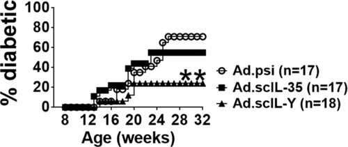
To examine the possible immune suppressive properties of scIL-Y, we utilized the NOD mouse model of T1D 23. T1D is a T-cell driven autoimmune disease that results in the destruction of insulin-producing β cells in the pancreatic islets of Langerhans 24. Eight-week-old female, prediabetic NOD mice were injected intravenously with 5 × 10⁸ infectious units of Ad.scIL-Y, Ad.scIL-35, or control Ad.psi5 virus and monitored for the onset of hyperglycemia over 6 months. As shown in Fig. 5, nearly 80% of the mice infected with Ad.scIL-Y (n = 17) were protected from developing diabetes. In contrast, treatment with Ad.scIL-35 (n = 18) was less effective in preventing diabetes with approximately 50% of the mice remaining diabetes free. The reduction in frequency as well as the delay in the time of onset of hyperglycemia in NOD mice following Ad.scIL-Y treatment further supports an immunosuppressive function for scIL-Y.
Immunosuppression associated with scIL-Y is not mediated through regulatory T cells
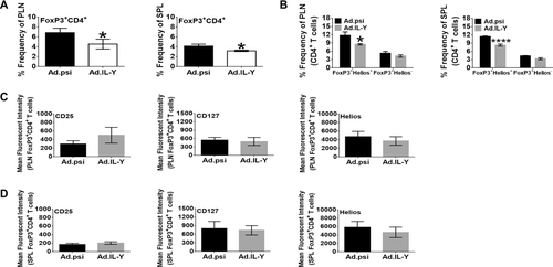
To determine the mechanism of suppression mediated by scIL-Y, we initially examined the possible role of CD4+ Treg cells in conferring protection from T1D. In T1D, studies have shown deficiencies in Treg cells in both their numbers and function 25-27 whereas replacement therapy or reagents that activate Treg cells protect against the development of T1D 28, 29. Eight-week-old female NOD mice were infected with Ad.scIL-Y or control Ad.psi5 virus. After four weeks, spleen (SPL) and pancreatic lymph node (PLN) cells were harvested and the frequency of Treg cells determined. A significant decrease in the percentage of Treg cells from Ad.scIL-Y treated mice in both the SPL and PLN (Fig. 6A) was observed when gating on the CD4+FoxP3+ population. The reduction of Treg cells was seen predominantly with native Treg cells (FoxP3+Helios+), but not the inducible subset (FoxP3+Helios−), as shown in Fig. 6B for both PLN and SPL. Within the CD4+FoxP3+ Treg cell population, analysis of the level of CD25, CD127, and Helios expression showed no significant differences in PLN or SPL Treg cells (Fig. 6C and D). Analysis of mice at 2-week postinfection showed no differences between treatment groups (data not shown). Thus, treatment of NOD mice with Ad.scIL-Y induces a protective mechanism that prevents the onset of diabetes. However, this mechanism does not appear to enhance the frequency or activation of CD4+FoxP3+ Treg cells.
Restimulation of T cells reveals suppression following Ad.scIL-Y treatment
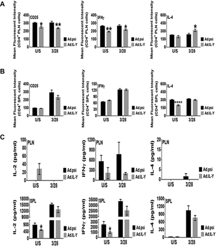
To evaluate further the effect of scIL-Y on effector T cells, we performed in vitro restimulation assay on whole SPL and PLN cells from Ad.scIL-Y treated NOD mice, which were activated through the TCR. Four weeks following infection, cells were prepared, transferred into wells previously coated with anti-CD3 mAb and cultured with anti-CD28 mAb for 2 days. Cells were probed for the expression of activation markers and for the production of cytokines such as IFN-γ and IL-4. PLN CD4+ T cells showed a significant reduction in the expression of both CD25 and IFN-γ in mice infected with Ad.scIL-Y if cells were either untreated or activated through the TCR (Fig. 7A). IL-4 expressing T cells also were elevated in the PLN of Ad.scIL-Y treated mice only following activation through the TCR. Splenic CD4+ T cells did not exhibit these changes; however, unstimulated IL-4 expressing cells were significantly reduced (Fig. 7B). We also examined the T cells for production of IL-10 and IL-17, markers of Tr1 and Th17 cells, respectively, and were unable to detect these cytokines (data not shown). The data from this experiment suggest that the treatment of NOD mice with Ad.scIL-Y antagonized the proinflammatory response (IFN-γ) while simultaneously inducing an anti-inflammatory (IL-4) response at least in the PLN.
Activation of APCs following treatment of NOD mice with Ad.scIL-Y
To test the effect of scIL-Y on APCs, NOD mice were infected with Ad.scIL-Y or Ad.psi5 and after 4 weeks, SPL and PLN were harvested and prepared for FACS analysis. The frequency of APC subsets (DCs, MΦ, and MDSCs) in the SPL did not differ between the treatment groups (Supporting Information Fig. 2A). However, the expression of CD86, a marker of activation, was elevated in splenic DCs (CD11c+CD11b+) following Ad.scIL-Y treatment. In addition, the frequencies of DCs and MΦ from Ad.scIL-Y infected mice were reduced while MDSCs remained at similar levels between each group (Supporting Information Fig. 2B). The activation of these APC subsets did not differ between each group of mice.
Discussion
The IL-12 family of heterodimeric cytokines, consisting of IL-12, IL-23, IL-27, and IL-35, has important roles in regulating the immune response. Given the promiscuous interactions between the IL-12 family subunits and with their heterodimeric receptors, it is possible that there are additional functional interactions between the IL-12 family of subunits. Thus, we generated adenoviral vectors expressing two additional hypothetical IL-12 family heterodimers not yet found naturally, termed IL-Y (p40 and p28) and IL-X (Ebi3 and p19), as single chain molecules. Other studies have shown that single chain fusions of G-CSF and GM-CSF and G-CSF and IL-3 are biologically active and that single chain IL-12 and Il-23 are as or more active than the normal dimeric cytokine 19-21. Our analysis suggests that IL-Y in particular is able to stimulate a specific subset of cytokines/chemokines, distinct from the IL-12 family members. For example, scIL-Y stimulated expression of MIP1α (CCL3), a chemokine involved in the recruitment of mononuclear cells, most notably neutrophils to the site of inflammation or injury 30, 31 and CCR5+ Treg cells 32. scIL-Y also specifically stimulated expression of the chemokine MIG (CXCL9), a T-cell chemoattractant, and the cytokines G-CSF, able to stimulate granulocytes and certain stem cell populations, IL-5 and IL-15. However, despite the ability of scIL-Y to stimulate expression of a specific subset of chemokines/cytokines, we have been unable to detect the secretion of endogenous IL-Y from stimulated mouse splenocytes.
The fact that IL12p40 binds to the IL-12Rβ1 whereas IL-27p28 binds primarily to IL-27Rα subunit suggests that the IL-Y receptor could consist of a heterodimer between IL-12Rβ1 and IL-27Rα. IL-27Rα is expressed on CD4+ T cells, NK cells, NKT cells, and macrophages, but also is expressed on activated B cells, mast cells, monocytes, dendritic cells, and polarized Th cells 33-35. Activation of IL-27Rα drives the phosphorylation of JAK1 and to a lesser extent JAK2 and TyK2, resulting in induction of STAT3 and STAT4 1, 33, 35. Our analysis in splenocytes demonstrated that scIL-Y stimulated phosphorylation of STAT3 and that the level of pSTAT3 induction by scIL-Y was reduced, but not completely eliminated, by KO of IL-27Rα. These observations suggest that scIL-Y signals through binding to the IL-27R, but that other interactions, such as with IL-12Rβ1, also could be important.
Given the genes stimulated by scIL-Y in splenocytes including G-CSF, IL-5, IL-15, MIP1α, and MIG, we initially hypothesized that localize, intratumoral expression would lead to induction of an anti-tumor immune response, similar to those observed with intratumoral injection of IL-12 and IL-23. However, tumor-bearing mice treated intratumorally with Ad.scIL-Y had enhanced tumor growth, compared to the control, suggesting that localized scIL-Y expression inhibits the anti-tumor response in vivo, similar to IL-35. Thus we explored the effects of transient expression of scIL-Y in the well-characterized NOD mouse model of autoimmune diabetes.
Members of the IL-12 cytokine family have been linked to autoimmune diseases 33. IL-12 is noted for its ability to skew the development of naïve CD4+ T cells toward a Th1 phenotype. Also, NOD mice injected with recombinant IL-12 during the early prediabetic stage developed diabetes at an accelerated rate 36-38 whereas injection of IL-12 late during the prediabetic stage had no effect. In addition, ectopic expression of IL-35 under the control of the rat insulin promoter in NOD mice prevents the development of diabetes 39. Interestingly, our results demonstrate that following intravenous injection of the adenoviral vectors expressing scIL-Y and scIL-35, transient expression of scIL-Y was more effective than scIL-35 in suppressing the onset of hyperglycemia in NOD mice. scIL-Y expression reduced the percentage of Treg cells in both the PLN and SPL while maintaining a consistent level of expression for Treg cell associated markers (CD25, Helios, and CD127). Furthermore, the analysis of splenocytes from Ad.scIL-Y treated mice showed a reduction of IL-2 production following TCR activation, similar to what has been shown for IL-27. Since the prevention of diabetes conferred by scIL-Y appears not to be mediated through Treg cells, we analyzed the functional profile of effector T cells. Here TCR stimulation showed a significant reduction in the expression of CD25 and IFN-γ by CD4+ T cells in the PLN from scIL-Y treated mice. In addition, IL-4 expression was increased in PLN CD4+ T cells. Furthermore, ex vivo analysis of splenic and PLN T cells showed no effect of Ad.scIL-Y on IFN-γ or IL-4 production by effector T cells (data not shown). Additionally, we were unable to detect IL-10 (Tr1 cells) or IL-17 (Th17 cells) expressing T cells (data not known). These data suggest that scIL-Y prevents the onset of hyperglycemia in NOD, in part, through the suppression of effector T cells function, either directly or indirectly.
IL-Y is structurally similar to IL-27, sharing the IL-27p28 subunit, and thus potentially may share similar functions. IL-27 is a pleotropic cytokine, which links it to various conditions ranging from cancer to autoimmunity 1, 6, 35. IL-27 was initially considered a proinflammatory cytokine since it induces the activation of STAT1 and T-bet and expression of IFN-γ 8, 40-42. However, subsequent studies showed that the proinflammatory phenotype was the result of effective suppression of the anti-inflammatory arm of the immune response (Th2 and Treg cells) 9, 43, 44. Similar to IL-27, Ad.scIL-Y treatment of NOD mice reduced the percentage of Treg cells and the number of IL-4 expressing CD4+ T cells in the spleen. However, unlike IL-27, scIL-Y reduces the level of IFN-γ, possibly making it a more immunosuppressive.
Our results demonstrate that not only does scIL-Y exhibit biological activity, but scIL-Y functions as a suppressive cytokine to limit autoimmunity. Our data are similar to those obtained in a recent study examining the biologically effects of a recombinant IL-12p40/IL-27p28 heterodimeric protein 45. In this study, the recombinant protein was injected into an animal model of uveitis where it prevented the destructive autoimmune response. However, unlike the reduction of Treg cells we observed in our diabetes model, they observed an increase in Treg cells along with a significant suppression of Th17, both of which are active participants in the pathogenesis of uveitis. We saw no effect of scIL-Y on the generation of Th17 cells both in ex vivo and in vitro assays (data not shown). However, in both studies, the expression of IFN-γ was significantly reduced. This difference in results could reflect differences between disease models or differences in the delivery method for IL-Y. Taken together, these results suggest that scIL-Y may serve as a useful therapeutic for reducing inflammation and ameliorating autoimmune diseases.
Materials and methods
Mouse models
All of the female C57BL/6, NOD ShijL, and IL-27Rα (C57BL/6 background) knockout mice were obtained from The Jackson Laboratory (Bar Harbor, ME) 46. Six- to seven-week-old C57BL/6 mice were used for the tumor experiments whereas for the type 1 diabetes analysis, the NOD mouse model was utilized. Animals were maintained under pathogen-free conditions at the animal facility at the University of Pittsburgh School of Medicine and The Scripps Research Institute, Scripps Florida. All procedures performed were approved by the University of Pittsburgh and Scripps Florida Institutional Animal Care and Use Committee.
Cell lines
MCA205 fibrosarcoma cells were maintained in RPMI supplemented with 10% fetal bovine serum (Invitrogen, Carlsbad, CA), and 1% penicillin/streptomycin (Gibco, Carlsbad, CA) and L-glutamine (Gibco, Carlsbad, CA).
Adenovirus
Adenoviruses expressing single-chain versions of IL-12 and IL-23 have been described previously 17. Ad.scIL-27 was constructed using the full-length clone of the murine EBI3 gene (GenBank NP056581, amino acids 1–228) linked to a truncated version of the IL-27p28 gene (GenBank NP663611, amino acids 29–234) using a 15 amino acid (Gly4Ser)3 spacer with a Bam HI restriction enzyme site (GGATCC, GlySer). The scIL-27 clone was codon-optimized for expression in mammalian cells using the UpGene codon optimization algorithm 47 and synthesized by GenScript, Inc (Piscataway, New Jersey). The scIL-27 fusion gene was inserted into the shuttle vector, pAd.Lox (GenBank U62024), at Sal I/Not I sites. For the construction of pAd.scIL-35, the p35 gene was amplified with p35-S (5′-GGTGGATCCCGGGTGATTCCGGTGTCCGGT-3′) and Ad-R (5′-GTAACCATTATAAGCTGC-3′) primers off pAd.scIL-12, digested with BamH I and Not I restriction enzymes and then inserted into the pAd.scIL-27 thus replacing p28. For the construction of pAd.scIL-X, the p19 gene was amplified with p19-S (5′-GGTAGATCT GTACCTAGATCCTCCAGCCCC-3′) and Ad-R primers using pAd.scIL-23 as a template and cloned into Bam HI and Not I sites of pAd.scIL-27, replacing p28. For the construction of pAd.scIL-Y, the p40 gene was amplified with Ad-S (5′-CAAGTCTCCACCCCATTG-3′) and p40-AS (5′-TACGGATCCACCCCCGCCCGAGCCTCCTCCACCGGAGCC-3′) primers using pAd.scIL-12 as a template, digested with Sal I and Bam HI restriction enzymes and then inserted into the Sal 1/Bam HI site of pAd.scIL-27, replacing the EBI3 gene. All the adenoviral shuttle plasmid constructs were confirmed by sequencing. The recombinant adenoviruses were propagated on HEK-293 cells and purified by CsCl banding, followed by dialysis in 3% sucrose solution. Particle titer of purified virus and MOI was determined as previously described 17, 48. Viruses were aliquoted and stored at −80°C until use. The expression and secretion of scIL-Y by recombinant adenoviruses was confirmed by ELISA. Ninety-six-well plates were coated overnight with (1 μg/well) anti-p40 monoclonal antibodies (mAb; Pharmingen) in carbonate coating buffer (pH 9.5) and blocked with PBS containing 2% BSA and 0.05% Tween20 for 1 h. The supernatant from A549 cells infected with Ad.scIL-Y (10 MOI) was added and incubated for 2 h. After the plates were washed, biotin-conjugated anti-p28 mAb (100 ng/well; R&D systems) was added to each well and incubated for 1 h. For detection of p40, 96-well plates were coated overnight with 200 μL of the supernatant from A549 cells infected with Ad.scIL-Y (10 MOI) in carbonate coating buffer (1:3) followed by biotin-conjugated anti-p40 mAb (50 ng/well; R&D systems) and avidin-HRP (1:500, Pharmingen) were added to each well and incubated for 1 h. The plates were washed three times and developed with 3,3’5,5’-tetramethylbenzidine, and the reaction was stopped with 1 M H2SO4 and absorbance at 450 nm was determined using an ELISA reader (BIO-TEK instruments).
Adenovirus functional assay
Relative expression of each cytokine from the adenoviral preparation (MOI 500) was analyzed 72 h postinfection of 4 × 10⁴ MCA205 cells. IL-12 and IL-23 levels were analyzed using the mouse IL-12 p70 and mouse IL-23 p19/p40 ELISA kits (eBioscience, San Diego, CA), respectively, following the manufacturers’ instructions. Biological activity of Ad.scIL-23 was assayed by infecting MCA205 cells with Ad.scIL-23 or Ad.psi5 (1000 MOI) and the supernatants collected 72 h later. To test the supernatants, spleens were then harvested from C57BL/6 mice, converted into a single-cell suspension, and red blood cells (RBC) were depleted using RBC lysis buffer (150 mM ammonia chloride, 1 mM sodium bicarbonate, and 0.1 mM EDTA at pH 7.7). Splenocytes were plated (2 × 10⁶ cells per well) in a 24-well plate and 24 h later treated with infected MCA205 conditioned media. Forty-eight hours later the supernatants were collected and analyzed for induction of cytokines with the Luminex® Multiplex mouse 32 cytokines/chemokines panel (Millipore, Billerica, MA).
In vivo tumor assay
Mice were inoculated with 1 × 10⁵ MCA tumor cells via subcutaneous injection into the abdomen. A total of 5 × 10⁸ I.U. of Ad.scIL-12, Ad.scIL-23, Ad.scIL-Y, or Ad.psi5 was injected intratumorally on days 7, 9, and 11 postinoculation. Tumor volume was monitored using a metric caliper every 3 to 4 days or until mice were sacrificed due to excessive tumor size or tumor ulceration.
Diabetes study
At eight weeks of age, NOD mice were treated with 5 × 10¹⁰ v.p. per mouse administered intravenously. Blood glucose monitoring was carried out once per week by tail vein bleeds using a FreeStyle Lite glucometer and test strips (Abbott Laboratory, Abbott Park, IL USA).
Flow cytometric analysis of pancreatic lymph nodes and spleen
Tissues were harvested, converted into single-cell suspension, and washed with 1 × PBS. Following depletion of RBCs, the cells were washed extensively and resuspended in FACS buffer (2% FBS, 1 × PBS, 2 mM EDTA, and 0.04% sodium azide) at a concentration of 3.75 × 10⁶ cells per mL. A 200 μL aliquot of each sample was then transferred into 96-well round-bottom plates (BD Bioscience San Jose, CA). To minimize background staining, Fc receptors were blocked using anti-CD16/CD32 mAb (1:600 dilution, purchased from BD Pharmagin) for 20 min at 10°C. Afterwards, the cells were stained with fluorochrome conjugated mAb (purchased from either BD Pharmagin or eBioscience) for 45 min at 10°C 49. The cells were washed twice with FACS buffer and then fixed with 2% paraformaldehyde. For intracellular staining, the cells were stained using a mouse FoxP3 staining buffer kit (BD Pharmingen, San Diego, CA USA) and used according to the manufacturer's instructions. All flow cytometry samples were processed through a BD LSR II flow cytometer (BD Bioscience San Jose, CA) and analyzed using Flowjo software (Tristar, Inc. Ashland, OR). A gating scheme for all flow cytometry data is shown in Supporting Information Fig. 3.
In vitro stimulation of wild type and IL-27Rα KO splenocytes with conditioned media
All of the T-cell assays are in compliance with MIATA guidelines. Conditioned media was generated using HEK-293 cells transfected with a plasmid encoding scIL12, scIL23, scIL-27, scIL-35, or scIL-Y for 48 h. For the in vitro stimulation assay, splenocytes were converted into a single-cell suspension and depleted of RBCs, washed extensively and then resuspended at 2 × 10⁶ per well prior to transfer into a 96-well plate. CM then was added at a 1:2 dilution. The cells were cultured for 48 h at which point supernatants and cells were harvested. The supernatants were assayed specifically for the chemokine MIP1α (CCL3) by ELISA (Sigma-Aldrich, St. Louis, MO) or by ELISA for IL-2 and IFN-γ (BD Pharmingen). The splenocytes were directly assayed for the production of cytokine by intracellular staining by flow cytometry 50. Splenocytes from wild type and IL-27Rα KO mice also were analyzed for the phosphorylation of either STAT3 or STAT4 following stimulation with the CM. For phospho-STAT (pSTAT) analysis, single cell suspended splenocytes were washed with PBS and then fixed in 2% paraformaldehyde. Subsequently, the cells were washed twice with PBS and permeabilized with 100% cold methanol for 45 min. The cells then were stained with anti-mouse pSTAT3 (pY705) and anti-mouse pSTAT4 (pY693) (BD Biosciences, San Jose, CA) and T-cell marker CD4 for one hour at 10°C 51. For Western blot analysis, splenocytes from wild-type mice (2 × 10⁶ cells/well) were stimulated with CM for 10 min, 1 h, and 3 h in 96-well round bottom plates. Subsequently, the cells were washed twice with PBS and then resuspended in lysis buffer (1 × protease inhibitor (Sigma-Aldrich Inc.), 1 × phosphatase inhibitor (Thermo Scientific), and 1 × cells lysis buffer (Cell Signaling)) and incubated on ice for 30 min. The extracts were centrifuged at 13 000 g for 20 min and the supernatants saved. The protein content for each extract was quantified and then prepared for Western blot analysis. The level of pSTAT3 was analyzed using an anti-phosphoSTAT3 (Tyr 705) mAb purchased from Cell Signaling while total STAT3 was measured using an anti-STAT3 mAb purchased from Santa Cruz, Inc.
In vitro restimulation assay
The effect of Ad.scIL-Y on effector T cells was assessed using splenocytes from spleens and peripheral lymph nodes of NOD mice one month postinfection. The splenocytes were assayed on an individual basis with multiple replicates whereas the PLNs for each treatment group were pooled together with multiple replicates. The cells were stimulated through the T-cell receptor using plate bound anti-CD3 mAb (1 μg/mL) and soluble anti-CD28 mAb (0.5 μg/mL) for two days. The supernatants were saved and analyzed by ELISA (IL-2, IL-4, and IFN-γ) while the cells were processed for intracellular cytokine FACS analysis (IL-4 and IFN-γ).
Acknowledgments
The authors would like to thank Dewayne Faulkner (University of Pittsburgh) for his assistance with flow cytometry, Joan Nash (University of Pittsburgh) for her administrative support, and Sara McGowan for providing animal husbandry services (The Scripps Research Institute-Florida). This work was supported by grants AG043376, AG024827, and AR051456 to P.D.R. from the National Institutes of Health and a program grant from the Juvenile Diabetes Research Foundation (JDRF) to PDR. R.R.F. was supported by a T32 grant from NIH on Autoimmunity and Immunopathology. This project used the University of Pittsburgh Cancer Institute (UPCI) Vector Facilities supported by the University of Pittsburgh's National Institutes of Health (NIH) Cancer Center Support Grant (CCSG) P30 CA047904.
Conflict of interest
The authors declare no financial or commercial conflict of interest.
References
Abbreviations
-
- CM
-
- conditioned media
-
- MΦ
-
- macrophages
-
- PLN
-
- pancreatic lymph node
-
- SPL
-
- spleen
-
- sc
-
- single chain
-
- T1D
-
- type 1 diabetes


