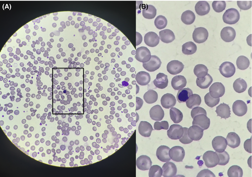
Hemoglobin SC smear recorded with a smartphone
Key Clinical Message
Taking photographs of microscope slides generally requires expensive equipment and a lot of effort. We demonstrate a technique to take clear, focused pictures of microscope slides with a smartphone. This could facilitate exchange of information between providers, allow for quick second opinions, and improve patient care.
A twenty-eight-year-old inpatient with SC disease was seen with symptoms of a painful crisis, and a peripheral smear was obtained to confirm the process. It shows partially and fully sickled cells, target cells, “pita bread” cells and a lymphocyte. The image was obtained with a smartphone aimed at the microscope eyepiece and slowly advanced toward the light source in a manner analogous to a fundoscopic eye examination; no special equipment was used. This technique can facilitate sharing of vital information in the community setting, where an expert opinion is not always readily available, and improve patient care (Fig. 1).

Conflict of Interest
None declared.
Authorship
All authors have made equal contributions and have approved the final version of this paper.


