Wunderlich Syndrome Caused by Spontaneous Rupture of Renal Angiomyolipoma in the Third Trimester of Pregnancy: Endovascular Management With Fetal Preservation—A Case Report and Literature Review
Funding: The authors received no specific funding for this work.
ABSTRACT
Renal angiomyolipoma (AML) are benign tumors incidentally diagnosed during routine radiologic images; nonetheless, AML rupture is uncommon and has been associated with fatal consequences, demanding prompt intervention, especially during pregnancy with symptoms of Wunderlich's syndrome (WS) and atrophied contralateral kidney. We report a 30-year-old pregnant woman who presented with gross hematuria and hypovolemic shock in her seventh month of pregnancy. Magnetic resonance imaging revealed a 15 × 13.5 × 16 cm AML mass in the left kidney, a 5 × 6 cm intrarenal pseudoaneurysm, and a massive retroperitoneal hematoma with an atrophied contralateral right kidney. After achieving hemodynamic stability, selective renal arterial embolization was performed with a successful outcome. She subsequently delivered her baby via cesarean section without complications. In conclusion, although WS due to AML rupture is rare during pregnancy, accurate diagnosis and prompt endovascular treatment should be pursued in cases involving a solitary kidney and associated comorbid conditions to safeguard the lives of both the patient and the baby, stabilize renal function, and prevent catastrophic complications.
Summary
- Even though Wunderlich's syndrome resulting from renal angiomyolipoma rupture is rare during pregnancy, accurate diagnosis and prompt endovascular treatment should be pursued in cases involving a solitary kidney and associated comorbid conditions.
- This approach aims to protect the lives of both the patient and the baby, stabilize renal function, and prevent catastrophic complications.
1 Introduction
Angiomyolipoma (AML) is a rare benign tumor that affects approximately 0.2%–0.6% of the global population. A significant risk associated with AML is the potential for rupture, which can lead to retroperitoneal bleeding, occurring in about 15% of cases [1-3]. Pregnancy increases the likelihood of rupture due to hormonal influences. Generally, non-pregnant patients with AML are asymptomatic, while pregnant patients often experience complications, primarily rupture [4].
Conservative management, selective renal arterial embolization, and nephrectomy are the primary treatments for complicated AML [3, 5]. Collaborative involvement from various medical specialties is essential for determining the optimal treatment strategy [5]. Considerations include maternal and fetal health, gestational age, facility availability, and patient delivery preferences [1]. There are few reports of renal AML rupture during pregnancy presenting with Wunderlich's syndrome (WS) symptoms and an atrophied contralateral kidney [4, 5]. This report discusses a case involving a young female in her 28th week of pregnancy who experienced a rapid progression of abdominal pain leading to hypovolemic shock. Prompt radiologic diagnosis and urgent intervention were undertaken to preserve the lives of both the patient and the fetus.
2 Case Presentations
2.1 Patient Information
A 30-year-old woman in her third trimester (28 weeks of pregnancy) (Gravida 4, Para 3) presented to our emergency department with acute generalized abdominal discomfort, left flank pain, and significant hematuria. She reported experiencing 8 h of acute, persistent pain that radiated to her left side and progressively worsened over time. The patient had no predisposing factors, a history of trauma, prior medical conditions, or current medications, and she was a non-smoker. She has been aware of her atrophic right kidney and renal angiomyolipoma (AML) for 7 years and has been undergoing regular perinatal follow-up.
2.2 Clinical Findings
Upon arrival, the patient was conscious (Glasgow Coma Scale [GCS]: 14) but in a state of shock. Her blood pressure measured 80/55 mmHg, pulse rate was 110 beats per minute, respiration rate was 18 breaths per minute, and she presented with a fever (oral temperature: 38.5°C). The patient's abdomen was soft and appropriately distended for her stage of pregnancy, and she reported left flank discomfort without any ecchymosis. However, a palpable mass was detected in the left flank during the abdominal examination.
2.3 Diagnostic Assessment
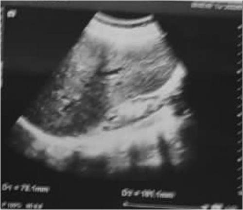
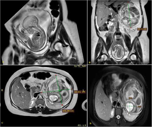
Blood tests indicated a total white blood cell count of 12 × 109/L and anemia, with a hemoglobin level of 6 g/dL, blood urea nitrogen of 14 mg/dL, and creatinine of 1.4 mg/dL. Urine sediment analysis revealed numerous red blood cells (RBCs), 12–14 leukocytes per microscopic field, and a few bacteria. Abdominal ultrasonography (US) showed a heterogeneous collection measuring 13 × 9 × 11 cm in the left retroperitoneum. The right kidney was relatively small, measuring 7.8 × 2.8 cm, with significantly reduced cortical thickness of up to 7 mm (Figure 1). Abdominal magnetic resonance imaging (MRI) revealed a large exophytic lesion (15 × 13.5 × 16 cm) with heterogeneous intensities arising from the anterior part of the left kidney. This lesion contained giant pseudoaneurysms measuring 5 × 6 cm, observed within the renal system as a retroperitoneal hematoma, along with tortuous, abnormal-appearing renal arteries consistent with the rupture of an angiomyolipoma (Figure 2A–D). The diagnosis was established as Wunderlich's syndrome (WS) due to the rupture of a left angiomyolipoma and pseudoaneurysms, accompanied by an atrophied right contralateral kidney. A fetal ultrasound was performed, documenting a live fetus with normal activity and an estimated gestational age of 28 weeks.
2.4 Therapeutic Intervention
Initially, fluid resuscitation and prophylactic antibiotics were administered without delay. Following stabilization and an improvement in hemoglobin levels, the patient was referred to a tertiary care unit for further evaluation. The decision was made to perform interventional radiological embolization to control the bleeding while preserving both renal function and the fetus.
2.5 Renal Arterial Embolization Procedure
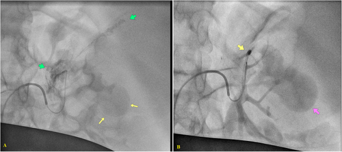
Firstly, a lead apron was used over her gravid uterus to reduce radiation exposure. Then, after light anesthesia and moderate sedation, a modified Allen's test was conducted to assess the patency of her femoral artery. The procedure started via US guidance and involved access to the left common femoral artery using a micropuncture kit and a 6 French Low Profile vascular sheath (Terumo, Tokyo, Japan). An infusion of verapamil (2.5 mg), heparin, (2000 units), and nitroglycerin (200 mcg) was administered via the sheath to mitigate arterial spasm and reduce the risk of arterial thrombosis. Angiographic procedures were performed utilizing the Cobra catheter (Boston Scientific), with diagnostic renal angiography revealing significant vascularity in the upper pole of the left kidney, suggesting renal AML and a significant intra-renal pseudoaneurysm measuring 5 × 6 cm (Figure 3A). A Progreat microcatheter was introduced to delineate the upper renal arteries and perform highly selective diagnostic angiography. Embolization was performed using PVA microparticles, but contrast flow persisted within the pseudoaneurysm. Consequently, a micro coil (3 mm; Boston Scientific) was deployed (Figure 3B), but severe vasospasm of the renal artery occurred, causing a reduction in contrast flow to peripheral branches which could potentially precipitate renal ischemia. For that, Nitroglycerin (50 mcg for three doses) was administered at five-minute intervals accompanied by meticulous blood pressure monitoring and the angiography showed a reduction of intra-renal arterial spasm and Doppler US studies also showed an enhancement in the kidney's vascularity (Figure 4). The procedure took approximately 2.5 h, with strict radiation exposure precautions in place. The procedure was prolonged due to colonic gas accumulation, which hindered the use of Digital Subtraction Angiography or Roadmap techniques. The patient's discomfort was further exacerbated by a lead apron and altered renal artery projections due to a ruptured vascularized mass, pseudoaneurysm, and gravid uterus.
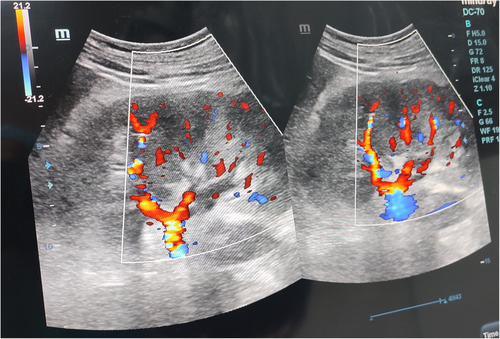
2.6 Outcome and Follow-Up
The patient was admitted to the intensive care unit (ICU) for observation and underwent a postoperative color Doppler ultrasound, which revealed preserved vascular flow with normal Doppler readings. She was discharged four days post-procedure, having experienced an uncomplicated antenatal course. The patient delivered via cesarean section at 39 weeks of gestation, as determined by her consulting obstetrician. Following the embolization procedure, the patient's creatinine level peaked at 2.2 mg/dL before gradually declining to 1.6 mg/dL and stabilizing between 1.4 and 1.5 mg/dL. Ultimately, her creatinine levels returned to pre-procedure values 6 months later.
3 Discussion
Wunderlich syndrome is primarily caused by tumors, which generally include both benign and malignant types (57%–61.5%), with renal angiomyolipoma (AML) being the most common cause (24%–31.5%) [6]. AML affects approximately 0.13% of the general population, with a higher incidence observed in women [4, 6]. Childbearing increases the risk of AML rupture, with contributing factors such as tumor size (greater than 4 cm), genetic abnormalities, and intra-tumoral aneurysms [7]. The occurrence of AML accompanied by spontaneous bleeding during pregnancy is a rare phenomenon [4, 8, 9].
Pregnancy significantly impacts AML by elevating estrogen and progesterone levels, which contribute to its etiology and development. Hormone receptors on smooth muscle cells slow ureteral movement, dilate the ureter, and cause moderate hydronephrosis. Consequently, increased blood volume, renal plasma flow, hemodynamic disruption, and elevated blood pressure may lead to aneurysm development and AML rupture [10]. In our case, tumor size and pregnancy were the primary factors associated with spontaneous AML rupture.
Even though symptoms such as hematuria and flank discomfort may occur, they are more frequently discovered incidentally during imaging for unrelated conditions [10]. The most common presenting symptom was abdominal discomfort (76.2%–88%), followed by shock (33%–42.1%), significant hematuria (19%–24%), and a palpable mass (10%) [9]. However, the percentage of patients presenting with symptoms varies widely in the literature (21%–100%) [4]. In our case, the patient exhibited abdominal pain, gross hematuria, and shock due to spontaneous rupture of an angiomyolipoma (AML), which resulted in a clinical presentation characterized by a triad of a palpable mass, flank pain, and hemorrhagic shock.
Computed tomography (CT) attenuation coefficients typically range around −20 Hounsfield Units (HU), which aids in the accurate identification of renal angiomyolipomas (AMLs) [4, 11]. With advancements in cross-sectional imaging techniques, even tumors with low fat content can now be effectively detected, achieving an impressive magnetic resonance imaging (MRI) specificity of up to 99% [1]. In our case, radiologic images confirmed the diagnosis of AML, showcasing all characteristic features. Consequently, it is crucial for individuals with a known history of AML to receive comprehensive pre-pregnancy contraceptive counseling. This counseling should address the potential risks associated with pregnancy in the context of AML, as well as the necessity for ongoing monitoring and management strategies to mitigate any complications that may arise during gestation. Such proactive measures are essential for ensuring the health and well-being of both the patient and the fetus. Regular follow-up appointments with healthcare providers are vital to assess the patient's condition and adapt treatment plans as necessary throughout the pregnancy [4].
The treatment goals for renal AMLs include preventing acute episodes, preserving renal parenchyma, and enhancing long-term renal function [4]. Before 1976, nearly 90% of sporadic renal AMLs were managed through total nephrectomy [4]. Therapeutic options for AML include conservative management, total or partial nephrectomy, and transcatheter selective arterial embolization [1-3]. Endovascular control achieved through angioembolization is a minimally invasive and effective technique with established efficacy in managing hemorrhagic urological emergencies [6]. Its applicability in critically ill patients makes it a preferred option for renal preservation, particularly in individuals with a solitary kidney [2, 3]. However, complications may occasionally arise following an embolization procedure, including the risk of unintended parenchymal infarction. Recent advancements in interventional radiology, such as improved imaging techniques and the introduction of smaller devices along with more precise embolic agents, have made super-selective embolization of the renal artery a viable strategy for diagnosing and treating renal hemorrhage [11]. Generally, this approach requires a short hospitalization, promotes rapid recovery, and is typically performed without general anesthesia, exhibiting low incidence rates of both early and late complications, thereby relegating traditional surgical methods like nephrectomy to rare occurrences [9, 11].
Various embolic agents have been documented in the literature for managing renal bleeding; however, most studies indicate that coils are the preferred technique for renal artery embolization across diverse clinical contexts [12]. This method was selected in the case outlined herein due to its advantages over alternative techniques. It allows for straightforward manipulation, with controlled detachment directed into the target vessel, thereby minimizing the extent of renal ischemia, as it does not impede distal microcirculation. The agent's dilution in iodinated contrast and the application of zoom capabilities during injection facilitate precise monitoring of the embolic agent; however, caution is warranted, as once deployed, it cannot be repositioned and obstructs access to more distal segments of the vessel into which it has been introduced [13]. In our case, due to hemodynamic instability and the presence of a solitary functional kidney at the 28th week of gestation, it was determined that an endovascular procedure utilizing transarterial embolization (TAE) would be performed. Compared to surgical interventions, TAE is characterized by its minimally invasive nature, eliminating the need for general anesthesia or open surgical techniques [1, 3].
4 Conclusion
Even though Wunderlich syndrome resulting from renal artery rupture is rare during pregnancy, accurate diagnosis and prompt endovascular treatment through angioembolization should be pursued in cases involving a solitary kidney and severe comorbid conditions. This approach aims to protect the lives of both the patient and the baby, maintain stable renal function, and prevent future catastrophic complications.
Author Contributions
Abdulqader Al-falahi: conceptualization, data curation, formal analysis, funding acquisition, investigation, methodology, project administration, resources, software, supervision, validation, visualization, writing – original draft, writing – review and editing. Maeen Al-Sayyadi: conceptualization, formal analysis, funding acquisition, investigation, resources, software, supervision, validation, visualization, writing – original draft. Bassam Al-Ozair: conceptualization, methodology, project administration, resources, supervision, visualization, writing – review and editing. Ali Alwah: formal analysis, investigation, project administration, resources, software, supervision, validation, writing – review and editing. Faisal Ahmed: conceptualization, data curation, formal analysis, funding acquisition, investigation, project administration, resources, software, supervision, validation, visualization, writing – original draft, writing – review and editing.
Acknowledgments
The authors have nothing to report.
Ethics Statement
The authors have nothing to report.
Consent
The patient gave written informed consent to publish this report in accordance with the journal's patient consent policy.
Conflicts of Interest
The authors declare no conflicts of interest.
Open Research
Data Availability Statement
The authors have nothing to report.


