Gastric volvulus: An uncommon and life threatening cause of acute gastric dilatation in a young male: A case report
Abstract
Gastric volvulus leading to acute gastric dilatation is a surgical emergency that should be considered as a differential in patients who present to the emergency department with severe epigastric pain and evidence of gastric outlet obstruction. This condition requires a high index of suspicion that enables early diagnosis and treatment.
1 INTRODUCTION
Acute gastric dilatation was first defined by Duplay in 1833. This entity may occur secondary to trauma, diabetes mellitus, electrolyte disturbances, gastric volvulus, infection, psychiatric or eating disorders, or in the immediate postoperative period.1 Gastric volvulus is a life-threatening condition, characterized by abnormal rotation of the stomach of more than 180°, to which prompt diagnosis and treatment are vitally important.2 It was first described in 1866 by Berti, based on the autopsy of a 61-year-old woman.3 The peak age group of incidence is in the fifth decade with children less than one-year-old making up 10%–20% of cases. No association with either sex or race has been reported. In 30% of cases, the volvulus occurs as a primary event, but it is more commonly secondary to another cause.2 The main consequence of the disorder is foregut obstruction that may be acute, recurrent, intermittent, or chronic. Furthermore, there is a risk of strangulation which may result in necrosis, perforation, and hypovolemic shock. The mortality rates for acute volvulus ranges from 30% to 50% which indicates the importance of early diagnosis and treatment.1 Diagnosis is usually based on radiological imaging, and surgical therapy is the treatment of choice.4
2 CASE PRESENTATION
A 21-year-old young male patient presented to the emergency department of our center, with a history of sudden onset of generalized abdominal pain for 1 day, which was constant, gradually increasing in intensity, and aggravated with a change in posture. He also had a few episodes of vomiting, which was non-foul smelling, non-projectile, and with liquid dark muddy content. There was abdominal distention which was more prominent toward the upper abdomen. He had normal bowel and bladder habits before. He had a prior history of admission, 1 year back with pain, and distension of the abdomen, which was diagnosed as a gastric volvulus and managed conservatively in the same hospital. He had no significant personal or family history. Physical examination revealed ill-looking status, with dehydrated and an anxious state. His pulse was 130 beats/min and regular, blood pressure was 90/60 mm of Hg, respiratory rate was 24 breaths/min, and the temperature 99°F. Per-abdominal examination revealed a tense and hugely distended abdomen with generalized tenderness more in the epigastric region. There was guarding and rebound tenderness in all regions of the abdomen. There was no appreciable organomegaly or any other palpable mass. Per-rectal examination revealed no significant abnormality. The provisional diagnosis was made as hollow viscus perforation peritonitis with hypovolemic shock. Laboratory results showed total count of 14,600, neutrophils 81%, hemoglobin 14.9 g%, platelets 291,000, U/C: 33/1.1 mg/dL, amylase 96u/L, Na/K: 139/3.8 mEq/L, S. lipase: 947u/L, PT/INR: 18.8/1.46, TB/DB: 0.4/0.1 mg%, SGPT:15, SGOT: 32, ALP: 107 U/L, T protein: 4.9, and albumin: 2.6 g%.
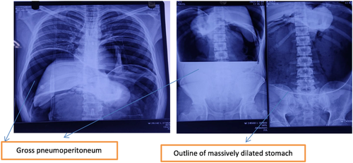
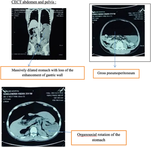
Radiological imaging was done. X-ray abdomen erect and supine showed gross pneumoperitoneum, and massively dilated stomach as shown in Figure 1. Contrast-enhanced CT scan (CECT) of the abdomen and pelvis was done, which revealed, a massively dilated stomach with loss of enhancement of gastric wall, gross pneumoperitoneum, and organo-axial rotation of the stomach as shown in Figure 2.
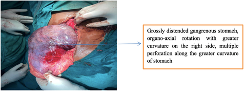
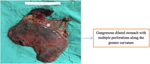
The patient was initially managed with IV fluids using an 18G IV cannula, antibiotic (Inj. Piperacillin + Tazobactum 4.5 g IV stat), nasogastric tube insertion, Foleys catheter insertion, and I/O charting. After initial resuscitation, the patient was rushed to the operating room and a laparotomy was done under general anesthesia. Per-operative findings were around 400 ml of the sanguineous collection along with vegetable matter in the peritoneal cavity, grossly distended gangrenous stomach rotated organoaxially (greater curvature towards the right side of midline more than 1800), a friable gastric wall with multiple perforations along the greater curvature, thrombosed splenic vessels with multiple necrotic areas over the lower pole of the spleen, surrounding bowel edematous with surrounding flakes attached to the small bowel wall. The findings are shown in Figure 3 and Figure 4.
Total gastrectomy and splenectomy with Roux-En-Y stapled esophagojejunostomy and jejuno-jejunostomy were done. Approximately 50 cm distal to duodenojejunal flexure, the jejunum limb was transected and brought near the distal end of the esophagus, and the anastomosis was done (Esophagojejunostomy) with 31 mm circular stapler. Also, the proximal end of the jejunum was end to side anastomosed with the distal end of the jejunum. Then after, a thorough peritoneal lavage was done with 10 L of normal saline, followed by drain placement of 28G in the pelvis.
The patient was extubated and shifted to the postoperative ward and continuous monitoring of vitals was done along with I/O charting.
Total parenteral nutrition (TPN) was started on the first post-operative day via central line, with resuming oral intake gradually from 3rd postoperative day, after the resumption of bowel sounds. Incentive spirometry was started from 1st postoperative day. Overall the postoperative period was uneventful. The patient was advised to take intermittent small meals, with less sugar and salt, in order to prevent dumping syndrome. The patient was discharged on the 5th postoperative day. He had no issues after 2 months of follow-up. The patient's party is satisfied with the treatment they received.
3 DISCUSSION
Gastric volvulus is a rare entity which is difficult to diagnose. When there is relaxation of gastric ligaments, any cause of stomach distention can predispose to excessive rotation, creating a favorable condition for volvulus.3 Other predisposing factors includes diaphragmatic trauma or surgery, congenital hernias, and diaphragmatic eventration which is associated with phrenic nerve paralysis.3 In some cases, adhesions may act as a rotational axis for the stomach, which may lead to volvulus. Gastric volvulus is characterized by the rotation of the stomach along its short or long axis, such that it leads to a variable degree of gastric inlet and outlet obstruction. Rotation of more than 180 degrees can lead to strangulation, necrosis, and finally perforation.5, 6 Hence, gastric volvulus is a true surgical emergency, which requires immediate management.
Gastric volvulus has been classified on the basis of both etiology, and the axis of rotation, but the most accepted classification of volvuli is based on the axis of rotation, as proposed by Singleton.1, 7 Type I is organo-axial (OA) volvulus, in which there is rotation around an axis connecting the pylorus and cardio-esophageal junction. Type II is mesentero-axial (MA) volvulus, in which there is rotation around the axis causing bisection of greater and lesser curvature of the stomach. The organo-axial type accounts for 59% of the cases, while mesentero-axial type accounts for 29% of the cases. Type III is a combination of OA and MA, and it occurs in 2% of cases, and type IV is unclassified, and it occurs in 10% of cases.1, 8
The classification of gastric volvulus can also be done based on etiology as primary/idiopathic volvulus and secondary volvulus.1 Primary volvulus are those which may be associated with adhesions, the or the defects in the normal ligamentous attachments of stomach. Secondary volvulus is those which may be associated with the disorders of gastric motility and structure, or with issues of surrounding structures such as the diaphragm and spleen.6 Most of the cases of gastric volvulus have a secondary cause. Diseases of the stomach, like peptic ulcers, cause retraction of the lesser curvature and predisposes the stomach to MA rotation. In adults, secondary gastric volvulus is usually seen in association with para-esophageal hernia and traumatic diaphragm injury.3, 6 The presentation of gastric volvulus can be acute, subacute, or chronic. The classical triad of retching, severe epigastric pain, and inability to pass NG tube (Borchardt triad) holds true for gastric volvulus, most of the time, especially when it presents acutely like in our case. Subacute presentation is usually associated with vague abdominal pain.1, 2
Diagnosis of gastric volvulus is difficult and is usually based on radiologic findings in combination with the clinical presentation.9 We may see two air-fluid levels, in the antrum and fundus, or a single air bubble with no additional luminal gas in the supine position, and a “beak” in the cardio-esophageal region, in a plain radiograph. Gastric volvulus is very accurately diagnosed on Abdominal CT scan where it shows an upside-down stomach with the pylorus lying higher than gastro-esophageal junction. The upper gastrointestinal series is also a useful tool for the diagnosis.8 Barium contrast studies are also helpful in the diagnosis of this condition.1 Since gastric volvulus is rarely suspected based on the clinical symptoms, barium studies are usually conducted as a first-line investigation, and most of the patients undergo an abdominal CT scan in order to confirm the diagnosis.1
Initial management of gastric volvulus is by nasogastric decompression, for decreasing the intragastric pressure. Then after, surgery is usually performed in order to check gastric viability of stomach, resect the gangrenous portion, and to perform de-rotation and gastropexy with or without gastrostomy with repair of secondary factors associated.6, 10 Laparotomy is still considered the most common surgical option for patients with gastric volvulus, though laparoscopic interventions have also been described. Surgical reduction of volvulus with or without gastropexy is the most commonly performed procedure.4, 6
4 CONCLUSION
Acute gastric volvulus is an emergency entity with high morbidity and mortality. It is an uncommon, and often unrecognized, surgical emergency that should be considered as a differential diagnoses in patients who present to the emergency department with severe epigastric pain and evidence of gastric outlet obstruction. A constellation of clinical symptoms along with findings in the radiological studies helps in making the diagnosis. Emergency laparotomy is needed to prevent serious complications such as stomach gangrene and perforation.
AUTHORS CONTRIBUTION
Naveen Chandra Bhatta (NCB) and Niliza Shakya (NS) contributed to concept of the study, study design, and surgical therapy for the patient. Karishma Kathayat (KK) and Sampada Dahal (SD) performed data collection and writing the manuscript. NCB, Bijay Shrivastava (BS), and Asish Shrestha (AS) are operating surgeon. Bikash Nepal (BN) and Rakesh Sthapit (RS) are the senior author and manuscript reviewer. All the authors individually did final proofreading of the manuscript before submission.
ACKNOWLEDGMENT
We would like to thank our patient for allowing us to share his case.
CONFLICT OF INTEREST
The authors declare that they have no competing interests.
ETHICAL APPROVAL
This article describes a case report. Therefore, no additional permission from our Ethics Committee was required as case reports are exempt from ethical approval in our institution.
CONSENT
Written informed consent was obtained from the patient for publication of this case report and accompanying images. A copy of the written consent is available for review by the Editor-in-Chief of this journal on request.
Open Research
DATA AVAILABILITY STATEMENT
All the data generated or analyzed during this study are included.


