Encephalopathy and brain atrophy during induction chemotherapy in acute lymphoblastic leukemia
Abstract
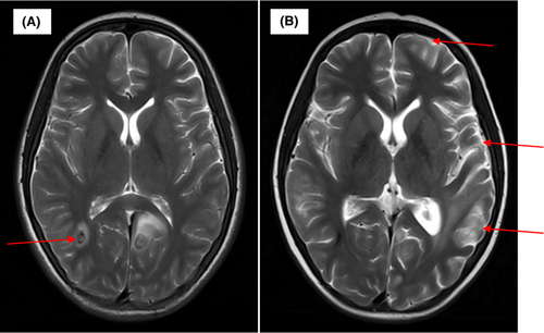
The MRI showed encephalopathy and brain atrophy of the left parietal lobe, occipital lobe and temporal lobe and decreased infiltration of the dura mater on T2-weighted imaging. But encephalopathy and brain atrophy could be improved with neurotrophic drugs and additional intelligence teaching.
1 INTRODUCTION
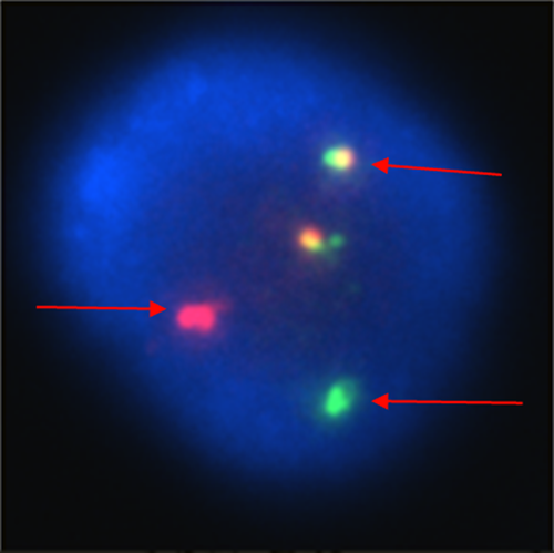
A 14-year-old girl presented with irregular fever and leukocytosis (white cell count 523.96 × 10⁹/L). Bone marrow examination demonstrated the immune-phenotype was common B cell acute lymphoblastic leukemia (B-ALL). FISH showed the lymphoblast cells with positive BCR-ABL1 fusion gene (Figure 1). The brain magnetic resonance imaging (MRI) revealed extensive infiltration of the dura mater and hemorrhage of the right parietal lobe (Figure 2A).The diagnosis of ALL was confirmed. During induction treatment with vincristine/ daunorubicin/ l-asparaginase/dexamethasone, the patient presented with memory loss and mental decline. She was sleepy and noncommunicative. In the same time, she had encephalopathy and her intelligence deteriorated. The MRI showed encephalopathy of the left parietal lobe, occipital lobe and temporal lobe and decreased infiltration of the dura mater (Figure 2B). Fourteen days later, the memory and intelligence markedly improved with neurotrophic drugs and additional intelligence teaching. Citicoline and nerve growth factor were used for her and teach her how to live.


Acknowledgments
This work was supported in part by the Science and Technology Planning Project of Guangdong Province, China (Grant No. 2015A020210041). Published with written consent of the patient.
CONFLICT OF INTEREST
None declared.
AUTHOR CONTRIBUTIONS
Y-L Tang: revised the manuscript critically. Q-R Li, S-M Fu, Y-L Mo, Y Li, C Liang, L-N Wang, W-Y Tang, X-Q Luo, and L-B Huang: wrote the manuscript. All authors read and approved the final manuscript.


