Intraoperative detection of second inferior vena cava during para-aortic lymphadenectomy for advanced-staged ovarian cancer: Lessons to be learned
Abstract
As the presence of second inferior vena cava may alter the extent of para-aortic lymphadenectomy, early preoperative imaging diagnosis is of great significance to avoid intraoperative difficulties.
1 INTRODUCTION
The presence of second (additional) vena cava is a described yet rare anatomic variation.1, 2 As such a variation may alter the level of left renal vein which is the upper limit of para-aortic lymphadenectomy in advanced-stage ovarian cancer, it worth highlights the significance of preoperative acknowledge of such an alteration in order to achieve uncomplicated completion of para-aortic lymphadenectomy.
2 CLINICAL IMAGE PRESENTATION
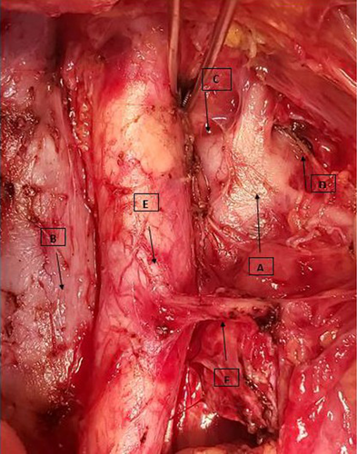
A 61-year-old patient was admitted for interval debulking after neoadjuvant chemotherapy for high-grade serous ovarian cancer. Preoperative imaging indicated the presence of bulky nodes, therefore necessitating para-aortic lymphadenectomy, while posed no suspicion for anatomic variations regarding inferior vena cava. The inability of finding left renal vein, which presents the upper surgical limit, posed the suspicion of its potential posterior origin from inferior vena cava. Cautious preparation of tissues leads finally to the discovery of an additional inferior vena cava from which left renal vein was originated, much lower that the anticipated surgical limit (Figure 1). Persistence of surgical preparation permitted the successful completion of para-aortic lymphadenectomy, which was extended lower than initially expected. Cautious preoperative imaging evaluation of great vessels is of remarkable significance in order to optimize surgery and avoid unnecessary extension of lymphadenectomy that may lead to severe intraoperative complications.

ACKNOWLEDGMENTS
Published with written consent of the patient.
CONFLICT OF INTEREST
None declared.
AUTHOR CONTRIBUTIONS
Stamatios Petousis, Chrysoula Margioula-Siarkou, and Frederic Guyon wrote the initial draft. Stamatios Petousis and Beatriz Navarro have edited the clinical image. Stamatios Petousis, Chrysoula Margioula-Siarkou, Beatriz Navarro, and Frederic Guyon performed the surgical operation. George Mavromatidis and Konstantinos Dinas significantly revised the draft.


