Managing cesarean section in a patient with rotated uterus due to a large myoma
Key Clinical Message
In pregnancies complicated by large myomas, obstetricians may face difficulties during cesarean section if they fail to notice the rotation of the uterus and could make an incorrect uterine incision. This error might cause massive intraoperative hemorrhage. Obstetricians must exercise extreme caution during cesarean sections complicated by large myomas.
Case
Although uterine rotation due to a myoma is a rare complication of pregnancy 1, obstetricians should be alerted to avoid making an incorrect uterine incision during cesarean section.
A 35-year-old woman (gravida 1, para 0) was referred to our hospital for delivery because her pregnancy was complicated by a left-sided myoma that measured 15 cm in diameter during ultrasonography. The patient's medical history included renal artery aneurysm, primary aldosteronism, and chronic hypertension.
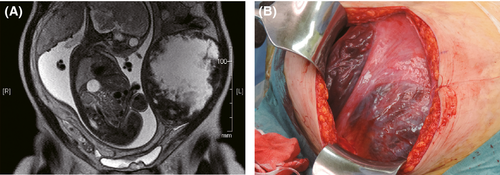
To determine the anatomical relationship between the uterus and the myoma, we performed magnetic resonance imaging (MRI) at 30 weeks' gestation (Fig. 1A). MRI revealed that the myoma was intramural and located on the left side. Notwithstanding the myoma, the pregnancy had not been complicated by abdominal pain, placental abruption, fetal growth restriction, or preterm labor and birth. Fetal growth was determined to be appropriate for the gestational age, and no major anomalies were revealed by ultrasound. Because of breech presentation, the patient was scheduled to undergo an elective cesarean section. Owing to her chronic hypertension, the cesarean section was scheduled for 37 weeks' gestation.

During the cesarean section, midline laparotomy showed multiple large vessels pressing on the uterus (Fig. 1B).
Question
How should the obstetrician manage a uterine myoma during cesarean section?
Answer
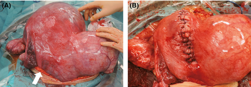
Prior to delivery, the obstetrician was unable to accurately understand the anatomical relationship between the myoma and the intra-abdominally placed uterus. Therefore, the abdominal incision was extended vertically around the umbilicus, and the uterus was brought outside the abdominal cavity (Fig. 2A). As a result, we were able to evaluate the spatial relationship between the tumor and the uterus. We then located the vessels belonging to the utero-ovarian ligament and the uterine parametrium; they were facing front because the myoma had rotated the uterus 90°. A vertical uterine incision was made to open the uterus (Fig. 2B). Intraoperative bleeding was 1600 mL. We managed the section successfully, and the patient delivered a healthy male infant, weighing 2538 g with Apgar scores of 4 and 9 at 1 and 5 min, respectively. The patient recovered well and was discharged 8 days after surgery. The neonate had no feeding-related complications; he gained sufficient weight and was discharged with his mother. Six months after delivery, the patient's myoma had decreased to approximately 11 cm in size.

In summary, if the obstetrician has difficulty in confidently identifying the anatomical landmarks required to properly orient the uterine incision during a cesarean section, he or she should bring the patient's uterus outside the abdomen. If an ill-placed uterine incision is made to deliver the child, massive intraoperative hemorrhaging can result.
Conflicts of Interest
The authors declare that they have no conflicts of interest or funding disclosures related to this study.


