Real-time 3D–3D image fusion of CTA/CBCT roadmap fluoroscopy in the transcatheter mitral intervention
Correspondence Jun Pu, MD, PhD, Department of Cardiology, Ren Ji Hospital, School of Medicine, Shanghai Jiao Tong University, 160 Pujian Rd, Shanghai 200127, China.
Email: [email protected]
Fuyu Cheng, Zhiqing Qiao, and Liang Zhao contributed equally to this study.
Abstract
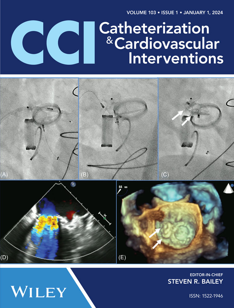
Absence of periprocedural visualization of three-dimensional (3D) left heart anatomy and its surrounding structures in fluoroscopy may reduce the rate of successful transcatheter mitral valve repair. We proposed a multimodal imaging strategy based on 3D computed tomography (CT) angiography and 3D cone beam CT fusion images, which enabled real-time visual inspection of 3D cardiac structures on fluoroscopy, to optimize transcatheter mitral intervention. This new image fusion technology, together with standard transesophageal echocardiography guidance, improved the efficiency and safety of the procedure, and could be considered as a new workflow for transcatheter mitral valve intervention.
CONFLICT OF INTEREST STATEMENT
The authors declare no conflict of interest.
Open Research
DATA AVAILABILITY STATEMENT
The data that support the findings of this study are available from the corresponding author upon reasonable request.