Atypical lymphocytes—From chickenpox to monkey pox
Atypical lymphocytes, also known as variant lymphocytes, are well recognized as a feature of viral infections, often but not always associated with lymphocytosis. Chickenpox, due to the varicella/zoster virus, is a well-established cause. Monkeypox, now renamed mpox, is the most recently recognized viral cause.1
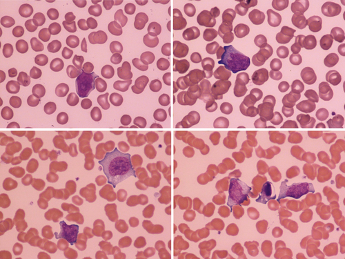
The upper two images (all images ×100 objective) show the blood film of 2-week-old, term baby born to a G1P1 mother, presenting with a skin rash that started on day 10 of life.2 The rash was pustular/vesicular on an erythematous base, affecting the limbs, buttocks, scalp, forehead, and dorsum of the hands and palms, and sparing mucus membranes. The baby then developed respiratory compromise requiring ventilation. Chest radiography showed right upper lobe pneumonia. The blood count showed white cell count 11.2 × 10⁹/L, hemoglobin concentration 107 g/L, platelet count 45 × 109/L, neutrophils 4.8 × 10⁹/L, and lymphocytes 5.5 × 10⁹/L. The blood film confirmed thrombocytopenia and showed normocytic normochromic red cells, left-shifted myeloid series, and reactive lymphocytes (images). PCR on swabs from skin lesions and throat swabs was positive for monkey pox (West African variant) and adenovirus. The baby was treated with dual antivirals (tecovirimat and cidofovir) and broad-spectrum antibacterials with subsequent improvement. Of note, as previously reported, both parents had a similar rash but had a milder clinical picture.2
The lower two images are from 10-year-old girl with a 1-week history of an itchy skin rash, starting on the lower limbs and then extending all over the body, including mucus membranes. Fever commenced at about day 7 of the rash, peaking at 39.5°C. The rash was umbilicated and vesicular on an erythematous base. PCR was positive for varicella/zoster. Her blood count and film showed lymphocytosis (5.4 × 10⁹/L) with atypical lymphocytes, some of which were plasmacytoid; apoptotic forms were common (images). The patient was treated with broad-spectrum antibacterials and aciclovir, which was poorly tolerated and hence switched to ganciclovir, with good effect.
In a small series of monkeypox infections previously reported, 6 of 11 patients investigated hematologically had atypical lymphocytes.2 Interestingly, the total lymphocyte count was increased in only one of these patients. In the baby reported here the lymphocyte count was also normal for age as it was in another reported patient.3
Not surprisingly, atypical lymphocytes are also a feature of small pox, having been reported almost a century ago.4
CONFLICT OF INTEREST STATEMENT
The authors declare no conflict of interest.


