Advance on fiber optic-based biosensors for precision medicine: From diagnosis to therapy
Abstract
Precision medicine that takes individual variability into account is a new way to minimize the individual variability for precise diagnosis and to establish the certain strategies of efficient therapy. Due to miniaturization sensing area, remoting detection, and flexible operation of light, fiber optics have been successfully used to develop in vitro, wearable, and implanted biosensors, exhibiting high potential in precision medicine from the diagnosis in vitro to therapy in vivo. When modified with the recognition element, the fiber optic-based biosensor enables the point of care testing for the detection of disease biomarkers. Meanwhile, the fiber optics packaged in a needle/catheter also monitor the medical Internet of things for the continuous assessment of human health. Moreover, the light from the cladding or leaky mode in the fiber optics excites the strong photothermal effect for therapy. The fiber optic-based biosensor addressed the drawbacks of plasmonic photothermal therapy that limited penetration depths and non-selectivity therapy. When integrated with a photothermal agent, the fiber optic-based biosensors provide in situ information for the diagnosis as well as specifically and efficiently kill the tumor cells in deep tissues. In this review, we summarized the advances of fiber optic-based biosensors in the diagnosis in vitro and therapy in vivo. The challenges and prospection of the fiber optic-based biosensor application in precision medicine were also explored in depth.
1 INTRODUCTION
In recent years, biosensors have made great progress in sensitivity, specificity, miniaturization, and versatility. The increasing interest focus on the high potential of the biosensor in the further progress toward clinical use.1 The current biosensors focus on the detection of one or more certain disease biomarkers that accurately reflect the state of the disease for in vitro diagnosis. The global epidemic of COVID together with other diseases of cancer and foodborne disease put in evidence the excellent demand for the rapid, accurate, simple, and large-scale scanning or diagnosis in vitro, also called “POCT”.2-4 it needs to be more accurate than clinical diagnosis relying on the bits of the biomarkers due to the significant individual variability. To address this problem, President Barack Obama of the USA launched a great project called Precision Medicine Initiative.5, 6 Precision medicine that provides the multi-dimensions of information of the human body is a new way to minimize the individual variability for precise diagnosis. Moreover, it is required to establish the certain therapy strategies that take individual variability into account. To meet the requirement of the precision medicine, it is urged to develop novel biosensors that broaden their detection ranging from the biomarkers to multi-dimensions of information and extend its application field from diagnosis in vitro to therapy in vivo.
In recent decades, numerous biosensors have been reported, such as test strip-based,7-10 microfluidic-based,2, 11 paper-based,12 smartphone-based,13-16 and fiber optic-based.15, 17, 18 Among them, fiber optic-based biosensors featuring miniaturization sensing area,19 remoting detection,20 and flexible operation of light,21 have been considered as a promising alternative for the biosensors. In commercial fiber optics, the light is limited to the fiber core and is hard to interact with the surrounding medium. Advanced techniques of mechanical process or optic engineering have been successfully used to fabricate the novel fiber optics with special structures, such as microfiber,22-24 long-period Bragg grating,25 bent fiber optic,26, 27 tapered fiber optic,28 and hetero-core fiber optic.29 The special structures change the propagation mode of light from the fundamental mode to high order mode, ensuring that the fiber optic is sensitive to the parameters, including refractive index, temperature, and strain. When modified with recognition elements, the fiber optics have been serviced as biosensors with high sensitivity and specificity for detection of biomarkers, showing promising potential for diagnosis in vitro.30, 31
Meanwhile, fiber optics were also devoted to the continuous assessment of human health. The fiber optics were packaged in a needle or catheter, allowing the production of wearable and implanted biosensors.32-34 The sensing area of the fiber optic-based biosensor can laminate onto the epidermis or be implanted into the body. The research interest of wearable and implanted biosensors has been extended from physical exercise activities to specialized healthcare, including breath,35 temperature,36 sport monitoring,37 pH,38 dissolved oxygen,39 and neurochemical.40 The critical information about our healthcare brings the important evidence for the clinical diagnosis and provides significant guidance for the therapy.
Fiber optic-based biosensor functionalized with the photothermal agents opens an opportunity for the therapy in vivo through the photothermal effect.41 Some photothermal agents absorb the energy from the photons and generate the local heat. A local temperature over 42°C results in irreversible tissue damage for tumor ablation, and the treatment is called plasmonic photothermal therapy (PPT). The integration with the fiber optic and PPT addresses some drawbacks of the traditional PPT, including limited penetration depths and non-selectivity toward the malignant cells or tissues.42, 43 The packaged fiber optics flexibly operate the light into the site of interest of our body. The light from the cladding or leaky mode in the fiber optics excites the strong photothermal effect, which makes it possible to develop the strategy of PPT in deep tissues.44, 45 With the development of optics engineering and nanomedicine, the concepts were demonstrated using the fiber optics to develop a biosensor with functions of photothermal effect.41, 46 The fiber optic-based biosensor has the potential to become an essential supplement to clinical instrumentation in the clinical laboratory for diagnosis. Still, it also improves the therapy efficiency and reduces the damage to normal tissues. The fiber optic-based biosensor ranges from point of care testing (POCT) in vitro, wearable detection, and implanted detection to therapy in vivo, showing a great potential for the precision medicine.
In this review, we focused on the emerging fiber optic-based biosensors and their application in clinical diagnosis and therapy, as shown in Figure 1. In the section on diagnosis, we discussed how to develop an in vitro, wearable, and implanted biosensor with high performance. Then, we attempted to summarize the milestones of the therapy in vivo based on the fiber optic-based biosensor. The challenges and prospects of the fiber optic-based biosensor were explored in depth. The review aimed to accelerate the development of the fiber optic-based biosensor and promote its application toward the precision medicine.
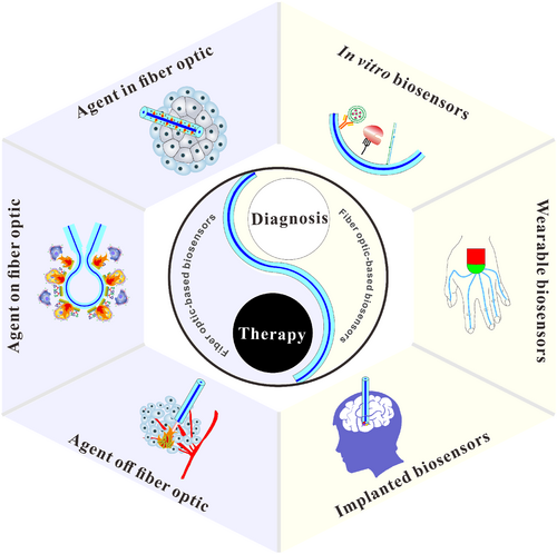
Summary of the fiber optic-based biosensors for the diagnosis and therapy.
2 DIAGNOSIS
2.1 In vitro
2.1.1 Immunosensor
Fiber optic-based immunosensors are based on highly specific binding of antibodies to antigens that occur on the fiber optic surface. Several strategies for the fiber optic-based immunosensors include direct, Sandwich, and competition methods. The direct approach is a common strategy in fiber optic-based immunosensors. Li’s group developed a fiber optic SPR-based immunoassay,47 as shown in Figure 2A. Unlike the traditional immunosensors, a half-antibody of goat anti-mouse Ig G was decorated on the gold surface of multimode fiber-single mode fiber-multimode fiber. In the heterocore fiber, the cladding modes of the single mode fiber were excited for strong SPR that is a sensitive response to the change in refractive index. The targets of mouse Ig G specifically bound with the half-antibody, leading to a strong SPR signal for sensing through changes in the refractive index. Due to their simple design, these strategies have been successfully used for the protein biomarker analysis in serum. The temperature fluctuation affects the accuracy of fiber optic-based biosensors through signal distribution. To address the problem, Ran et al. developed a temperature compensation method based on microfiber Bragg gating,48 as shown in Figure 2B. The fiber optic probe simultaneously measured the temperature and refractive index by changing the second and third harmonic resonances. It was found that temperature affected the activation of immunoreaction dynamics on the fiber optic surface between the antigen and antibody. Under the optimal temperature, the biosensor enabled the label-free and high-efficient detection of the cardiac biomarker of cTn-1. Besides, to reduce the interference of variability between the individual human serum sample, a commercial human serum from Sigma was serviced as the reference in the fiber optic-based biosensor.52 However, the direct immunoassay often suffers from inadequate sensitivity. Some novel strategies were developed to address the deficiency for highly sensitive detection. An effective means is to design the new structure fiber optics, such as D-shaped fiber53 and long-period fiber grating,25 to enhance the light-target interaction. Another is to couple the fiber optic-based biosensor with the advanced analytical techniques of the optofluidic waveguide.54
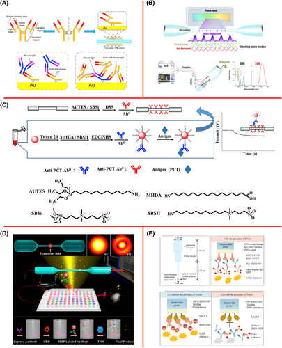
Three kinds of methods of the fiber optic-based immunosensors, namely direct, Sandwich and competition methods. (A) Half-antibody of goat anti-mouse Ig G enhances the sensitivity of direct method based on the heterocore fiber SPR.47 Copyright 2019, Elsevier. (B) Temperature compensation method based on microfiber Bragg gating for the of detection of cTn-1.48 Copyright 2021, Elsevier. (C) Sandwich assay based on antibody-modified AuNPs-target-antibody.49 Copyright 2020, Elsevier. (D) Sandwich assay based on HRP-antibody-C-reaction protein-antibody.50 Copyright 2019, American Chemical Society. (E) Competition method based on fiber optic-biolayer interferometry for the rapid detection of SARS-CoV-2 antibodies.51 Copyright 2022, Elsevier.
The Sandwich method based on immunoassay provides an effective strategy to amplify the signal of the fiber optic-based biosensor. The antibody on the fiber optic surface captured the targets. The labeled antibody was then specifically bound with targets to form the Sandwich of antibody-target-antibody for signal amplification. Gold nanoparticles (AuNPs) with strong plasmonic resonance, easy chemical modification, and well-stability have been considered ideal candidates to label with the antibody for the signal amplification. The diameter and concentration of AuNPs labels were considered as the key factors for the sensitivity of the Sandwich method.55 Besides, the nonspecific adsorption of the antibody-modified AuNPs caused a strong background signal to influence the detection accuracy. Chiang et al. developed a new signal enhancement method, namely fiber optic AuNPs-linked immunosorbent assay by improving the traditional Sandwich,49 as shown in Figure 2C. In the methods, the agents of sulfobetaine silane and sulfobetaine thiol were coated on the surface of fiber optic and AuNPs respectively to minimize the nonspecific interactions. The buffer pH and pI of the antibody were optimal to reduce the possible electrostatic interaction. The formation of the sandwich-like structure of antibody-modified AuNPs-target-antibody amplifies the LSPR signal. Due to the low background and high sensing signal, the modified Sandwich method is about five orders more sensitive than the direct one,49 showing a great potential to determine the procalcitonin in the blood plasma. Recently, the horseradish peroxidase that catalyzes the oxidation of 3,3′,5,5′-tetramethylbenzidine for visual quantification analysis was also employed to label with the antibody for the Sandwich immunoassay,50 as shown in Figure 2D. Under the formation of the Sandwich structure of the HRP-antibody-C-reaction protein-antibody, the microfiber acted as a reader, showing a strong response to the concentration of the C reaction protein. In summary, two antibodies are needed to bind with a target at different binding sites, limiting its application. A typical example is that some targets, such as small molecules, are hard to bind with two antibodies. Thus, it is not easy to apply the Sandwich method for the detection of the targets with small molecule weight.
The competition method is another common strategy for the small molecules in fiber optic-based biosensors. First, the targets of small molecules coupling with antibodies are modified on the surface of the fiber optics. Then, the presence of targets will competitively bind with the antibody, causing a change in the refractive index. The competition method was also coupled with the fiber optic-biolayer interferometry for the rapid detection of SARS-CoV-2 antibodies,51 as shown in Figure 2E. In the control group, the HRP-modified receptor binding domain was captured by the neutralizing antibodies on the surface of the fiber optics, where the generated 3,3′-diaminobenzidine tetrahydrochloride significantly improved the signal of fiber optic-biolayer interferometry. The neutralizing antibodies competitively bound with the HRP-modified receptor binding domain, hindering the signal generation. The method rapidly detected the SARS-CoV-2 neutralizing antibodies within 20 min.
Based on the above strategies, the immunoassay based on the fiber optic biosensor meets the requirements of high-speed, accuracy, anti-interference, and sensitivity for the application of the human serum samples. The immunoassay has been widely used in human serum, and urine, showing a promising potential for the clinical POCT diagnosis in vivo.
2.1.2 Aptasensors
Aptamers are short and single-stranded DNA or RNA obtained through the systematic evolution of ligands by exponential enrichment. Due to its advantages of high affinity, ease of synthesis, low cost, and structure stability,56 aptamer is also selected as a recognition element for the aptasensor based on fiber optics. Our previous work demonstrated that the density and orientation of the aptamers played a vital role in the aptasensor based on the Ω-shaped fiber optic.57, 58 Due to the bent area making the light propagation from the fundamental mode into the high order mode,59 the Ω-shaped fiber optic possessed high refractive index sensitivity for the LSPR-based aptasensor when the gold nanoparticles were coated on the fiber optic surface,60, 61 as shown in Figure 3A. Compared with the traditional thiol-modified aptamer, the ployA-modified aptamer adjusted the density on the fiber optic surface through the length of the ployA. The short ployA-modified aptamer with high density generated steric hindrance, which hindered the binding between the aptamer and target, while the long ployA-modified aptamer with low density also reduced the binding possibility. Besides, the single-strand aptamer with flexible construction and negative charge quickly absorbs on the citrate-wrapped AuNPs surface. To hinder the aptamer lying down on the AuNPs surface, a new T-shaped aptamer that contained the original aptamer, duplex-strand link domain, and ployA strand was designed,62 as shown in Figure 3B. The link domain with a rigid structure allowed the aptamer to stand on the AuNPs surface. Thus, the T-shaped aptamer-modified fiber optic LSPR exhibits high binding affinity and excellent specificity against the target N protein and has successfully been used to detect the SARS-CoV-2 from clinical samples and cold-chain foods.
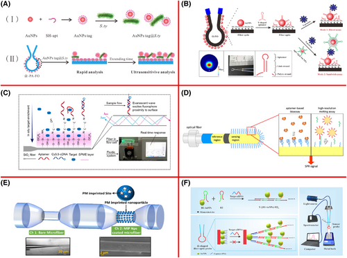
Fiber optic-based biosensor using the aptamer, molecularly imprinted nanocarrier and DNA-DNA hybridization as recognition elements. (A) PloyA-modified aptamer improved the sensitivity of the Ω-shaped fiber optic.57 Copyright 2022, Elsevier. (B) T-shaped aptamer enhanced binding affinity of fiber optic-based LSPR.62 Copyright 2022, American Chemical Society. (C) Nanoscale affinity double layer overcome the bottleneck of poor anti-interference capability of the fiber optic-based aptasensor.63 Copyright 2021, American Chemical Society. (D) A dual-region self-referencing fiber-optic SPR.64 Copyright 2022, American Chemical Society. (E) Microfiber interferometry modified with molecular imprinted nanocarrier for the detection of parathion methyl.65 Copyright 2019, Elsevier. (F) Hybridization chain reaction and nanomaterials for highly sensitive fiber optic-based LPSR.66 Copyright 2023, Elsevier.
Since the aptamer structure is vulnerable to the solution environment, including ion concentration, pH, and temperature, the poor anti-matrix interference is a great obstacle to the aptasensor based on the fiber optics. Zhao et al. devised a nanoscale affinity double layer on the fiber optic surface to overcome the bottleneck,63 as shown in Figure 3C. A nanoscale layer of solid-phase microextraction was fabricated on the fiber optic surface to rapidly enrich the targets. Then, the aptamer layer was immobilized on the layer of solid-phase microextraction to purify the target from the matrix. Finally, the binding interaction between the targets and aptamer is greatly enhanced for strong fluorescence excited by the evanescent wave. Besides, the dual region self-referencing fiber optic SPR was also used to avoid the matrix interference,64 as shown in Figure 3D. One region was serviced as a reference channel to compensate for the external interference signal, the others were designed to monitor the targets in the samples. Sample dilution is also adopted to solve the problem of anti-matrix interference. In this method, it needs to improve the sensitivity of the aptamer. The Sandwich method that amplifies the signal of the aptasensor was also designed to improve the accuracy indirectly.67, 68
2.1.3 Others
Recently, many novel recognition elements that specifically captured the targets from the biological matrix were used for the fiber optic-based biosensors, including bacteriophage,69 imprinting molecular,65 DNA-DNA hybridization,66 and peptides.70 Bacteriophages such as T467 and MS2 bacteriophages,71 are a kind of virus that specifically recognizes the host bacteria. Up to now, different kinds of fiber optics, including microcavity-based fiber optic,71 microfiber,69 long-period grating,72 and U-shaped,73 have been integrated with the bacteriophages for the specific detection of bacteria in a complex matrix. However, the application field of bacteriophage-based fiber optic biosensors is limited to bacteria. The molecularly imprinted polymers have complementary memory scaffolds of the target of the template molecule. Thus, molecularly imprinted polymers can recognize various targets in theory. Recently, Shrivastav et al. synthesized a molecular imprinted nanocarrier to develop microfiber interferometry for the detection of parathion methyl,65 as shown in Figure 3E. The biosensor distinguished the various other targets belonging to the parathion methyl family. DNA-DNA hybridization was based on the principles of Watson-Crick base pairing. The design in length and sequences adjusted the secondary structure of the captured DNA. The molecular beam shows strong ability to distinguish the complementary targets of microRNA from the mismatch microRNAs even the single-base mismatch microRNA.74 The molecular beam was also integrated with the hybridization chain reaction and nanomaterials for highly sensitive LPSR based on fiber optics in our recent work,66 as shown in Figure 3F. In a one-base mismatch hybridization chain reaction, one of the hairpins was modified on the gold nanoparticles. The target circulating cell-free DNA was hybridized with the captured DNA and one of hairpins of hybridization chain reaction, simultaneously. Then, the hybridization chain reaction was triggered to generate the duplex-stranded DNA concatemer linked with numerous gold nanoparticles. The biosensor exhibited high sensitivity and specificity for DNA detection. The captured DNA endows high selectivity for the detection of nucleotide acid in the fiber optic-based biosensor.74, 75 In summary, the biosensors with different fiber optic probes exhibit high sensitivity and wide linear range for the detection of various targets of small molecules, nucleic acid, proteins, exosomes, cells and bacteria, as shown in Table S1. In fact, the reported biosensors, including fluorescent,76, 77 electrochemical,78 and SERs-based sensors,79 also possess high performance on the sensitivity, linear range, and selectivity. Compared with those biosensors, the fiber optic-based biosensor exhibits a unique advantage that directly analyze the interaction between the target and fiber optic probe without the assistance of the label. The characteristic of label-free analysis makes the operation simple, which is more suitable for POCT diagnosis.
2.2 Wearable and implanted biosensors
The wearable and implanted biosensors open an opportunity for continuous monitoring of the state of human health. The fiber optic featuring a thin and soft body flexibly operated the light and brought the wearable and implanted biosensor into reality.80, 81 The fiber optic sensor is a powerful tool for sensing humidity, pressure, and strain, showing promising potential for wearable breath,35 temperature,36 and sport monitoring.37 Bao et al. fabricated an eccentric fiber Bragg grating inscribed single mode fiber optic,35 as shown in Figure 4A. The cladding mode was highly sensitive to the change in refractive index induced by humidity, and the core mode maintained stable spectral feature. The fiber optic-based biosensor rapidly responded and recovered from breathing within 92 and 100 ms, respectively. The biosensors also recognized different breathing patterns, including normal, slow, fast, and random. Besides, the silica-based distributed fiber optic sensor distinguished the location, magnitudes, and the modes of mechanical deformation,34 as shown in Figure 4B. After being encapsulated with different dyes in the different regions, the fiber optic detected the behaviors of undeformed, stretched, bent, and pressed. When the fiber optic was integrated with a soft glove, the three finger joints' movement in real time were reconstructed. Guan’s group also developed a flexible wearable optical sensor based on a microfiber Bragg grating,83 as shown in Figure 4C. In the biosensor, the core of the microfiber Bragg grating was encapsulated into a ductile PDMS film. The biosensor was sensitive to the changes in pressure, bending angle and temperature, so it can monitor several health parameters, including wrist pulses, finger knuckle bending, and human body temperature. Apart from the detection of physical parameters, the wearable fiber optic-based biosensor was also integrated with a fluorescent freeze-dried, cell-free device for the direct detection of nucleic acid,82 as shown in Figure 4D. However, sweat and sunlight induced degradation of the wearable sensors. When the fiber optic biosensors were employed for the physical parameter, the packaging enabled the interaction between the sensing area of fiber optic with sweat and sunlight. For the biomarker detection, a suitable fluid sampling system, such as microfluidics, is recommended to be integrated with fiber optic-based biosensor to eliminate the erosion from sweat.84 The sampling system rapidly transports biofluid rather than the sweat over the sensors, ensuring the long-term usage of the biosensors. In conclusion, the fiber optic-based biosensor is an innovative device to smartly monitor the healthcare information for the medical Internet of things.81

The wearable biosensors based on the fiber optic. (A) The eccentric fiber Bragg grating inscribed single mode fiber optic for the recognition of the different breathing patterns.35 Copyright 2021, Elsevier. (B) Fiber optic integrated with soft glove for the three finger joints' movement.34 Copyright 2020, The American Association for the Advancement of Science. (C) Wearable fiber optic-based biosensor for the detection of nucleic acid.82 Copyright 2021, Springer Nature.
To address this fragility, the fiber optic packaging is vital for the implanted biosensor. The packaged fiber optic immunosensor coupling with the TFBG-based SPR technology accurately detected the cytokeratin 17 protein,85 as shown in Figure 5A. The packaging made of a biocompatible polymer provided enough stiffness to penetrate human lung biopsy, where the cytokeratin 17 protein specifically bound with the fiber optics. The packaged fiber optic immunosensor provides a milestone for in situ detection of biomarkers in the tissues.88 The needle/catheter was also used as a package for the implanted fiber optics for the physiological oxygen detection in the deep tissue.89 In fact, the needle/catheter was implanted into the blood vein for real-time monitoring of the biomarker using fiber optic-based photoacoustic sensor.90 When the catheter helped the fiber optic-based biosensor implant into the simulated model of human arm vein, the photoacoustic sensor measured the concentration of heparin absorbed on the fiber optic surface. The needle-packaged fiber optic sensor provides an imaging depth, spatial resolution and low-cost instrument for the biomarker sensing in deep tissue and biofluids.91
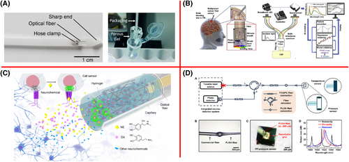
Implanted biosensors based on fiber optics. (A) Packaged fiber optic immunosensor.85 Copyright 2017, Elsevier. (B) Multiplexed fiber optic sensor coupling with artificial intelligence platform.86 Copyright 2022, Elsevier. (C) A fiber-optic probe embedded with engineering cells and hydrogels enabled for the real-time detection of neurochemicals in the freely moving animals.40 Copyright 2023, The American Association for the Advancement of Science. (D) Bioresorbable fiber optic based on poly(lactic-co-glycolic acid) for implanted biosensor.87 Copyright 2019, The American Association for the Advancement of Science.
When packaged with suitable materials, the silica-fiber optics is also a popular technique for the dynamic monitoring of the brain targets. Zhang et al. developed a multiplexed fiber optic sensor coupling with an artificial intelligence platform,86 as shown in Figure 5B. The end-face of the fiber optic was fixed with the sensing films of the four biomarkers, namely pH, temperature, dissolved oxygen, and glucose levels. The medicine-learning algorithm eliminated the cross-interferences in reflection spectra for the quantitative and accurate monitoring of the four biomarker levels in brain artificial cerebrospinal fluid. Recently, fiber-optic probes embedded with engineering cells and hydrogels enabled the real-time detection of neurochemicals in the freely moving animals,40 as shown in Figure 5C. The porous structure in hydrogels allowed the neurochemical molecules of dopamine, norepinephrine, and adenosine triphosphate rapidly diffused into the polymeric networks. When the neurochemical molecules connected with the engineering cells, the fluorescent proteins in the cells were expressed. The fiber optic-based biosensors were adopted to dynamically monitor neurochemicals in various vertebrates under various physiological and pathological conditions. The silica-fiber optics is non-resorbable and could be surgically extracted when used, which is a limitation in the continuous measurement within a long-time. The bioresorbable fiber optic made of poly(lactic-co-glycolic acid) bypassed the problem and eliminated the surgery extraction,87 as shown in Figure 5D. The reflection spectrum of the Fabry-Pérot interferometer based on the bioresorbable fiber was employed to accurately monitor the pressure and temperature inside the brain. Besides, the fiber optic probes were also used to design the endomicroscopy for brain imaging.92, 93 The fiber optic enables multiplex, long-time measurement, or imaging of the targets in the brain, showing a promising prospect for the clinical application of the brain.
Recently, the fiber optic-based implanted biosensors have also been used to assess tumors accurately and timely. Wu et al. designed a fluorescent biosensor based on the tapered fiber optics for the monitoring of the hypoxia gradient of tumor.39 After optimization of the fiber head structure, end diameter, excitation light power, and probe concentration, the biosensor realized in situ and rapid detection of the nitroreductase, an indicator of the hypoxia in the solid tumor, within 20 s. The biosensor enabled the potential of margin identification and guided the clinical surgery.
3 THERAPY
Some photo-sensitive agents transform the light energy into local heat through the photothermal effect. Three main strategies were developed to be integrated with the fiber optic and photothermal effect for therapy, including photosensitive materials off, on and in the fiber optics. When the NIR laser with high power propagated along the fiber optics, the local temperature of fiber optic surface was generated by the photothermal effect. When the cancer cells were captured on the surface of fiber optic with a surface temperature higher than 41.4°C, the localized high temperature could induce the cell apoptosis or necrocytosis.94 To be specific, the phospholipid molecules from the lipid bilayer disintegrated, which causes the cell apoptosis or necrocytosis.95, 96 Up to now, many photothermal therapies using fiber optic-based biosensors have been successfully developed, as shown in Table S2. The performance of photothermal therapy is discussed in depth in the following description.
3.1 Photosensitive materials off the fiber optic
In the first strategy, the photo-sensitive materials were enriched in the cancer cells or solid tumors, and the irradiated light from the fiber optic induced the temperature elevation for the therapy. An inserting fiber-optic was designed to induce the photothermal ablation.97 The gold nanorod-based theragnostic nanoparticles are efficient and hepatic site-selective delivery for optical/magnetic resonance/X-ray imaging. Under the imaging guidance, the fiber optic packaged with the catheter manipulated the laser light with a wavelength of 808 nm into the solid tumor for the photothermal ablation. To smartly recognize the tumor tissue, Zhu’s group synthesized a novel temperature-adaptive hydrogel fiber optics that was fabricated by the interaction of the functional monomer of N-isopropylacrylamide and the hydrophilic component of N-dimethylacrylamide,98 as shown in Figure 6A. The fiber optics showed the characteristic of a temperature-gated light propagation effect. The scattered light from the fiber optics heated the pre-injected photothermal nanoparticles in the solid tumor. The fiber optic regulated the temperature around the tumor, avoiding the side-effects on the normal tissue caused by overtemperature. To improve the efficiency, the fiber optic meshes coupling with silica rods were designed as near infrared red (NIR)illumination devices,99 as shown in Figure 6B. The device is a possible alternative strategy for the in vivo thermographic profiling of orthotopic ovarian tumors. Engineered nanomaterials hold significant promise to delivery photo-sensitive materials to specific organelles and tissues.101 The emerging lipid-based, polymeric, and inorganic nanoparticles with the personalized manner enter the era of precision medicine, which guarantees the safe and effective delivery of photosensitive materials. With the help of engineered nanomaterials, fiber optics exhibit promising applications in intelligent photomedicine.
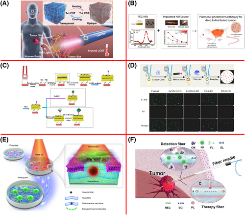
Photothermal effect based on nanoparticles off, on and in the fiber optic. (A) The temperature-adaptive hydrogel fiber optic made of functional monomer of N-isopropylacrylamide and hydrophilic component of N-dimethylacrylamide for interventional photomedicine.98 Copyright 2022, Springer Nature. (B) Fiber optic meshes coupling with silica rods for NIR illumination devices.99 Copyright 2013, American Chemical Society. (C) Integration of the cytosensor with the plasmonic photothermal therapy (PPT) using the microfiber.46 Copyright 2019, The American Association for the Advancement of Science. (D) Ω-shaped fiber-optic LSPR for the cytosensor and PPT.100 Copyright 2021, Elsevier. (E) Iron oxide nanoparticle-embedded nanofiber optics for photothermal effects.96 Copyright 2021, Springer Nature. (F) Rare-earth-doped fiber-optic theragnostic packaged with needle for the integration of biosensor with therapy.41 Copyright 2022, Wiley-VCH GmbH.
3.2 Photosensitive materials on the fiber optic
The cladding and leaky mode of the fiber optic excites the strong the photothermal effect of photo-sensitive materials, such as gold nanorods,102 graphene oxide supported Cu2-xS nanoplates,103 and gold nanostar,104 on the fiber optic surface. As discussed above, a fiber optic with a novel structure is a powerful technique to develop a cytosensor when the suitable recognition elements are modified on the surface of fiber optics. Thus, it is possible to integrate the cytosensor with the PPT. The concept was demonstrated using the microfiber-based SPR in 2019,46 as shown in Figure 6C. The black phosphorus-supported Au nanohybrids as coating on the fiber optic surface are not only used to modify the antibody of ErbB2 for cytosensor, but also absorbed the pump light with a wavelength of 980 nm for the PPT. The gold nanorods-black phosphorous nanointerface also achieved a similar function for integration with cytosensor and PPT,105 as shown in Figure 6D. Recently, our group further verified the concept using the Ω-shaped fiber-optic LSPR rather than SPR.100 The hybridized nanolayer of gold nanoparticles/gold nanorods together with Ω-shaped fiber-optic enhanced the refractive index sensitivity and sensitively captured the target cancer cell using the aptamer. Then, strong NIR light was leaked from the bending area of the Ω-shaped fiber-optic and excited the photothermal effect of the hybridized nanolayer, causing a sharp increase in temperature. Due to the superiority of localized and shape-depended heat distribution, Ω-shaped fiber-optic specifically killed the cancer cells captured on the fiber optic surface, providing a new way to minimize the side effect of PPT. Regrettably, the concept is not achieved in the animal mode.
3.3 Photosensitive materials in the fiber optic
It is concerned about the safety of the presence of the photo-sensitive nanoparticles off and on the fiber optic. The nanoparticles in the fiber optic ensure the indirect contact of the tissue with the nanoparticles. In the recent work, Xiong et al. fabricated an iron oxide nanoparticle-embedded nanofiber optic functionalized with photothermal effects,96 as shown in Figure 6E. The collagen was coated on the fiber optic surface to facilitate cell-adhesive. When the NIR laser continuously radiated on the fiber optic, iron oxide nanoparticles in the nanofiber optic generated the local heat to form the pores on the cell membrane. With the increase in laser energy, the increase in pores also led to the apoptosis/necrosis of cancer cells. Further, Ran et al. reported a rare-earth-doped fiber-optic theragnostic packaged with a needle for the integration with biosensor and therapy,41 as shown in Figure 6F. In the fiber optic-based biosensor, the fluorescent switch method was used to detect nitroeductase for fast and accurate tumor recognition and diagnosis. The rare earth was employed for the photoheating mediated by the NIR laser. The inscribed fiber Bragg grating was exploited for the real-time feedback on the potential of the generated temperature. The fiber optic fiber integrated diagnosis and treatment through a “plug and play” paradigm, minimizing the body invasion and normal tissue damage.
4 CHALLENGES AND CURRENT STRATEGIES
The fiber optic featuring the merits of small volume, flexibility to light operation, and low-cost was easy to fabricate various structures, including microfibers, U-shaped fiber, Ω-shaped fiber-optics, microfiber Bragg gratings, tapered fiber, fiber with heterogeneous cores, and D-shaped fiber. The novel structures of fiber optic probes endowed great ability to develop biosensors. When modified with the recognition element, the fiber optic probes have been successfully used to develop the immunosensors, aptasensors, and other biosensors. Further, the optimal packaging and design of the analysis method extended the biosensors from the in vitro detection to wearable and implanted biosensors. Interestingly, photo-sensitive agents off, on, and in the fiber optic probes excited the strong photothermal effect for therapy. Thus, the fiber optic probes could be integrated with biosensors and therapy, showing great potential for sensitive diagnosis and efficient therapy. Due to the huge individual variability, precision medicine depended on the precise diagnosis, providing an efficient therapeutic strategy for everyone. However, the current fiber optic-based biosensor did not meet the requirement of precision medicine. Some challenges are eager to be addressed:
Firstly, unlike the diagnosis in traditional medicine relying on one or two key biomarkers, the diagnosis of precision medicine assessed the state of the disease under the sufficient understanding of the expression level of a set of biomarkers or physiological indexes. It is required to develop a fiber optic-based biosensor with high throughput. The present fiber optic-based biosensor focused on the improvement of the performance for a single biomarker. It is still a challenge to construct a fiber optic-based biosensor that can detect multiple biomarkers simultaneously. The smartphone camera integrated with the gating collected the light signal from the seven fiber optic probes in our previous work.106 The medicine learning algorithm sensitively and specifically recognized the individual from large-scale clinical cases. Introduction of the above techniques will be a possible way to design the fiber optic-based biosensors with high throughput.
Secondly, some physiological indexes in precision medicine should be monitored long-term through the wearable or implanted biosensors. To avoid the influence on the behavior of human beings, the miniaturization setup of fiber optic-based wearable biosensors is required. Although the fiber optic has a diameter in micrometer scale, the detector and light source in the setup was cumbersome. The emerging the micro-electromechanical systems (MEMS)technology could integrate large-scale devices into a system within a micrometer level. The introduction of MEMS will further develop the miniature fiber optic-based biosensor. The implanted fiber optic-based biosensors ensure the long-term monitoring of biomarkers or physiological indexes. Traditional silica-based fiber optics are very fragile and stable. Once snapped, the fiber optics in the body could induce inflammation and other abnormal physiological reactions, bringing unknown damage to our health. The new fiber optic with high biocompatibility, degradability, and flexibility are urgently needed to be synthesized. The new fiber optic together with the miniaturization setup is an alternative scheme to design a wearable/implanted biosensor for the diagnosis of personality medicine.
Thirdly, high efficiency and low side-effects are the basic indices of the therapy for the personality medicine. Current therapies based on fiber optic probes focus on the photothermal effect. Therapy based on a single strategy could not eliminate the solid tumor thoroughly. Several other therapy strategies, including photodynamic treatment, chemotherapy, and optogenetic therapies, were combined into a system that efficiently improved the curative effect. Integration with diagnosis and treatment provided significant guidance for the treatment of precision medicines to reduce the side-effects. Although it was carried out that fiber optic-based biosensor can be integrated with diagnosis and treatment, the biosensor provided the concentration of the biomarkers on a dot rather than space distribution. The space distribution of the biomarker concentration brings more significant information, such as the boundary of the solid tumor, to guide the therapy. The lack of space station information inhibited the development of fiber optic-based biosensors for precision medicine. Recently, Piosano et al. exploited the modal characteristic of the tapered fiber for the multisite photometry along the cone area.107 The generated fluorescence signal that was confined around the cone area was collected through a two-photon scanning system. The tapered fiber optic with a collection system was engineered to obtain the concentration of dopamine at multiple sites in the deep brain, even extended to the biological system or organ. The advanced technique based on optical engineering will address the issue of the space station information of biosensors to minimize the side-effects of the therapy.
5 CONCLUSIONS
The fiber optic has been successfully applied in the fields of biosensors in vitro, wearable/implanted biosensors, and therapy in vivo. Firstly, there are several recognition elements for fiber optic-based biosensors in vitro, including antibodies, aptamers, bacteriophages, imprinting molecules, DNA-DNA hybridization, and peptides. Among them, antibody and aptamer are two kinds of common recognition elements for the design of the immunoassay and aptasensor, respectively. It is flexible to design an immunoassay based on the fiber optic for the detection of protein biomarkers in human serum through several strategies of direct, Sandwich, and competition methods. It is also found that the density and orientation of the aptasensor on the AuNPs play a vital role in the sensitivity of the aptasensor. The techniques of solid-phase microextraction and self-referencing region improve the anti-interference of the aptasensor, making it possible for the applications in human serum.
Furthermore, fiber optics are powerful materials for developing wearable and implanted biosensors with a thin and soft body can flexibly operate the light. A fiber optic sensor that is sensitive to the changes in humidity, pressure, and strain has been considered as a promising candidate for breath, temperature, and sport monitoring. When the fragile fiber optics were encapsulated with a needle or catheter, the biosensor long-time monitored the biomarkers in the brain, blood vessels, and deep tissue.
Finally, some photo-sensitive material off, on and in fiber optics can excite strong photothermal effect for cancer therapy. Interestingly, the implanted biosensor was also coupled with the therapy to achieve the goal of integration of diagnosis and treatment. Thus, the obtained information from the biosensor provides a significant guide for the therapy, showing a promising potential for smart medicine.
In order to promote its application in the precision medicine, we discussed the challenges of the current fiber optic-based biosensor, including low throughput, bulky volume of setup, potential damage to the human body, and low therapy efficiency. With the advance of cut-edging techniques of medicine learning algorithm, MEMS, emerging materials and optical engineering, the novel fiber optic-based biosensors with excellent performance in diagnosis and therapy can better meet the requirements of the precision medicine.
ACKNOWLEDGMENTS
This work was supported by the National Natural Science Foundation of China (No. 62305235, No. 82373633, and No. 22274104), Fundamental Research Funds for the Central Universities (YJ202291), the Key R&D project of Science and Technology Department of Sichuan Province (2023YFS0400), and Open Fund of the Guangdong Provincial Key Laboratory of Optical Fiber Sensing and Communications (Jinan University).
CONFLICT OF INTEREST STATEMENT
The authors declare no conflicts of interest.
ETHICS STATEMENT
Not applicable.
Biographies

Zewei Luo received his Ph.D. degree in biochemistry and molecular biology from Sichuan University in 2019 and then joined Northwest University as a postdoctor. He presently is a distinguished associate researcher in the Research Center of Analytical Instrumentation, School of Mechanical Engineering of Sichuan University. His current research interest is focusing on the development of fiber optic-based biosensors in the field of medicine diagnosis and treatment.

Mingyue Li received his bachelor's degree at Sichuan University in 2023. He is now studying for his masters degree at Sichuan University. His current research major is fiber optic-based biosensors.

Xinyu Kong is a graduate student in the School of Chemistry and Materials Science, Northwest University. Her main research interest is fiber optic biosensors. Graduate Tutoris Jiwei Shen associate professor.

Yu Li will receive her Ph.D in Septemder from College of Life Science, Sichuan University. She is engaged in the research of optical fiber LSPR biosensors for bacterial detection.

Wenwen Li is currently an assistant professor at West China School of Public Health of Sichuan University. She received her Ph.D. degree in Analytical Chemistry from Sichuan University in 2018. Her research focuses on metabolic VOCs analysis and disease diagnosis biomarkers.

Ze Tian graduated from Sichuan University in 2019 with his bachelors degree. He is now studying for his Ph.D. degree at the Sichuan University. His research interests include the laser-induced breakdown spectroscopy and optical design.

Qun Cao is a Postdoctoral Scholar at the David H. Koch Institute for Integrative Cancer Research at the Massachusetts Institute of Technology. He received his B.S. in Chemistry in 2016 from the University of Science and Technology of China (USTC) and his Ph.D. in 2020 from the University of Virginia at Charlottesville. During his Ph.D., he worked with Professor B. Jill Venton to develop carbon electrodes for neurotransmitter sensing. After a postdoc with Professor X. Tracy Cui at the University of Pittsburgh, he joined Professor Michael J. Cima's lab at MIT in 2023 to work on microsamples of brain interstitial fluid to determine soluble and membrane bound protein.

Muhammad Haider Zaman received his Master's degree (M. Phil.) from the University of Agriculture Faisalabad, Pakistan, in 2021. He is now studying for his Ph.D. degree at Sichuan University, China. His research area is laser-matter interaction, such as LIBS, surface mapping, laser ablation, biomedical, laser propulsion, and AAS.

Yongxin Li is an associate professor in West China School of Public Health, Sichuan University. She is mainly engaged in the study of novel biosensors, microfluidic chip and their application in public health, particularly in foodborne-pathogenic bacteria, biomarkers, viruses and chemical detection.

Wei Xiao received his Ph.D. degree from Jinan University in 2020. He joined the department of laboratory medicine at Guangdong Second Provincial General Hospital as a researcher since 2020. His research interest mainly focusses on biosensor in the diagnosis of infectious diseases and tumors, specifically in the immunosensors, paper-based microfluidic analytical devices and smartphone-based sensors.

Yixiang Duan is currently a National Special Professor and Director of Research Center of Analytical Instrumentation, Sichuan University, China. His current research interests include fiber optic-based biosensors, novel analytical instrumentation, noninvasive medical diagnostics, new ionization source for mass spectrum, molecule spectrometry, as well as new biosensors design and development, and various applications.


