Standard Article

Acetone Carboxylase

Alexander B Alleman

Alexander B Alleman

Institute of Biological Chemistry, Washington State University, Pullman, WA, 99164 USA

Search for more papers by this author
Gregory A Prussia

Gregory A Prussia

Institute of Biological Chemistry, Washington State University, Pullman, WA, 99164 USA

Search for more papers by this author
Florence Mus

Florence Mus

Institute of Biological Chemistry, Washington State University, Pullman, WA, 99164 USA

Search for more papers by this author
John W Peters

John W Peters

Institute of Biological Chemistry, Washington State University, Pullman, WA, 99164 USA

Search for more papers by this author

Abstract

Carboxylases incorporate CO2, or its hydrated form HCO3, into organic substrates. Assimilatory carboxylases combine CO2 to a diversity of carbon sources allowing for growth on intransigent, poorly activated organic compounds such as acetone. Acetone carboxylase catalyzes the adenosine triphosphate-dependent carboxylation of acetone to acetoacetate. Acetone carboxylase undergoes a large conformational shift to allow phosphorylated intermediates, carboxyphosphate and phosphenolacetone to interact with the Mn bound active site. The coordinated Mn orients the intermediates for the carboncarbon bond formation producing acetoacetate. Biochemical, molecular, structural, and spectroscopic studies have shown how acetone carboxylase functions and gives clues on how this unique biotin-independent carboxylation mechanism couples ATP hydrolysis to bicarbonate-dependent carboxylation. This article highlights the body of biochemical, spectroscopic, and structural work leading to insights into the catalytic mechanism of acetone carboxylase.

3D Structure
Description unavailable

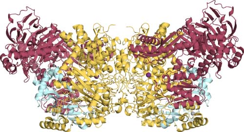
The overall structure of the (αβγ)2 heterohexameric enzyme with the α-subunit (yellow) forming the dimeric core, β-subunit (red) and γ-subunit (light blue) located on the exterior of the dimeric core. The Mn2+ ion (purple) in the middle of the α-subunit and the Zn2+ ion (grey) in the γ-subunit. Figure was made with PyMOL1 with PDB code: 5M45.

The full text of this article hosted at iucr.org is unavailable due to technical difficulties.