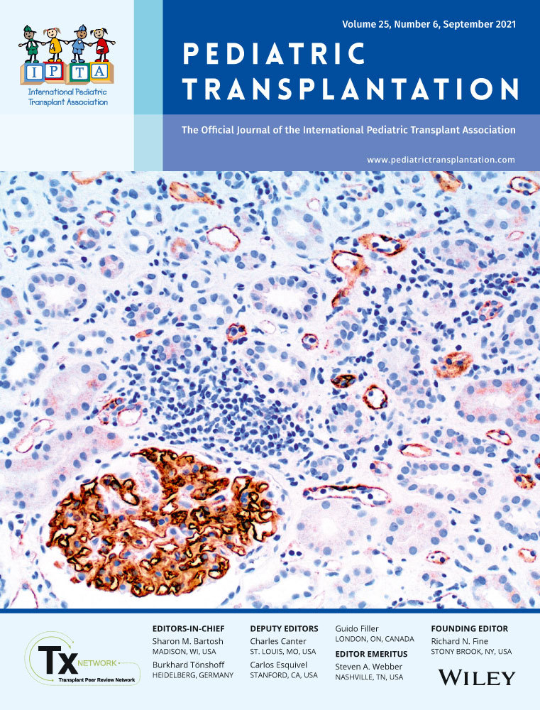
Antithymocyte globulin induction therapy and myocardial complement deposition in pediatric heart transplantation
Abstract
Background
Antithymocyte globulin (ATG) consists of polyclonal antibodies directed primarily against human T lymphocytes but may contain antibodies with affinity for other tissues in the transplanted organ, resulting in complement (C4d) deposition. This phenomenon has been demonstrated in endomyocardial biopsies (EMBs) of adult cardiac transplants. We examined the relationship of induction immunosuppression with ATG and C4d deposition in EMB of pediatric cardiac transplants.
Methods
Results of C4d immunohistochemistry were available from all EMB of patients transplanted at our center between June 2012 and April 2018 (n = 48) who received induction immunosuppression with either ATG (n = 20) or basiliximab (n = 28) as the standard of care.
Results
C4d deposition in the first year post–heart transplant was more commonly seen among patients who received ATG induction (20% of EMBs in ATG group vs 1% of EMBs in basiliximab group; p < .0001). C4d deposition related to ATG was observed early post-transplant (50% ATG vs 0% basiliximab on first EMB; p < .0001 and 35% ATG vs 0% basiliximab on the second EMB; p = .0012). While this difference waned by the third EMB (5% ATG vs 0% basiliximab; p = .41), positive C4d staining persisted to the sixth EMB in the ATG group only (6%).
Conclusion
C4d deposition is common on EMB up to 1 year post–pediatric cardiac transplant following ATG induction. This high rate of positive C4d staining in the absence of histologic AMR after ATG induction therapy must be accounted for in making clinical decisions regarding cardiac allograft rejection diagnosis and treatment.
CONFLICT OF INTEREST
None of the authors has a financial relationship with a commercial entity that has an interest in the subject of the presented manuscript or other conflicts of interest to disclose. The other authors report no conflicts of interest in this study.
Open Research
DATA AVAILABILITY STATEMENT
The data that support the findings of this study are available from the corresponding author upon reasonable request.


