Volume 25, Issue 5 pp. 807-812
Clinical Investigative Study

Optic Neuritis and the Visual Pathway: Evaluation of Neuromyelitis Optica Spectrum by Resting-State fMRI and Diffusion Tensor MRI

Fernanda Cristina Rueda Lopes MD, MSc

Corresponding Author

Fernanda Cristina Rueda Lopes MD, MSc

Federal University of Rio de Janeiro, Radiology, Rua Presidente João Pessoa, Niterói, Rio de Janeiro, Brazil

Correspondence: Address correspondence to Fernanda C Rueda Lopes, Federal University of Rio de Janeiro, Radiology, Rua Presidente João Pessoa, 385/704, Niterói, Rio de Janeiro, Brazil, 24220-330. E-mail: [email protected].Search for more papers by this author
Soniza Vieira Alves-Leon MD, PhD

Soniza Vieira Alves-Leon MD, PhD

Federal University of Rio de Janeiro, Neurology, Rio de Janeiro, Rio de Janeiro, Brazil

Search for more papers by this author
Jose Mauricio Godoy MD

Jose Mauricio Godoy MD

State University of Rio de Janeiro, Neurology, Rio de Janeiro, Rio de Janeiro, Brazil

Search for more papers by this author
Simone de Souza Batista Scherpenhuijzen MD

Simone de Souza Batista Scherpenhuijzen MD

State University of Rio de Janeiro, Neurology, Rio de Janeiro, Rio de Janeiro, Brazil

Search for more papers by this author
Leticia Fezer MD

Leticia Fezer MD

State University of Rio de Janeiro, Neurology, Rio de Janeiro, Rio de Janeiro, Brazil

Search for more papers by this author
Emerson Leandro Gasparetto MD, PhD

Emerson Leandro Gasparetto MD, PhD

Federal University of Rio de Janeiro, Radiology, Rua Presidente João Pessoa, Niterói, Rio de Janeiro, Brazil

Search for more papers by this author
First published: 15 January 2015
Citations: 13

ABSTRACT

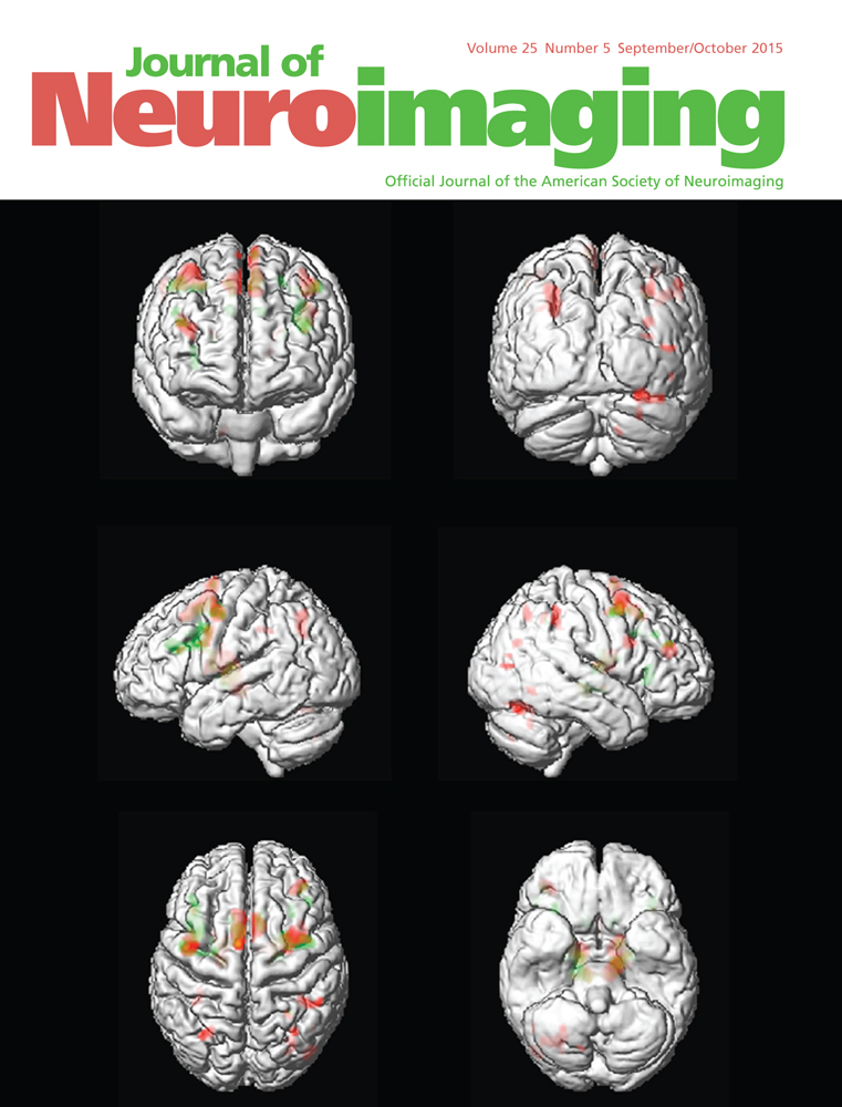
BACKGROUND AND PURPOSE

Optic neuritis (ON) is an acute episode of inflammation in the visual pathway (VP). It may occur as part of a demyelinating disease, which can affect white matter (WM) throughout the VP. Compensatory cortical adaptations may occur following WM damage to maintain visual integrity. Our aim was to investigate whether resting-state functional MRI (rsfMRI) can detect cortical adaptations following ON attacks and to correlate rsfMRI with diffusion tensor imaging (DTI) of WM within the VP.

MATERIALS AND METHODS

Neuromyelitis optica spectrum patients were compared to healthy controls at least 6 months after ON onset. DTI and rsfMRI were performed and post-processed using FSL tools (TBSS for DTI and MELODIC for fMRI).

RESULTS

Ptients had higher synchronization values than controls in the visual network (3.48 vs. 2.12, P < .05). A weak trend of correlation was revealed between fMRI and structural analysis by DTI using fractional anisotropy (right side: R = −.36, P < .08; left side: R = .075, P < .73).

CONCLUSIONS

The rsfMRI detected cortical reorganization following ON attack, but WM was considerably preserved in the posterior VP.

The full text of this article hosted at iucr.org is unavailable due to technical difficulties.