Cerebriform plantar hyperplasia: the major cutaneous feature of Proteus syndrome
Abstract
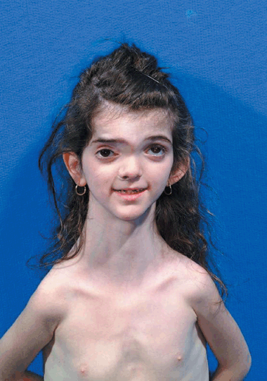
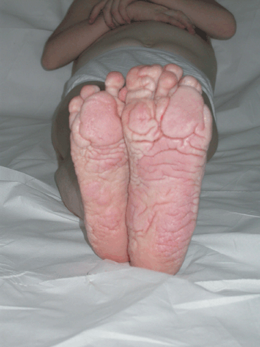
A 6-year-old girl was referred for clinical evaluation. Her phenotype was characterized by an asymmetric face, dysmorphic skull with frontal-parietal hyperostosis, hypertelorism, strabismus, long neck, and low-set antiverted ears (Fig. 1). Her thorax was also dysmorphic with dropped shoulders because of the abnormal orientation of the scapulae and scoliosis. Moreover, the following features were evident: multiple abdominal lipomatous formations, muscle hypotonia and hypotrophia, lower limb heterometria, severe limitation of spinal articulation movements, congenital papular-nodular nevus over the posterior axillary line, macrodactyly of the third and fourth right fingers, left hand smaller than the right, and thickening of the cutaneous tissues of the plantar surface of the foot with a gyrate and cerebriform aspect (Fig. 2). In addition, simple hypermetropic astigmatism of both eyes, exotropia and hypertrophy of the left eye, and dental caries were found.

Cranial and facial asymmetry with fronto-parietal exostosis on the right side

Cerebriform bilateral plantar hyperplasia
All routine blood examinations were within normal limits; electromyography disclosed a primary muscle disease. Thyroid ultrasound showed a dyshomogeneous structure with normal dimensions. Abdominal ultrasound showed a normal liver, gall bladder, and spleen; however, the pyelic cavities of the kidney were enlarged. Finally, molecular genetic analysis of the PTEN gene was negative.
The clinical and instrumental features and the presence of significant cerebriform plantar hyperplasia allowed us to propose a diagnosis of Proteus syndrome.


