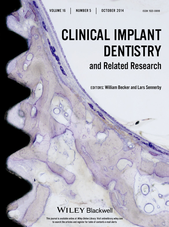
In Vivo and In Vitro Studies of Epithelial Cell Behavior around Titanium Implants with Machined and Rough Surfaces
Corresponding Author
Ikiru Atsuta DDS, PhD
Assistant professor
Section of Implant and Rehabilitative Dentistry, Division of Oral Rehabilitation, Faculty of Dental Science, Kyushu University, Fukuoka, Japan
Center for Craniofacial Molecular Biology, University of Southern California School of Dentistry, Los Angeles, California, USA
Reprint requests: Dr. Ikiru Atsuta, Section of Implant and Rehabilitative Dentistry, Division of Oral Rehabilitation, Faculty of Dental Science, Kyushu University, 3-1-1 Maidashi, Higashi-ku, Fukuoka 812-8582, Japan; e-mail: [email protected]Search for more papers by this authorYasunori Ayukawa DDS, PhD
assistant professor
Section of Implant and Rehabilitative Dentistry, Division of Oral Rehabilitation, Faculty of Dental Science, Kyushu University, Fukuoka, Japan
Search for more papers by this authorAkihiro Furuhashi DDS, PhD
clinical fellow
Section of Implant and Rehabilitative Dentistry, Division of Oral Rehabilitation, Faculty of Dental Science, Kyushu University, Fukuoka, Japan
Search for more papers by this authorYoichiro Ogino DDS, PhD
assistant professor
Section of Implant and Rehabilitative Dentistry, Division of Oral Rehabilitation, Faculty of Dental Science, Kyushu University, Fukuoka, Japan
Search for more papers by this authorYasuko Moriyama DDS, PhD
assistant professor
Section of Implant and Rehabilitative Dentistry, Division of Oral Rehabilitation, Faculty of Dental Science, Kyushu University, Fukuoka, Japan
Search for more papers by this authorYoshihiro Tsukiyama DDS, PhD
associate professor
Section of Implant and Rehabilitative Dentistry, Division of Oral Rehabilitation, Faculty of Dental Science, Kyushu University, Fukuoka, Japan
Search for more papers by this authorKiyoshi Koyano DDS, PhD
professor
Section of Implant and Rehabilitative Dentistry, Division of Oral Rehabilitation, Faculty of Dental Science, Kyushu University, Fukuoka, Japan
Search for more papers by this authorCorresponding Author
Ikiru Atsuta DDS, PhD
Assistant professor
Section of Implant and Rehabilitative Dentistry, Division of Oral Rehabilitation, Faculty of Dental Science, Kyushu University, Fukuoka, Japan
Center for Craniofacial Molecular Biology, University of Southern California School of Dentistry, Los Angeles, California, USA
Reprint requests: Dr. Ikiru Atsuta, Section of Implant and Rehabilitative Dentistry, Division of Oral Rehabilitation, Faculty of Dental Science, Kyushu University, 3-1-1 Maidashi, Higashi-ku, Fukuoka 812-8582, Japan; e-mail: [email protected]Search for more papers by this authorYasunori Ayukawa DDS, PhD
assistant professor
Section of Implant and Rehabilitative Dentistry, Division of Oral Rehabilitation, Faculty of Dental Science, Kyushu University, Fukuoka, Japan
Search for more papers by this authorAkihiro Furuhashi DDS, PhD
clinical fellow
Section of Implant and Rehabilitative Dentistry, Division of Oral Rehabilitation, Faculty of Dental Science, Kyushu University, Fukuoka, Japan
Search for more papers by this authorYoichiro Ogino DDS, PhD
assistant professor
Section of Implant and Rehabilitative Dentistry, Division of Oral Rehabilitation, Faculty of Dental Science, Kyushu University, Fukuoka, Japan
Search for more papers by this authorYasuko Moriyama DDS, PhD
assistant professor
Section of Implant and Rehabilitative Dentistry, Division of Oral Rehabilitation, Faculty of Dental Science, Kyushu University, Fukuoka, Japan
Search for more papers by this authorYoshihiro Tsukiyama DDS, PhD
associate professor
Section of Implant and Rehabilitative Dentistry, Division of Oral Rehabilitation, Faculty of Dental Science, Kyushu University, Fukuoka, Japan
Search for more papers by this authorKiyoshi Koyano DDS, PhD
professor
Section of Implant and Rehabilitative Dentistry, Division of Oral Rehabilitation, Faculty of Dental Science, Kyushu University, Fukuoka, Japan
Search for more papers by this authorAbstract
Background
The surface roughness of a dental implant affects the epithelial wound healing process and may significantly enhance implant prognosis.
Purpose
We explored the influence of surface roughness on peri-implant epithelium (PIE) sealing and down-growth by comparing machine-surfaced (Ms) and rough-surfaced (Rs) implants.
Materials and Methods
(1) Maxillary first molars were extracted from rats and replaced with Ms or Rs implants. (2) We also compared changes in the morphology of cultured rat oral epithelial cells (OECs) grown on Ms or Rs titanium (Ti) plates.
Results
(1) After 4 weeks, the PIE around Ms and Rs implants showed a similar structure to junctional epithelium (JE). At 16 weeks, Rs implants appeared to form a weak epithelial seal at the tissue-implant interface and exhibited markedly less PIE down-growth than Ms implants but was deeper than that observed in natural teeth. (2) We observed less expression of adhesion proteins in OECs cultured on Rs plates than in cells grown on Ms plates. Additionally, cell adherence, migration, and proliferation on Rs plates were lower, whereas apoptosis was reduced on Ms plates.
Conclusion
Ms implants are a better choice for integration with an epithelial wound healing process.
References
- 1 Brånemark PI, Adell R, Albrektsson T, Lekholm U, Lundkvist S, Rockler B. Osseointegrated titanium fixtures in the treatment of edentulousness. Biomaterials 1983; 4: 25–28.
- 2 Chehroudi B, Gould TR, Brunette DM. A light and electron microscopic study of the effects of surface topography on the behavior of cells attached to titanium-coated percutaneous implants. J Biomed Mater Res 1991; 25: 387–405.
- 3 Eisenbarth E, Meyle J, Nachtigall W, Breme J. Influence of the surface structure of titanium materials on the adhesion of fibroblasts. Biomaterials 1996; 17: 1399–1403.
- 4 Mustafa K, Silva Lopez B, Hultenby K, Wennerberg A, Arvidson K. Attachment and proliferation of human oral fibroblasts to titanium surfaces blasted with TiO2 particles. A scanning electron microscopic and histomorphometric analysis. Clin Oral Implants Res 1998; 9: 195–207.
- 5 Ellingsen JE, Thomsen P, Lyngstadaas SP. Advances in dental implant materials and tissue regeneration. Periodontol 2000 2006; 41: 136–156.
- 6 Wennerberg A, Sennerby L, Kultje C, Lekholm U. Some soft tissue characteristics at implant abutments with different surface topography. A study in humans. J Clin Periodontol 2003; 30: 88–94.
- 7 Orsini G, Assenza B, Scarano A, Piattelli M, Piattelli A. Surface analysis of machined versus sandblasted and acid-etched titanium implants. Int J Oral Maxillofac Implants 2000; 15: 779–784.
- 8 Albrektsson TO, Johansson CB, Sennerby L. Biological aspects of implant dentistry: osseointegration. Periodontol 2000 1994; 4: 58–73.
- 9 Ikeda H, Yamaza T, Yoshinari M, et al. Ultrastructural and immunoelectron microscopic studies of the peri-implant epithelium-implant (Ti-6Al-4V) interface of rat maxilla. J Periodontol 2000; 71: 961–973.
- 10 Borradori L, Sonnenberg A. Hemidesmosomes: roles in adhesion, signaling and human diseases. Curr Opin Cell Biol 1996; 8: 647–656.
- 11 Atsuta I, Yamaza T, Yoshinari M, et al. Ultrastructural localization of laminin-5 (gamma2 chain) in the rat peri-implant oral mucosa around a titanium-dental implant by immuno-electron microscopy. Biomaterials 2005; 26: 6280–6287.
- 12 Ikeda H, Shiraiwa M, Yamaza T, et al. Difference in penetration of horseradish peroxidase tracer as a foreign substance into the peri-implant or junctional epithelium of rat gingivae. Clin Oral Implants Res 2002; 13: 243–251.
- 13 Berglundh T, Gotfredsen K, Zitzmann NU, Lang NP, Lindhe J. Spontaneous progression of ligature induced peri-implantitis at implants with different surface roughness: an experimental study in dogs. Clin Oral Implants Res 2007; 18: 655–661.
- 14 Atsuta I, Yamaza T, Yoshinari M, et al. Changes in the distribution of laminin-5 during peri-implant epithelium formation after immediate titanium implantation in rats. Biomaterials 2005; 26: 1751–1760.
- 15 Furuhashi A, Ayukawa Y, Atsuta I, Okawachi H, Koyano K. The difference of fibroblast behavior on titanium substrata with different surface characteristics. Odontology 2012; 100: 199–205.
- 16 Atsuta I, Ayukawa Y, Ogino Y, Moriyama Y, Jinno Y, Koyano K. Evaluations of epithelial sealing and peri-implant epithelial down-growth around “step-type” implants. Clin Oral Implants Res 2012; 23: 459–466.
- 17 Shiraiwa M, Goto T, Yoshinari M, Koyano K, Tanaka T. A study of the initial attachment and subsequent behavior of rat oral epithelial cells cultured on titanium. J Periodontol 2002; 73: 852–860.
- 18 Yan T, Sun R, Deng H, Tan B, Ao N. The morphological and biomechanical changes of keratocytes cultured on modified p (HEMA-MMA) hydrogel studied by AFM. Scanning 2009; 31: 246–252.
- 19 Li Y, Lin JL, Reiter RS, Daniels K, Soll DR, Lin JJ. Caldesmon mutant defective in Ca(2+)-calmodulin binding interferes with assembly of stress fibers and affects cell morphology, growth and motility. J Cell Sci 2004; 117: 3593–3604.
- 20 Métrailler-Ruchonnet I, Pagano A, Carnesecchi S, Ody C, Donati Y, Barazzone Argiroffo C. Bcl-2 protects against hyperoxia-induced apoptosis through inhibition of the mitochondria-dependent pathway. Free Radic Biol Med 2007; 42: 1062–1074.
- 21 Berglundh T, Lindhe J. Dimension of the periimplant mucosa. Biological width revisited. J Clin Periodontol 1996; 23: 971–973.
- 22 Cochran DL, Hermann JS, Schenk RK, Higginbottom FL, Buser D. Biologic width around titanium implants. A histometric analysis of the implanto-gingival junction around unloaded and loaded nonsubmerged implants in the canine mandible. J Periodontol 1997; 68: 186–198.
- 23 Grunder U. Stability of the mucosal topography around single-tooth implants and adjacent teeth: 1-year results. Int J Periodontics Restorative Dent 2000; 20: 11–17.
- 24 Tarnow DP, Wallace SS, Froum SJ, Rohrer MD, Cho SC. Histologic and clinical comparison of bilateral sinus floor elevations with and without barrier membrane placement in 12 patients: part 3 of an ongoing prospective study. Int J Periodontics Restorative Dent 2000; 20: 117–125.
- 25 Jansen VK, Conrads G, Richter EJ. Microbial leakage and marginal fit of the implant-abutment interface. Int J Oral Maxillofac Implants 1997; 12: 527–540.
- 26 Tarnow D, Stahl SS, Magner A, Zamzok J. Human gingival attachment responses to subgingival crown placement. Marginal remodelling. J Clin Periodontol 1986; 13: 563–569.
- 27 Schroeder HE. Healing and regeneration following periodontal treatment. Dtsch Zahnarztl Z 1986; 41: 536–538.
- 28 Michelson PH, Tigue M, Jones JC. Human bronchial epithelial cells secrete laminin 5, express hemidesmosomal proteins, and assemble hemidesmosomes. J Histochem Cytochem 2000; 48: 535–544.
- 29 Sieg DJ, Hauck CR, Schlaepfer DD. Required role of focal adhesion kinase (FAK) for integrin-stimulated cell migration. J Cell Sci 1999; 112(Pt 16): 2677–2691.
- 30 Diener A, Nebe B, Luthen F, et al. Control of focal adhesion dynamics by material surface characteristics. Biomaterials 2005; 26: 383–392.
- 31 Eisenbarth E, Velten D, Schenk-Meuser K, et al. Interactions between cells and titanium surfaces. Biomol Eng 2002; 19: 243–249.
- 32 Chu CL, Reenstra WR, Orlow DL, Svoboda KK. Erk and PI-3 kinase are necessary for collagen binding and actin reorganization in corneal epithelia. Invest Ophthalmol Vis Sci 2000; 41: 3374–3382.
- 33 Rezniczek GA, Janda L, Wiche G. Plectin. Methods Cell Biol 2004; 78: 721–755.
- 34 Nobes CD, Hall A. Rho GTPases control polarity, protrusion, and adhesion during cell movement. J Cell Biol 1999; 144: 1235–1244.
- 35 Okumura A, Goto M, Goto T, et al. Substrate affects the initial attachment and subsequent behavior of human osteoblastic cells (Saos-2). Biomaterials 2001; 22: 2263–2271.
- 36 Wang XJ, Maier K, Fuse S, et al. Thrombospondin-1-induced migration is functionally dependent upon focal adhesion kinase. Vasc Endovascular Surg 2008; 42: 256–262.
- 37 Lademann J, Weigmann H, Rickmeyer C, et al. Penetration of titanium dioxide microparticles in a sunscreen formulation into the horny layer and the follicular orifice. Skin Pharmacol Appl Skin Physiol 1999; 12: 247–256.
- 38 Roehlecke C, Kuhnt AK, Fehrenbach H, Werner C, Funk RH, Kasper M. Resistance of L132 lung cell clusters to glyoxal-induced apoptosis. Histochem Cell Biol 2000; 114: 283–292.


