Effects of paeoniflorin-6′-O-benzene sulfonate on the pharmacokinetics, excretion and tissue distribution of leflunomide in rats
Ning Xiao
Institute of Clinical Pharmacology, Key Laboratory of Anti-inflammatory and Immune Medicine, Ministry of Education, Anti-inflammatory Immune Drugs Collaborative Innovation Center, Anhui Medical University, Hefei, China
Search for more papers by this authorFeng Xiao
Institute of Clinical Pharmacology, Key Laboratory of Anti-inflammatory and Immune Medicine, Ministry of Education, Anti-inflammatory Immune Drugs Collaborative Innovation Center, Anhui Medical University, Hefei, China
Search for more papers by this authorJinzhang Gao
Institute of Clinical Pharmacology, Key Laboratory of Anti-inflammatory and Immune Medicine, Ministry of Education, Anti-inflammatory Immune Drugs Collaborative Innovation Center, Anhui Medical University, Hefei, China
Search for more papers by this authorZhengkun Xu
Institute of Clinical Pharmacology, Key Laboratory of Anti-inflammatory and Immune Medicine, Ministry of Education, Anti-inflammatory Immune Drugs Collaborative Innovation Center, Anhui Medical University, Hefei, China
Search for more papers by this authorQianlei Wang
Institute of Clinical Pharmacology, Key Laboratory of Anti-inflammatory and Immune Medicine, Ministry of Education, Anti-inflammatory Immune Drugs Collaborative Innovation Center, Anhui Medical University, Hefei, China
Search for more papers by this authorJiajie Kuai
Institute of Clinical Pharmacology, Key Laboratory of Anti-inflammatory and Immune Medicine, Ministry of Education, Anti-inflammatory Immune Drugs Collaborative Innovation Center, Anhui Medical University, Hefei, China
Search for more papers by this authorCorresponding Author
Wei Wei
Institute of Clinical Pharmacology, Key Laboratory of Anti-inflammatory and Immune Medicine, Ministry of Education, Anti-inflammatory Immune Drugs Collaborative Innovation Center, Anhui Medical University, Hefei, China
Correspondence
Wei Wei and Chun Wang, Institute of Clinical Pharmacology, Key Laboratory of Anti-inflammatory and Immune Medicine, Ministry of Education, Anti-inflammatory Immune Drugs Collaborative Innovation Center, Anhui Medical University, Hefei, Anhui 230032, China.
Email: [email protected];[email protected]
Search for more papers by this authorCorresponding Author
Chun Wang
Institute of Clinical Pharmacology, Key Laboratory of Anti-inflammatory and Immune Medicine, Ministry of Education, Anti-inflammatory Immune Drugs Collaborative Innovation Center, Anhui Medical University, Hefei, China
Correspondence
Wei Wei and Chun Wang, Institute of Clinical Pharmacology, Key Laboratory of Anti-inflammatory and Immune Medicine, Ministry of Education, Anti-inflammatory Immune Drugs Collaborative Innovation Center, Anhui Medical University, Hefei, Anhui 230032, China.
Email: [email protected];[email protected]
Search for more papers by this authorNing Xiao
Institute of Clinical Pharmacology, Key Laboratory of Anti-inflammatory and Immune Medicine, Ministry of Education, Anti-inflammatory Immune Drugs Collaborative Innovation Center, Anhui Medical University, Hefei, China
Search for more papers by this authorFeng Xiao
Institute of Clinical Pharmacology, Key Laboratory of Anti-inflammatory and Immune Medicine, Ministry of Education, Anti-inflammatory Immune Drugs Collaborative Innovation Center, Anhui Medical University, Hefei, China
Search for more papers by this authorJinzhang Gao
Institute of Clinical Pharmacology, Key Laboratory of Anti-inflammatory and Immune Medicine, Ministry of Education, Anti-inflammatory Immune Drugs Collaborative Innovation Center, Anhui Medical University, Hefei, China
Search for more papers by this authorZhengkun Xu
Institute of Clinical Pharmacology, Key Laboratory of Anti-inflammatory and Immune Medicine, Ministry of Education, Anti-inflammatory Immune Drugs Collaborative Innovation Center, Anhui Medical University, Hefei, China
Search for more papers by this authorQianlei Wang
Institute of Clinical Pharmacology, Key Laboratory of Anti-inflammatory and Immune Medicine, Ministry of Education, Anti-inflammatory Immune Drugs Collaborative Innovation Center, Anhui Medical University, Hefei, China
Search for more papers by this authorJiajie Kuai
Institute of Clinical Pharmacology, Key Laboratory of Anti-inflammatory and Immune Medicine, Ministry of Education, Anti-inflammatory Immune Drugs Collaborative Innovation Center, Anhui Medical University, Hefei, China
Search for more papers by this authorCorresponding Author
Wei Wei
Institute of Clinical Pharmacology, Key Laboratory of Anti-inflammatory and Immune Medicine, Ministry of Education, Anti-inflammatory Immune Drugs Collaborative Innovation Center, Anhui Medical University, Hefei, China
Correspondence
Wei Wei and Chun Wang, Institute of Clinical Pharmacology, Key Laboratory of Anti-inflammatory and Immune Medicine, Ministry of Education, Anti-inflammatory Immune Drugs Collaborative Innovation Center, Anhui Medical University, Hefei, Anhui 230032, China.
Email: [email protected];[email protected]
Search for more papers by this authorCorresponding Author
Chun Wang
Institute of Clinical Pharmacology, Key Laboratory of Anti-inflammatory and Immune Medicine, Ministry of Education, Anti-inflammatory Immune Drugs Collaborative Innovation Center, Anhui Medical University, Hefei, China
Correspondence
Wei Wei and Chun Wang, Institute of Clinical Pharmacology, Key Laboratory of Anti-inflammatory and Immune Medicine, Ministry of Education, Anti-inflammatory Immune Drugs Collaborative Innovation Center, Anhui Medical University, Hefei, Anhui 230032, China.
Email: [email protected];[email protected]
Search for more papers by this authorNing Xiao and Feng Xiao contributed equally to this work.
Funding information: Project for Basic and Clinical Cooperative Research in Anhui Medical University, Grant/Award Number: 2019xkjT016; Key Projects of Anhui Province University Outstanding Youth Talent Support Program, Grant/Award Number: gxyqZD2019017; Natural Science Foundation of Anhui Province, Grant/Award Number: 2008085QH402; National Natural Science Foundation of China, Grant/Award Number: 81973332
Abstract
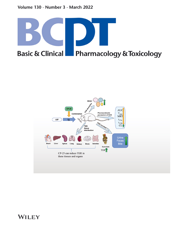
Paeoniflorin-6′-O-benzene sulfonate (CP-25) is a novel ester derivative of paeoniflorin, which has been shown to have synergistic pharmacodynamic effects with leflunomide (LEF). To determine the effects of CP-25 on the pharmacokinetics of LEF in rats, we developed a ultra-high performance liquid chromatography-tandem mass spectrometry (UPLC-MS/MS)-based method for the determination of levels of teriflunomide (TER, an active metabolite of LEF). This method was used to determine TER concentrations in the plasma, urine, faeces and bile; heart, liver, spleen, lung, kidney, intestinal, brain and synovial tissues; and peripheral blood mononuclear cells (PBMCs) of rats in the control (LEF [10 mg/kg]) and combined (CP-25 [50 mg/kg × 7d] plus LEF [10 mg/kg]) groups. TER area under the curve [AUC], Tmax, mean residence time (MRT), t1/2α and t1/2β were significantly lower, and clearance (CL) was significantly higher in the combined group than in the control group. Oral CP-25 administration in combination with LEF was found to promote TER excretion in urine, faeces and bile and to reduce its contents in most tissues and organs, especially in the liver, which may reduce LEF-induced liver injury. CP-25 also increased TER exposure in the synovium and its absorption by PBMCs, and this could explain the synergistic effects of CP-25 and LEF.
CONFLICT OF INTEREST
The author reports no conflicts of interest in this work.
Supporting Information
| Filename | Description |
|---|---|
| bcpt13685-sup-0001-Supporting Information.docxWord 2007 document , 833.4 KB |
Table 1S The elution gradient of Samples. Table 2S The optimized parameters for TER and IS. Figure 1S Linearity of TER in plasma samples. X represents the concentration of TER (μg/mL), and Y represents the ratio of the peak area of TER to the peak area of IS. Figure 2S Linearity of TER in faeces samples. X represents the concentration of TER (ng/mL), and Y represents the ratio of the peak area of TER to the peak area of IS. Figure 3S Linearity of TER in urine samples. X represents the concentration of TER (ng/mL), and Y represents the ratio of the peak area of TER to the peak area of IS. Figure 4S Linearity of TER in bile samples. X represents the concentration of TER (μg/mL), and Y represents the ratio of the peak area of TER to the peak area of IS. Figure 5S Linearity of TER in heart samples. X represents the concentration of TER (ng/mL), and Y represents the ratio of the peak area of TER to the peak area of IS. Figure 6S Linearity of TER in liver samples. X represents the concentration of TER (μg/mL), and Y represents the ratio of the peak area of TER to the peak area of IS. Figure 7S Linearity of TER in spleen samples. X represents the concentration of TER (ng/mL), and Y represents the ratio of the peak area of TER to the peak area of IS. Figure 8S Linearity of TER in lung samples. X represents the concentration of TER (ng/mL), and Y represents the ratio of the peak area of TER to the peak area of IS. Figure 9S Linearity of TER in kidney samples. X represents the concentration of TER (μg/mL), and Y represents the ratio of the peak area of TER to the peak area of IS. Figure 10S Linearity of TER in intestine samples. X represents the concentration of TER (μg/mL), and Y represents the ratio of the peak area of TER to the peak area of IS. Figure 11S Linearity of TER in brain samples. X represents the concentration of TER (ng/mL), and Y represents the ratio of the peak area of TER to the peak area of IS. Figure 12S Linearity of TER in synovium samples. X represents the concentration of TER (ng/mL), and Y represents the ratio of the peak area of TER to the peak area of IS. Figure 13S Linearity of TER in PBMC samples. X represents the concentration of TER (ng/mL), and Y represents peak area of TER. |
Please note: The publisher is not responsible for the content or functionality of any supporting information supplied by the authors. Any queries (other than missing content) should be directed to the corresponding author for the article.
REFERENCES
- 1Breedveld FC, Dayer JM. Leflunomide: mode of action in the treatment of rheumatoid arthritis. Ann Rheum Dis. 2000; 59(11): 841-849. https://doi.org/10.1136/ard.59.11.841
- 2Siemasko KF, Chong AS, Williams JW, Bremer EG, Finnegan A. Regulation of B cell function by the immunosuppressive agent leflunomide. Transplantation. 1996; 61(4): 635-642. https://doi.org/10.1097/00007890-199602270-00020
- 3Chong AS, Finnegan A, Jiang X, et al. Leflunomide, a novel immunosuppressive agent. The mechanism of inhibition of T cell proliferation. Transplantation. 1993; 55(6): 1361-1366. https://doi.org/10.1097/00007890-199306000-00028
- 4Pinto P, Dougados M. Leflunomide in clinical practice. Acta Reumatol Port. 2006; 31(3): 215-224.
- 5Fragoso YD, Brooks JB. Leflunomide and teriflunomide: altering the metabolism of pyrimidines for the treatment of autoimmune diseases. Expert Rev Clin Pharmacol. 2015; 8(3): 315-320. https://doi.org/10.1586/17512433.2015.1019343
- 6Wiacek R, Kolossa K, Jankowski T, et al. The efficacy and safety of leflunomide in patients with active rheumatoid arthritis. Adv Clin Exp Med. 2012; 21(3): 337-342.
- 7Nguyen M, Kabir M, Ravaud P. Short-term efficacy and safety of leflunomide in the treatment of active rheumatoid arthritis in everyday clinical use: open-label, prospective study. Clin Drug Investig. 2004; 24(2): 103-112. https://doi.org/10.2165/00044011-200424020-00005
- 8Curtis JR, Beukelman T, Onofrei A, et al. Elevated liver enzyme tests among patients with rheumatoid arthritis or psoriatic arthritis treated with methotrexate and/or leflunomide. Ann Rheum Dis. 2010; 69(1): 43-47. https://doi.org/10.1136/ard.2008.101378
- 9Bilasy SE, Essawy SS, Mandour MF, Ali EA, Zaitone SA. Myelosuppressive and hepatotoxic potential of leflunomide and methotrexate combination in a rat model of rheumatoid arthritis. Pharmacol Rep. 2015; 67(1): 102-114. https://doi.org/10.1016/j.pharep.2014.08.009
- 10Bae J, Park JW. Topical delivery of leflunomide for rheumatoid arthritis treatment: evaluation of local tissue deposition of teriflunomide and its anti-inflammatory effects in an arthritis rat model. Drug Dev Ind Pharm. 2016; 42(2): 254-262. https://doi.org/10.3109/03639045.2015.1044906
- 11Rozman B. Clinical pharmacokinetics of leflunomide. Clin Pharmacokinet. 2002; 41(6): 421-430. https://doi.org/10.2165/00003088-200241060-00003
- 12Wu YJ, Zhao MY, Wang J, et al. Absorption and efflux characteristics of CP-25 in plasma and peripheral blood mononuclear cells of rats by UPLC-MS/MS. Biomed Pharmacother. 2018; 108: 1651-1657. https://doi.org/10.1016/j.biopha.2018.09.156
- 13Zhao M, Zhou P, Yu J, et al. The tissue distribution and excretion study of paeoniflorin-6'-O-benzene sulfonate (CP-25) in rats. Inflammopharmacology. 2019; 27(5): 969-974. https://doi.org/10.1007/s10787-018-0463-3
- 14Wang C, Yuan J, Zhang LL, Wei W. Pharmacokinetic comparisons of Paeoniflorin and Paeoniflorin-6'O-benzene sulfonate in rats via different routes of administration. Xenobiotica. 2016; 46(12): 1142-1150. https://doi.org/10.3109/00498254.2016.1149633
- 15Yang XD, Wang C, Zhou P, et al. Absorption characteristic of paeoniflorin-6'-O-benzene sulfonate (CP-25) in in situ single-pass intestinal perfusion in rats. Xenobiotica. 2016; 46(9): 775-783. https://doi.org/10.3109/00498254.2015.1121553
- 16Zhang F, Shu JL, Li Y, et al. CP-25, a novel anti-inflammatory and immunomodulatory drug, inhibits the functions of activated human B cells through regulating BAFF and TNF-alpha signaling and comparative efficacy with biological agents. Front Pharmacol. 2017; 8:933. Published 2017 Dec 22. https://doi.org/10.3389/fphar.2017.00933
- 17Wang Y, Han CC, Cui D, et al. Correction to: Immunomodulatory Effects of CP-25 on Splenic T Cells of Rats with Adjuvant Arthritis. Inflammation. 2018; 41(3): 1064. https://doi.org/10.1007/s10753-018-0770-2
- 18Jia XY, Chang Y, Wei F, et al. CP-25 reverses prostaglandin E4 receptor desensitization-induced fibroblast-like synoviocyte dysfunction via the G protein-coupled receptor kinase 2 in autoimmune arthritis. Acta Pharmacol Sin. 2019; 40(8): 1029-1039. https://doi.org/10.1038/s41401-018-0196-2
- 19Wang DD, Jiang MY, Wang W, et al. Paeoniflorin-6'-O-benzene sulfonate down-regulates CXCR4-Gβγ-PI3K/AKT mediated migration in fibroblast-like synoviocytes of rheumatoid arthritis by inhibiting GRK2 translocation. Biochem Biophys Res Commun. 2020; 526(3): 805-812. https://doi.org/10.1016/j.bbrc.2020.03.164
- 20Yang X, Zhao Y, Jia X, et al. CP-25 combined with MTX/LEF ameliorates the progression of adjuvant-induced arthritis by the inhibition on GRK2 translocation. Biomed Pharmacother. 2019; 110: 834-843. https://doi.org/10.1016/j.biopha.2018.12.040
- 21Tveden-Nyborg P, Bergmann TK, Jessen N, Simonsen U, Lykkesfeldt J. BCPT policy for experimental and clinical studies. Basic Clin Pharmacol Toxicol. 2021; 128(1): 4-8.
- 22Xuan J, Ren Z, Qing T, et al. Mitochondrial dysfunction induced by leflunomide and its active metabolite. Toxicology. 2018; 396–397: 33-45. https://doi.org/10.1016/j.tox.2018.02.003
- 23Mehta V, Kisalay S, Balachandran C. Leflunomide. Indian J Dermatol Venereol Leprol. 2009; 75(4): 422-424. https://doi.org/10.4103/0378-6323.53157
- 24Keen HI, Conaghan PG, Tett SE. Safety evaluation of leflunomide in rheumatoid arthritis. Expert Opin Drug Saf. 2013; 12(4): 581-588. https://doi.org/10.1517/14740338.2013.798299
- 25Bao C, Chen S, Gu Y, et al. Leflunomide, a new disease-modifying drug for treating active rheumatoid arthritis in methotrexate-controlled phase II clinical trial. Chin Med J (Engl). 2003; 116(8): 1228-1234.
- 26Maddison P, Kiely P, Kirkham B, et al. Leflunomide in rheumatoid arthritis: recommendations through a process of consensus. Rheumatology (Oxford). 2005; 44(3): 280-286. https://doi.org/10.1093/rheumatology/keh500
- 27Kremer J, Genovese M, Cannon GW, et al. Combination leflunomide and methotrexate (MTX) therapy for patients with active rheumatoid arthritis failing MTX monotherapy: open-label extension of a randomized, double-blind, placebo controlled trial. J Rheumatol. 2004; 31(8): 1521-1531.
- 28Chen J, Wang Y, Wu H, Yan S, Chang Y, Wei W. A modified compound from paeoniflorin, CP-25, suppressed immune responses and synovium inflammation in collagen-induced arthritis mice. Front Pharmacol. 2018; 9:563. Published 2018 Jun 7. https://doi.org/10.3389/fphar.2018.00563
- 29Zhang L, Wei W. Anti-inflammatory and immunoregulatory effects of paeoniflorin and total glucosides of paeony. Pharmacol Ther. 2020; 207:107452. https://doi.org/10.1016/j.pharmthera.2019.107452
- 30Hu Z, Qin F, Gao S, Zhen Y, Huang D, Dong L. Paeoniflorin exerts protective effect on radiation-induced hepatic fibrosis in rats via TGF-β1/Smads signaling pathway. Am J Transl Res. 2018; 10(3): 1012-1021. Published 2018 Mar 15
- 31Ma Z, Chu L, Liu H, et al. Beneficial effects of paeoniflorin on non-alcoholic fatty liver disease induced by high-fat diet in rats. Sci Rep. 2017; 7:44819. Published 2017 Mar 16. https://doi.org/10.1038/srep44819
- 32Liu DF, Wei W, Song LH. Protective effect of paeoniflorin on immunological liver injury induced by bacillus Calmette-Guerin plus lipopolysaccharide: modulation of tumour necrosis factor-alpha and interleukin-6 MRNA. Clin Exp Pharmacol Physiol. 2006; 33(4): 332-339. https://doi.org/10.1111/j.1440-1681.2006.04371.x
- 33Qin Y, Tian YP. Protective effects of total glucosides of paeony and the underlying mechanisms in carbon tetrachloride-induced experimental liver injury. Arch Med Sci. 2011; 7(4): 604-612. https://doi.org/10.5114/aoms.2011.24129
- 34Wang H, Wei W, Wang NP, et al. Effects of total glucosides of peony on immunological hepatic fibrosis in rats. World J Gastroenterol. 2005; 11(14): 2124-2129. https://doi.org/10.3748/wjg.v11.i14.2124
- 35Xiang N, Li XM, Zhang MJ, et al. Total glucosides of paeony can reduce the hepatotoxicity caused by Methotrexate and Leflunomide combination treatment of active rheumatoid arthritis. Int Immunopharmacol. 2015; 28(1): 802-807. https://doi.org/10.1016/j.intimp.2015.08.008
- 36Ma LL, Wu ZT, Wang L, et al. Inhibition of hepatic cytochrome P450 enzymes and sodium/bile acid cotransporter exacerbates leflunomide-induced hepatotoxicity. Acta Pharmacol Sin. 2016; 37(3): 415-424. https://doi.org/10.1038/aps.2015.157
- 37Zhao Y, He X, Ma X, et al. Paeoniflorin ameliorates cholestasis via regulating hepatic transporters and suppressing inflammation in ANIT-fed rats. Biomed Pharmacother. 2017; 89: 61-68. https://doi.org/10.1016/j.biopha.2017.02.025
- 38Liu YM, Chen JW, Chen LX, Xie X, Mao N. Overexpression of P-glycoprotein on fibroblast-like synoviocytes in refractory rheumatoid arthritis patients: a potential mechanism for multidrug resistance in rheumatoid arthritis treatment. Genet Mol Res. 2016; 15(2):gmr.15027927. Published 2016 Jun 17. https://doi.org/10.4238/gmr.15027927
- 39Kim KA, Joo HJ, Park JY. Effect of ABCG2 genotypes on the pharmacokinetics of A771726, an active metabolite of prodrug leflunomide, and association of A771726 exposure with serum uric acid level. Eur J Clin Pharmacol. 2011; 67(2): 129-134. https://doi.org/10.1007/s00228-010-0916-0
Citing Literature