Invited Commentary
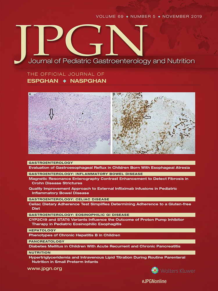
Intestinal Wall Texture Analysis
Finding Fibrosis in Pediatric Patients With Crohn Disease
Andrew Phelps,
Corresponding Author
Andrew Phelps
UCSF Benioff Children's Hospital San Francisco, San Francisco, CA
Address correspondence and reprint requests to Andrew Phelps, MD, UCSF Benioff Children's Hospital San Francisco, 1975 4th Street C1758N, San Francisco, CA 94158 (e-mail:
[email protected]).
Search for more papers by this author
First published: 01 November 2019
No abstract is available for this article.
REFERENCES
- 1.Tabari A, Kilcoyne A, Jeck WR, et al. Texture analysis of magnetic resonance enterography contrast enhancement can detect fibrosis in Crohn disease strictures. J Pediatr Gastroenterol Nutr 2019; 69: 533–538.
- 2.Vernier-Massouille G, Balde M, Salleron J, et al. Natural history of pediatric Crohn's disease: a population-based cohort study. Gastroenterology 2008; 135: 1106–1113.
- 3.Bettenworth D, Bokemeyer A, Baker M, et al. Assessment of Crohn's disease-associated small bowel strictures and fibrosis on cross-sectional imaging: a systematic review. Gut 2019; 68: 1115–1126.
- 4.Crohn BB, Ginzburg L, Oppenheimer GD. Regional ileitis: a pathologic and clinical entity. JAMA 1932; 99: 1323–1329.
- 5.Gore RM. Cross-sectional imaging of inflammatory bowel disease. Radiol Clin North Am 1987; 25: 115–131.
- 6.Li J, Qureshi M, Gupta A, et al. Quantification of degree of liver fibrosis using fibrosis area fraction based on statistical chi-square analysis of heterogeneity of liver tissue texture on routine ultrasound images. Acad Radiol 2019; 26: 1001–1007.
- 7.Phelps A. Liver ultrasound texture analysis: the computer finds more to quantify than meets the eye. Acad Radiol 2019; 26: 1008–1009.
- 8.Hosny A, Parmar C, Quackenbush J, et al. Artificial intelligence in radiology. Nat Rev Cancer 2018; 18: 500–510.