Long-term follow up of pure myxoid liposarcomas with special reference to local recurrence and progression to round cell lesions
Abstract
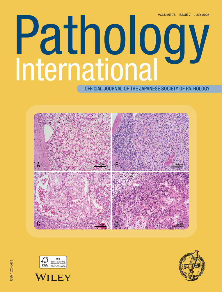
Myxoid and round cell liposarcomas cytogenetically share the t(12;16)(q13;p11) translocation, and represent a continuous morphological spectrum including pure myxoid liposarcoma (PML), mixed (myxoid/round cell) liposarcoma (MXL) and pure round cell liposarcoma (RCL). It has been documented that a critical amount of RC lesion in low-grade, MXL is associated with poor prognosis. The present long-term follow-up study examined the factors that influence the progression of PML to RCL and several other prognostic factors and compared the results with those of liposarcomas initially diagnosed as MXL. Using 34 primary PML and their local recurrences after curative resection, and two initially diagnosed MXL, the appearance of RC lesion in PML was examined, and the prognostic significance of the local recurrence rate (times/month), tumor size, infiltrative proliferation (defined as entrapping of striated muscle fibers in the tumor), tumor necrosis and mitotic figures. The follow-up period ranged from 19 to 378 months (mean, 173.4 months). Based on Cox’s proportional hazards regression model, a recurrence rate ≥ 0.01 in 24 PML with recurrences (P = 0.037) and the presence of infiltrative proliferation in 34 PML (P = 0.043) were significant independent factors associated with poor prognosis. Round cell lesions were detected in the last recurrences of two surviving PML. These results indicate that through their recurrence, PML might potentially become malignant, independent of the proportion of RC lesions in the tumor. The appearance of RC lesions in subsequent recurrences may indicate a progression from PML to MXL.