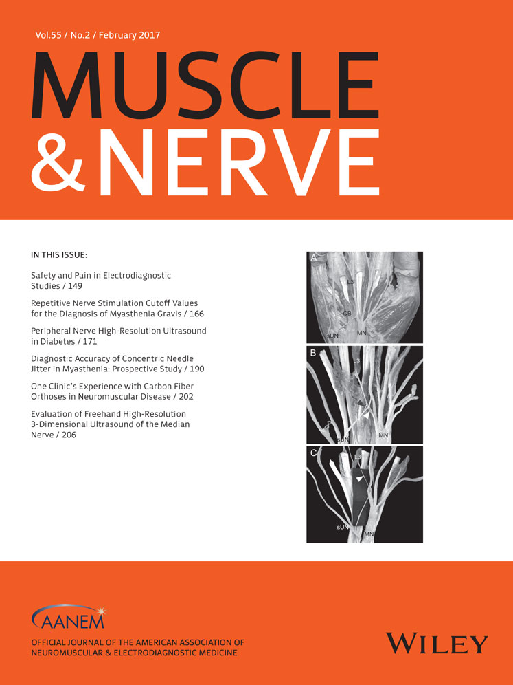
Variations of lumbrical muscle innervation patterns in the hand, focusing on the dual innervation of the third lumbrical muscle
Funding: This work was supported by research fund of Catholic Kwandong University (CKURF-201601820001).
Disclosure: The author has no relevant financial or conflict of interest disclosures.
ABSTRACT
Introduction: This study was conducted to clarify the innervation patterns of the lumbrical muscles by identifying the origin of the nerve fascicles innervating these muscles. Methods: The lumbricals in the hand were investigated in 50 specimens of embalmed Korean adult cadavers. Results: Dual innervation of the third lumbrical was most frequently observed in 64.0%. The third lumbrical was innervated by a branch arising from the median nerve (MN) distal to site at which the superficial branch of the ulnar nerve (sUN) joins the MN in 34%. When separating and tracing the nerve fascicles from the MN distal to the communicating branch from the sUN to MN, the fascicles contained parts of the MN and sUN in 18% and part of the MN in 16%. Conclusions: These results will be helpful for accurate diagnoses, surgical procedures, and electrophysiological examinations in lesions of the MN and ulnar nerve in the hand. Muscle Nerve, 2016 Muscle Nerve 55: 160–165, 2017