

Sulfated Hyaluronan Influences the Formation of Artificial Extracellular Matrices and the Adhesion of Osteogenic Cells
Alina Miron
Institute of Materials Science, Max Bergmann Center of Biomaterials, Technische Universität Dresden, 01069 Dresden, Germany
Search for more papers by this authorSandra Rother
Institute of Materials Science, Max Bergmann Center of Biomaterials, Technische Universität Dresden, 01069 Dresden, Germany
Search for more papers by this authorLinda Huebner
Institute of Materials Science, Max Bergmann Center of Biomaterials, Technische Universität Dresden, 01069 Dresden, Germany
Search for more papers by this authorUte Hempel
Institute of Physiological Chemistry, Technische Universität Dresden, 01307 Dresden, Germany
Search for more papers by this authorIris Käppler
Institute of Textile Machinery and High Performance Material Technology, Technische Universität Dresden, 01062 Dresden, Germany
Search for more papers by this authorStephanie Moeller
Biomaterials Department, INNOVENT e.V., 07745 Jena, Germany
Search for more papers by this authorMatthias Schnabelrauch
Biomaterials Department, INNOVENT e.V., 07745 Jena, Germany
Search for more papers by this authorDieter Scharnweber
Institute of Materials Science, Max Bergmann Center of Biomaterials, Technische Universität Dresden, 01069 Dresden, Germany
Search for more papers by this authorCorresponding Author
Vera Hintze
Institute of Materials Science, Max Bergmann Center of Biomaterials, Technische Universität Dresden, 01069 Dresden, Germany
Search for more papers by this authorAlina Miron
Institute of Materials Science, Max Bergmann Center of Biomaterials, Technische Universität Dresden, 01069 Dresden, Germany
Search for more papers by this authorSandra Rother
Institute of Materials Science, Max Bergmann Center of Biomaterials, Technische Universität Dresden, 01069 Dresden, Germany
Search for more papers by this authorLinda Huebner
Institute of Materials Science, Max Bergmann Center of Biomaterials, Technische Universität Dresden, 01069 Dresden, Germany
Search for more papers by this authorUte Hempel
Institute of Physiological Chemistry, Technische Universität Dresden, 01307 Dresden, Germany
Search for more papers by this authorIris Käppler
Institute of Textile Machinery and High Performance Material Technology, Technische Universität Dresden, 01062 Dresden, Germany
Search for more papers by this authorStephanie Moeller
Biomaterials Department, INNOVENT e.V., 07745 Jena, Germany
Search for more papers by this authorMatthias Schnabelrauch
Biomaterials Department, INNOVENT e.V., 07745 Jena, Germany
Search for more papers by this authorDieter Scharnweber
Institute of Materials Science, Max Bergmann Center of Biomaterials, Technische Universität Dresden, 01069 Dresden, Germany
Search for more papers by this authorCorresponding Author
Vera Hintze
Institute of Materials Science, Max Bergmann Center of Biomaterials, Technische Universität Dresden, 01069 Dresden, Germany
Search for more papers by this authorAbstract
The aim of this study is to compare differentially sulfated hyaluronan (sHA) derivatives and chondroitin sulfate (CS) with respect to their ability to influence the formation of artificial extracellular matrices (aECMs) during in vitro-fibrillogenesis of collagen type I at high- and low-ionic strength. Analysis is performed using turbidity, biochemical assays, atomic force (AFM), and transmission electron microscopy (TEM). In general, high-sulfated glycosaminoglycans (GAGs) associate to a higher amount with collagen than the low-sulfated ones. The addition of GAGs prior to fibrillogenesis at low-ionic strength results in a dose-dependent decrease in fibril diameter. At high-ionic strength these effects are only obtained for the sHA derivatives but not for CS. Likewise, increasing concentrations and degree of GAG sulfation strongly affected the kinetics of fibrillogenesis. The impact of sulfation degree on F-actin location and fiber formation in SaOS-2 cells implies that adhesion-related intracellular signaling is influenced to a variable extent.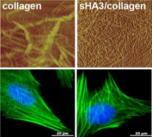
Supporting Information
As a service to our authors and readers, this journal provides supporting information supplied by the authors. Such materials are peer reviewed and may be re-organized for online delivery, but are not copy-edited or typeset. Technical support issues arising from supporting information (other than missing files) should be addressed to the authors.
| Filename | Description |
|---|---|
| mabi201400292-sm-0001-SuppData.docx598.8 KB | Supporting Data |
Please note: The publisher is not responsible for the content or functionality of any supporting information supplied by the authors. Any queries (other than missing content) should be directed to the corresponding author for the article.
References
- 1 K. E. Kadler, D. F. Holmes, J. A. Trotter, J. A. Chapman, Biochem. J. 1996, 316, 1.
- 2 E. G. Canty, K. E. Kadler, J. Cell Sci. 2005, 118, 1341.
- 3 G. C. Wood, Biochem. J. 1960, 75, 588.
- 4 D. E. Birk, F. H. Silver, Arch. Biochem. Biophys. 1984, 235, 178.
- 5 K. G. Danielson, H. Baribault, D. F. Holmes, H. Graham, K. E. Kadler, R. V. Iozzo, J. Cell Biol. 1997, 136, 729.
- 6 L. Svensson, A. Aszodi, F. P. Reinholt, R. Fassler, D. Heinegard, A. Oldberg, J. Biol. Chem. 1999, 274, 9636.
- 7 A. Corsi, T. Xu, X. D. Chen, A. Boyde, J. Liang, M. Mankani, B. Sommer, R. V. Iozzo, I. Eichstetter, P. G. Robey, P. Bianco, M. F. Young, J. Bone Miner. Res. 2002, 17, 1180.
- 8 S. Kalamajski, A. Oldberg, Matrix Biol. 2010, 29, 248.
- 9 I. M. Kuc, P. G. Scott, Connect Tissue Res 1997, 36, 287.
- 10 P. Sini, A. Denti, M. E. Tira, C. Balduini, Glycoconj. J. 1997, 14, 871.
- 11 S. Bierbaum, T. Douglas, T. Hanke, D. Scharnweber, S. Tippelt, T. K. Monsees, R. H. W. Funk, H. Worch, J. Biomed. Mater. Res. A 2006, 77, 551.
- 12 C. Ruehland, E. Schonherr, H. Robenek, U. Hansen, R. V. Iozz, P. Bruckner, D. G. Seidler, Febs J. 2007, 274, 4246.
- 13 M. Maccarana, S. Kalamajski, M. Kongsgaard, S. P. Magnusson, A. Oldberg, A. Malmstrom, Mol. Cell Biol. 2009, 29, 5517.
- 14 M. B. Mathews, L. Decker, Biochem. J. 1968, 109, 517.
- 15 B. Obrink, Eur. J. Biochem. 1973, 33, 387.
- 16 B. Obrink, Eur. J. Biochem. 1973, 34, 129.
- 17 S. D. Snowden, J. Mc,K Biopolymers, 1980, 19, 767.
- 18 D. A. Parry, M. H. Flint, G. C. Gillard, A. S. Craig, FEBS Lett. 1982, 149, 1.
- 19 T. Douglas, S. Heinemann, C. Mietrach, U. Hempel, S. Bierbaum, D. Scharnweber, H. Worch, Biomacromolecules. 2007, 8, 1085.
- 20 D. Stamov, M. Grimmer, K. Salchert, T. Pompe, C. Werner, Biomaterials 2008, 29, 1.
- 21 K. Stuart, A. Panitch, Biopolymers 2008, 89, 841.
- 22 D. D. Allison, K. J. Grande-Allen, Tissue Eng., 2006, 12, 2131.
- 23 J. Schiller, J. Becher, S. Moller, K. Nimptsch, T. Riemer, M. Schnabelrauch, Mini-Rev. Org. Chem. 2010, 7, 290.
- 24 V. Hintze, S. Moeller, M. Schnabelrauch, S. Bierbaum, M. Viola, H. Worch, D. Scharnweber, Biomacromolecules. 2009, 10, 3290.
- 25 V. Hintze, A. Miron, S. Moeller, M. Schnabelrauch, H. P. Wiesmann, H. Worch, D. Scharnweber, Acta Biomater. 2012, 8, 2144.
- 26 A. van der Smissen, V. Hintze, D. Scharnweber, S. Moeller, M. Schnabelrauch, A. Majok, J. C. Simon, U. Anderegg, Biomaterials 2011, 32, 8938.
- 27 U. Hempel, S. Moller, C. Noack, V. Hintze, D. Scharnweber, M. Schnabelrauch, P. Dieter, Acta Biomater. 2012, 8, 4064.
- 28 J. Salbach, S. Kliemt, M. Rauner, T. D. Rachner, C. Goettsch, S. Kalkhof, M. von Bergen, S. Moller, M. Schnabelrauch, V. Hintze, D. Scharnweber, L. C. Hofbauer, Biomaterials 2012, 33, 8418.
- 29 M. Nagahata, T. Tsuchiya, T. Ishiguro, N. Matsuda, Y. Nakatsuchi, A. Teramoto, A. Hachimori, K. Abe, Biochem. Biophys. Res. Commun. 2004, 315, 603.
- 30 R. Kunze, M. Rosler, S. Moller, M. Schnabelrauch, T. Riemer, U. Hempel, P. Dieter, Glycoconj. J. 2010, 27, 151.
- 31 S. Kliemt, C. Lange, W. Otto, V. Hintze, S. Moller, M. von Bergen, U. Hempel, S. Kalkhof, J. Proteome Res. 2013, 12, 378.
- 32 B. R. Williams, R. A. Gelman, D. C. Poppke, K. A. Piez, J. Biol. Chem. 1978, 253, 6578.
- 33 F. H. Silver, D. E. Birk, Coll. Relat. Res. 1983, 3, 393.
- 34 O. H. Lowry, N. J. Rosebrough, A. L. Farr, R. J. Randall, J. Biol. Chem. 1951, 193, 265.
- 35 U. Hempel, V. Hintze, S. Moller, M. Schnabelrauch, D. Scharnweber, P. Dieter, Acta Biomater. 2012, 8, 659.
- 36 V. Hintze, A. Miron, S. Moller, M. Schnabelrauch, S. Heinemann, H. Worch, D. Scharnweber, J. Tissue Eng. Regen. Med. 2012, 8, 314.
- 37 R. W. Farndale, D. J. Buttle, A. J. Barrett, Biochim. Biophys. Acta 1986, 883, 173.
- 38 J. E. Scott, Biochem. J. 1980, 187, 887.
- 39 J. E. Scott, Biochem. J. 1988, 252, 313.
- 40 J. A. Chapman, M. Tzaphlidou, K. M. Meek, K. E. Kadler, Electron Microsc. Rev. 1990, 3, 143.
- 41 M. R. van der Harst, P. A. Brama, C. H. van de Lest, G. H. Kiers, J. DeGroot, P. R. van Weeren, Osteoarthritis Cartilage 2004, 12, 752.
- 42 V. M. Mania, A. G. Kallivokas, C. Malavaki, A. P. Asimakopoulou, J. Kanakis, A. D. Theocharis, G. Klironomos, G. Gatzounis, A. Mouzaki, E. Panagiotopoulos, N. K. Karamanos, Iubmb Life 2009, 61, 447.
- 43 J. A. Ezzo, J. Anthropol. Archaeol. 1994, 13, 1.
- 44 C. Wolf-Brandstetter, A. Lode, T. Hanke, D. Scharnweber, H. Worch, J. Biomed. Mater. Res. A 2006, 79, 882.
- 45 H. Tian, C. Li, W. Liu, J. Li, G. Li, Colloids Surf. B Biointerfaces 2013, 105, 259.
- 46 P. Clark, P. Connolly, A. S. Curtis, J. A. Dow, C. D. Wilkinson, Development 1987, 99, 439.
- 47 I. Mercier, J. P. Lechaire, A. Desmouliere, F. Gaill, M. Aumailley, Exp. Cell. Res. 1996, 225, 245.

