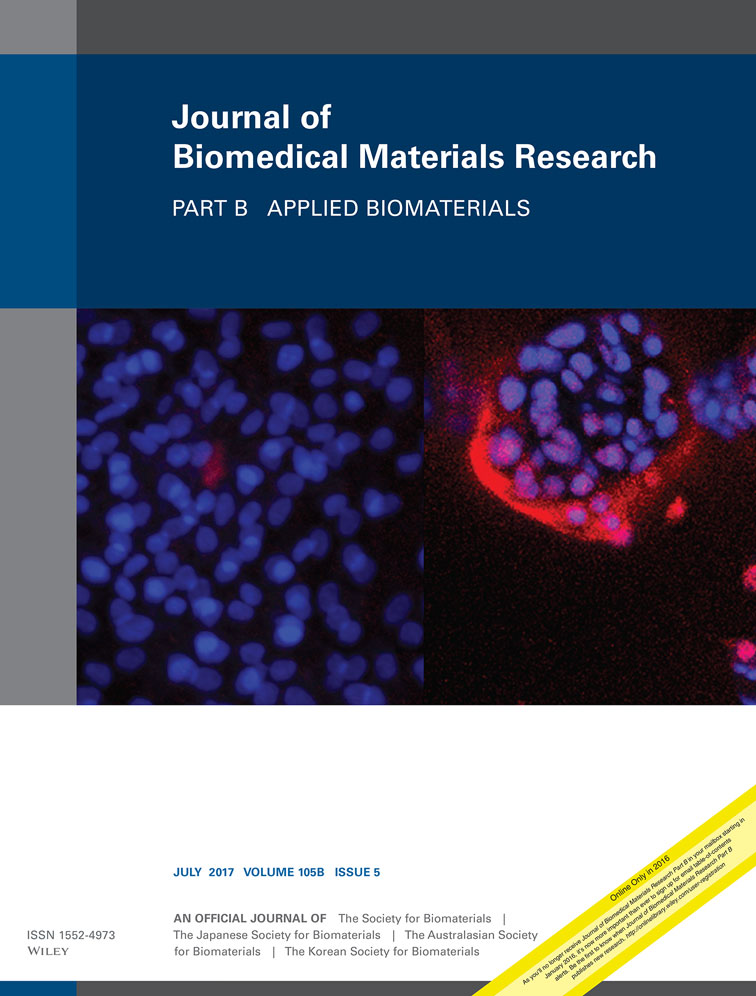
Development of a 3D cell printed structure as an alternative to autologs cartilage for auricular reconstruction
Ju Young Park
Division of Integrative Biosciences and Biotechnology, Pohang University of Science and Technology (POSTECH), Pohang, Korea
Both authors contributed equally to this work.
Search for more papers by this authorYeong-Jin Choi
Division of Integrative Biosciences and Biotechnology, Pohang University of Science and Technology (POSTECH), Pohang, Korea
Both authors contributed equally to this work.
Search for more papers by this authorJin-Hyung Shim
Department of Mechanical Engineering, Korea Polytechnic University, Siheung, Korea
Search for more papers by this authorJeong Hun Park
Department of Mechanical Engineering, Pohang University of Science and Technology (POSTECH), Pohang, Korea
Search for more papers by this authorCorresponding Author
Dong-Woo Cho
Department of Mechanical Engineering, Pohang University of Science and Technology (POSTECH), Pohang, Korea
Correspondence to: D.-W. Cho; e-mail: [email protected]Search for more papers by this authorJu Young Park
Division of Integrative Biosciences and Biotechnology, Pohang University of Science and Technology (POSTECH), Pohang, Korea
Both authors contributed equally to this work.
Search for more papers by this authorYeong-Jin Choi
Division of Integrative Biosciences and Biotechnology, Pohang University of Science and Technology (POSTECH), Pohang, Korea
Both authors contributed equally to this work.
Search for more papers by this authorJin-Hyung Shim
Department of Mechanical Engineering, Korea Polytechnic University, Siheung, Korea
Search for more papers by this authorJeong Hun Park
Department of Mechanical Engineering, Pohang University of Science and Technology (POSTECH), Pohang, Korea
Search for more papers by this authorCorresponding Author
Dong-Woo Cho
Department of Mechanical Engineering, Pohang University of Science and Technology (POSTECH), Pohang, Korea
Correspondence to: D.-W. Cho; e-mail: [email protected]Search for more papers by this authorAbstract
Surgical technique using autologs cartilage is considered as the best treatment for cartilage tissue reconstruction, although the burdens of donor site morbidity and surgical complications still remain. The purpose of this study is to apply three-dimensional (3D) cell printing to fabricate a tissue-engineered graft, and evaluate its effects on cartilage reconstruction. A multihead tissue/organ building system is used to print cell-printed scaffold (CPS), then assessed the effect of the CPS on cartilage regeneration in a rabbit ear. The cell viability and functionality of chondrocytes were significantly higher in CPS than in cell-seeded scaffold (CSS) and cell-seeded hybrid scaffold (CSHS) in vitro. CPS was then implanted into a rabbit ear that had an 8 mm-diameter cartilage defect; at 3 months after implantation the CPS had fostered complete cartilage regeneration whereas CSS and autologs cartilage (AC) fostered only incomplete healing. This result demonstrates that cell printing technology can provide an appropriate environment in which encapsulated chondrocytes can survive and differentiate into cartilage tissue in vivo. Moreover, the effects of CPS on cartilage regeneration were even better than those of AC. Therefore, we confirmed the feasibility of CPS as an alternative to AC for auricular reconstruction. © 2016 Wiley Periodicals, Inc. J Biomed Mater Res Part B: Appl Biomater, 105B: 1016–1028, 2017.
REFERENCES
- 1 Brent B. Auricular repair with autogenous rib cartilage grafts: Two decades of experience with 600 cases. Plast Reconstr Surg 1992; 90: 355–374; discussion 375–376.
- 2 Siegert R. Combined reconstruction of congenital auricular atresia and severe microtia. Laryngoscope 2003; 113: 2021–2027; discussion 2028–2029.
- 3 Brent B. Technical advances in ear reconstruction with autogenous rib cartilage grafts: Personal experience with 1200 cases. Plast Reconstr Surg 1999; 104: 319–334; discussion 335–338.
- 4 Yu DS, Jiang HY, Yang QH, Pan B, Lin L, Wang TL, Wang YM, Qin X, Zhuang HX. Technical innovations in ear reconstruction using a skin expander with autogenous cartilage grafts. J Plast Reconstr Aes 2008; 61: S59–S69.
- 5 Moon BJ, Lee HJ, Jang YJ. Outcomes following rhinoplasty using autologous costal cartilage. Arch Facial Plast Surg 2012; 14: 175–180.
- 6 Uppal RS, Sabbagh W, Chana J, Gault DT. Donor-site morbidity after autologous costal cartilage harvest in ear reconstruction and approaches to reducing donor-site contour deformity. Plast Reconstr Surg 2008; 121: 1949–1955.
- 7 Craigmyle MB. Studies of cartilage autografts and homografts in the rabbit. Br J Plast Surg 1955; 8: 93–100.
- 8 Davis WB, Gibson T. Absorption of autogenous cartilage grafts in man. Br J Plast Surg 1956; 9: 177–185.
- 9 Lattyak BV, Maas CS, Sykes JM. Dorsal onlay cartilage autografts: comparing resorption in a rabbit model. Arch Facial Plast Surg 2003; 5: 240–243.
- 10 Khan IM, Gilbert SJ, Singhrao SK, Duance VC, Archer CW. Cartilage integration: Evaluation of the reasons for failure of integration during cartilage repair. A review. Eur Cells Mater 2008; 16: 26–39.
- 11 Theodoropoulos JS, De Croos JN, Park SS, Pilliar R, Kandel RA. Integration of tissue-engineered cartilage with host cartilage: An in vitro model. Clin Orthop Relat Res 2011; 469: 2785–2795.
- 12 Otto IA, Melchels FP, Zhao X, Randolph MA, Kon M, Breugem CC, Malda J. Auricular reconstruction using biofabrication-based tissue engineering strategies. Biofabrication 2015; 7: 032001.
- 13 Bichara DA, O'Sullivan NA, Pomerantseva I, Zhao X, Sundback CA, Vacanti JP, Randolph MA. The tissue-engineered auricle: past, present, and future. Tissue Eng Part B: Rev 2012; 18: 51–61.
- 14 Zhou L, Pomerantseva I, Bassett EK, Bowley CM, Zhao X, Bichara DA, Kulig KM, Vacanti JP, Randolph MA, Sundback CA. Engineering ear constructs with a composite scaffold to maintain dimensions. Tissue Eng Part A 2011; 17: 1573–1581.
- 15 Xue J, Feng B, Zheng R, Lu Y, Zhou G, Liu W, Cao Y, Zhang Y, Zhang WJ. Engineering ear-shaped cartilage using electrospun fibrous membranes of gelatin/polycaprolactone. Biomaterials 2013; 34: 2624–2631.
- 16 Zhang L, He A, Yin Z, Yu Z, Luo X Liu W, Zhang W, Cao Y, Liu Y, Zhou G. Regeneration of human-ear-shaped cartilage by co-culturing human microtia chondrocytes with BMSCs. Biomaterials 2014; 35: 4878–4887.
- 17 Reiffel AJ, Kafka C, Hernandez KA, Popa S, Perez JL, Zhou S, Pramanik S, Brown BN, Ryu WS, Bonassar LJ. High-fidelity tissue engineering of patient-specific auricles for reconstruction of pediatric microtia and other auricular deformities. PloS one 2013; 8: e56506.
- 18 Rosa RG, Joazeiro PP, Bianco J, Kunz M, Weber JF, Waldman SD. Growth factor stimulation improves the structure and properties of scaffold-free engineered auricular cartilage constructs. PLoS One 2014; 9: e105107.
- 19 Chang SC, Tobias G, Roy AK, Vacanti CA, Bonassar LJ. Tissue engineering of autologous cartilage for craniofacial reconstruction by injection molding. Plast Reconstr Surg 2003; 112: 793–799.
- 20 Bichara DA, Zhao X, Hwang NS, Bodugoz-Senturk H, Yaremchuk MJ, Randolph MA, Muratoglu OK. Porous poly (vinyl alcohol)-alginate gel hybrid construct for neocartilage formation using human nasoseptal cells. J Surg Res 2010; 163: 331–336.
- 21 Markstedt K, Mantas A, Tournier I, Martínez Ávila HC, Hägg D, Gatenholm P. 3D bioprinting human chondrocytes with nanocellulose–alginate bioink for cartilage tissue engineering applications. Biomacromolecules 2015; 16: 1489–1496.
- 22 Mannoor MS, Jiang Z, James T, Kong YL, Malatesta KA, Soboyejo WO, Verma N, Gracias DH, McAlpine MC. 3D printed bionic ears. Nano Lett 2013; 13: 2634–2639.
- 23 Kamil S, Vacanti M, Aminuddin B, Jackson M, Vacanti C, Eavey R. Tissue engineering of a human sized and shaped auricle using a mold. Laryngoscope 2004; 114: 867–870.
- 24 Saim AB, Cao Y, Weng Y, Chang CN, Vacanti MA, Vacanti CA, Eavey RD. Engineering autogenous cartilage in the shape of a helix using an injectable hydrogel scaffold. Laryngoscope 2000; 110: 1694–1697.
- 25 Lee SJ, Broda C, Atala A, Yoo JJ. Engineered cartilage covered ear implants for auricular cartilage reconstruction. Biomacromolecules 2010; 12: 306–313.
- 26 Neumeister MW, Wu T, Chambers C. Vascularized tissue-engineered ears. Plast Reconstr Surg 2006, 117: 116–122.
- 27 Gerard C, Catuogno C, Amargier-Huin C, Grossin L, Hubert P, Gillet P, Netter P, Dellacherie E, Payan E. The effect of alginate, hyaluronate and hyaluronate derivatives biomaterials on synthesis of non-articular chondrocyte extracellular matrix. J Mater Sci: Mater Med 2005; 16: 541–551.
- 28 Marijnissen WJ, van Osch GJ, Aigner J, Verwoerd-Verhoef HL, Verhaar JA. Tissue-engineered cartilage using serially passaged articular chondrocytes. Chondrocytes in alginate, combined in vivo with a synthetic (E210) or biologic biodegradable carrier (DBM). Biomaterials 2000; 21: 571–580.
- 29 Marijnissen WJCM, van Osch GJVM, Aigner J, van der Veen SW, Hollander AP, Verwoerd-Verhoef HL, Verhaar JAN. Alginate as a chondrocyte-delivery substance in combination with a non-woven scaffold for cartilage tissue engineering. Biomaterials 2002; 23: 1511–1517.
- 30 Atala A, Kim W, Paige KT, Vacanti CA, Retik AB. Endoscopic treatment of vesicoureteral reflux with a chondrocyte-alginate suspension. J Urol 1994; 152(2 Pt 2); 641–643; discussion 644.
- 31 Paige KT, Cima LG, Yaremchuk MJ, Vacanti JP, Vacanti CA. Injectable cartilage. Plast Reconstr Surg 1995; 96: 1390–1398; discussion 1399–1400.
- 32 Das S, Pati F, Choi YJ, Rijal G, Shim JH, Kim SW, Ray AR, Cho DW, Ghosh S. Bioprintable, cell-laden silk fibroin-gelatin hydrogel supporting multilineage differentiation of stem cells for fabrication of three-dimensional tissue constructs. Acta Biomater 2015; 11: 233–246.
- 33 Lee JS, Hong JM, Jung JW, Shim JH, Oh JH, Cho DW. Three-dimensional printing of composite tissue with complex shape applied to ear regeneration. Biofabrication 2014; 6: 024103.
- 34 Park JY, Choi JC, Shim JH, Lee JS, Park H, Kim SW, Doh J, Cho DW. A comparative study on collagen type I and hyaluronic acid dependent cell behavior for osteochondral tissue bioprinting. Biofabrication 2014; 6(3): 035004.
- 35 Shim JH, Lee JS, Kim JY, Cho DW. Bioprinting of a mechanically enhanced three-dimensional dual cell-laden construct for osteochondral tissue engineering using a multi-head tissue/organ building system. J Micromech Microeng 2012; 22(8): 085014.
- 36 Kundu J, Shim JH, Jang J, Kim SW, Cho DW. An additive manufacturing-based PCL-alginate-chondrocyte bioprinted scaffold for cartilage tissue engineering. J Tissue Eng Regen Med 2015; 9: 1286–1297.
- 37 Schuurman W, Khristov V, Pot MW, van Weeren PR, Dhert WJ, Malda J. Bioprinting of hybrid tissue constructs with tailorable mechanical properties. Biofabrication 2011; 3(2): 021001.
- 38 Murphy SV, Atala A. 3D bioprinting of tissues and organs. Nature Biotechnol 2014; 32: 773–785.
- 39 Woodfield TB, Malda J, de Wijn J, Peters F, Riesle J, van Blitterswijk CA. Design of porous scaffolds for cartilage tissue engineering using a three-dimensional fiber-deposition technique. Biomaterials 2004; 25: 4149–4161.
- 40 Shim JH, Kim MJ, Park JY, Pati RG, Yun YP, Kim SE, Song HR, Cho DW. Three-dimensional printing of antibiotics-loaded poly-epsilon-caprolactone/poly(lactic-co-glycolic acid) scaffolds for treatment of chronic osteomyelitis. Tissue Eng Regen Med 2015; 12: 283–293.
- 41 Rowley JA, Madlambayan G, Mooney DJ. Alginate hydrogels as synthetic extracellular matrix materials. Biomaterials 1999; 20: 45–53.
- 42 Furukawa KS, Suenaga H, Toita K, Numata A, Tanaka J, Ushida T, Sakai Y, Tateishi T. Rapid and large-scale formation of chondrocyte aggregates by rotational culture. Cell Transplant 2003; 12: 475–479.
- 43 Gigout A, Jolicoeur M, Buschmann MD. Low calcium levels in serum-free media maintain chondrocyte phenotype in monolayer culture and reduce chondrocyte aggregation in suspension culture. Osteoarthr Cartil 2005; 13: 1012–1024.
- 44 Zhang Z, McCaffery JM, Spencer RG, Francomano CA. Hyaline cartilage engineered by chondrocytes in pellet culture: Histological, immunohistochemical and ultrastructural analysis in comparison with cartilage explants. J Anat 2004; 205: 229–237.
- 45 von der Mark K, Gauss V, von der Mark H, Muller P. Relationship between cell shape and type of collagen synthesised as chondrocytes lose their cartilage phenotype in culture. Nature 1977; 267(5611): 531–532.
- 46 Yaeger PC, Masi TL, de Ortiz JL, Binette F, Tubo R, McPherson JM. Synergistic action of transforming growth factor-beta and insulin-like growth factor-I induces expression of type II collagen and aggrecan genes in adult human articular chondrocytes. Exp Cell Res 1997; 237: 318–325.
- 47 Ippolito E, Pedrini VA, Pedrini-Mille A. Histochemical properties of cartilage proteoglycans. J Histochem Cytochem 1983; 31: 53–61.
- 48 Chang CH, Liu HC, Lin CC, Chou CH, Lin FH. Gelatin-chondroitin-hyaluronan tri-copolymer scaffold for cartilage tissue engineering. Biomaterials 2003; 24: 4853–4858.
- 49 Frenkel SR, Bradica G, Brekke JH, Goldman SM, Ieska K, Issack P, Bong MR, Tian H, Gokhale J, Coutts RD, Kronengold RT. Regeneration of articular cartilage—Evaluation of osteochondral defect repair in the rabbit using multiphasic implants. Osteoarthr Cartil 2005; 13: 798–807.
- 50 Anderson JM, Rodriguez A, Chang DT. Foreign body reaction to biomaterials. Sem Immunol 2008; 20: 86–100.
- 51 Brodkin KR, Garcia AJ, Levenston ME. Chondrocyte phenotypes on different extracellular matrix monolayers. Biomaterials 2004; 25: 5929–5938.
- 52 Chaipinyo K, Oakes BW, van Damme MP. Effects of growth factors on cell proliferation and matrix synthesis of low-density, primary bovine chondrocytes cultured in collagen I gels. J Orthop Res 2002; 20: 1070–1078.
- 53 Frondoza C, Sohrabi A, Hungerford D. Human chondrocytes proliferate and produce matrix components in microcarrier suspension culture. Biomaterials 1996; 17: 879–888.
- 54 Benya PD, Shaffer JD. Dedifferentiated chondrocytes reexpress the differentiated collagen phenotype when cultured in agarose gels. Cell 1982; 30: 215–224.
- 55 Aulthouse AL, Beck M, Griffey E, Sanford J, Arden K, Machado MA, Horton WA. Expression of the human chondrocyte phenotype in vitro. In Vitro Cell Dev Biol1989; 25: 659–668.
- 56 Gagne TA, Chappell-Afonso K, Johnson JL, McPherson JM, Oldham CA, Tubo RA, Vaccaro C, Vasios GW. Enhanced proliferation and differentiation of human articular chondrocytes when seeded at low cell densities in alginate in vitro. J Orthop Res 2000; 18: 882–890.
- 57 Bonaventure J, Kadhom N, Cohen-Solal L, Ng KH, Bourguignon J, Lasselin C, Freisinger P. Reexpression of cartilage-specific genes by dedifferentiated human articular chondrocytes cultured in alginate beads. Exp Cell Res 1994; 212: 97–104.
- 58 Beekman B, Verzijl N, Bank RA, von der Mark K, TeKoppele JM. Synthesis of collagen by bovine chondrocytes cultured in alginate; posttranslational modifications and cell-matrix interaction. Exp Cell Res 1997; 237: 135–141.
- 59 Song TH, Jang J, Choi YJ, Shim JH, Cho DW. 3D printed drug/cell carrier enabling effective release of cyclosporin A for xenogeneic cell-based therapy. Cell Transplant 2015; 24: 2513–2525.
- 60 Pati F, Ha DH, Jang J, Han HH, Rhie JW, Cho DW. Biomimetic 3D tissue printing for soft tissue regeneration. Biomaterials 2015; 62: 164–175.
- 61 Costa PF, Dias AF, Reis RL, Gomes ME. Cryopreservation of cell/scaffold tissue-engineered constructs. Tissue Eng Part C 2012; 18: 852–858.
- 62 Kang KS, Lee SI, Hong JM, Lee JW, Cho HY, Son JH, Paek SH, Cho DW. Hybrid scaffold composed of hydrogel/3D-framework and its application as a dopamine delivery system. J Controlled Release2014; 175: 10–16.
- 63 Chan BP, Leong KW. Scaffolding in tissue engineering: general approaches and tissue-specific considerations. Eur Spine J 2008; 17(Suppl 4): 467–479.


