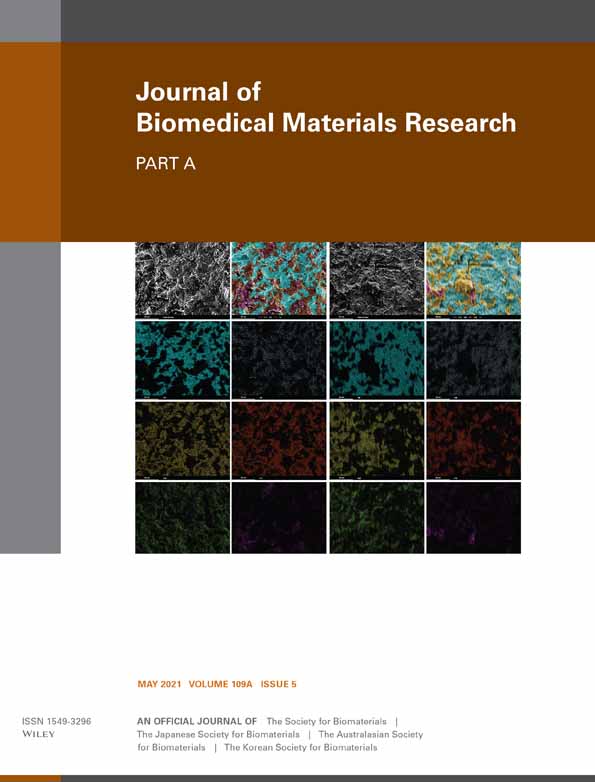
Olfactory ensheathing cells seeded decellularized scaffold promotes axonal regeneration in spinal cord injury rats
Fangzheng Yu
Department of Hand Surgery and Peripheral Neurosurgery, The First Affiliated Hospital of Wenzhou Medical University, Wenzhou, Zhejiang, China
Search for more papers by this authorPeifeng Li
Department of Hand Surgery and Peripheral Neurosurgery, The First Affiliated Hospital of Wenzhou Medical University, Wenzhou, Zhejiang, China
Search for more papers by this authorShenghu Du
Department of Hand Surgery and Peripheral Neurosurgery, The First Affiliated Hospital of Wenzhou Medical University, Wenzhou, Zhejiang, China
Search for more papers by this authorKoonHei W. Lui
Department of Plastic Surgery, The First affiliated hospital of Sun Yat-Sen University, Guangdong, China
Search for more papers by this authorYutian Lin
Department of Hand Surgery and Peripheral Neurosurgery, The First Affiliated Hospital of Wenzhou Medical University, Wenzhou, Zhejiang, China
Search for more papers by this authorJian Wang
Department of Hand Surgery and Peripheral Neurosurgery, The First Affiliated Hospital of Wenzhou Medical University, Wenzhou, Zhejiang, China
Search for more papers by this authorJin Mei
Institute of Neuroscience, Wenzhou Medical University, Wenzhou, China
Search for more papers by this authorCorresponding Author
Jian Xiao
School of Pharmaceutical Science, Wenzhou Medical University, Wenzhou, Zhejiang, China
Correspondence
Junyi Zhu, Department of Hand Surgery and Peripheral Neurosurgery, The First Affiliated Hospital of Wenzhou Medical University, Wenzhou, Zhejiang 325035, China.
Email: [email protected]
Jian Xiao, School of Pharmaceutical Science, Wenzhou Medical University, Wenzhou, Zhejiang, China.
Email: [email protected]
Search for more papers by this authorCorresponding Author
Junyi Zhu
Department of Hand Surgery and Peripheral Neurosurgery, The First Affiliated Hospital of Wenzhou Medical University, Wenzhou, Zhejiang, China
Correspondence
Junyi Zhu, Department of Hand Surgery and Peripheral Neurosurgery, The First Affiliated Hospital of Wenzhou Medical University, Wenzhou, Zhejiang 325035, China.
Email: [email protected]
Jian Xiao, School of Pharmaceutical Science, Wenzhou Medical University, Wenzhou, Zhejiang, China.
Email: [email protected]
Search for more papers by this authorFangzheng Yu
Department of Hand Surgery and Peripheral Neurosurgery, The First Affiliated Hospital of Wenzhou Medical University, Wenzhou, Zhejiang, China
Search for more papers by this authorPeifeng Li
Department of Hand Surgery and Peripheral Neurosurgery, The First Affiliated Hospital of Wenzhou Medical University, Wenzhou, Zhejiang, China
Search for more papers by this authorShenghu Du
Department of Hand Surgery and Peripheral Neurosurgery, The First Affiliated Hospital of Wenzhou Medical University, Wenzhou, Zhejiang, China
Search for more papers by this authorKoonHei W. Lui
Department of Plastic Surgery, The First affiliated hospital of Sun Yat-Sen University, Guangdong, China
Search for more papers by this authorYutian Lin
Department of Hand Surgery and Peripheral Neurosurgery, The First Affiliated Hospital of Wenzhou Medical University, Wenzhou, Zhejiang, China
Search for more papers by this authorJian Wang
Department of Hand Surgery and Peripheral Neurosurgery, The First Affiliated Hospital of Wenzhou Medical University, Wenzhou, Zhejiang, China
Search for more papers by this authorJin Mei
Institute of Neuroscience, Wenzhou Medical University, Wenzhou, China
Search for more papers by this authorCorresponding Author
Jian Xiao
School of Pharmaceutical Science, Wenzhou Medical University, Wenzhou, Zhejiang, China
Correspondence
Junyi Zhu, Department of Hand Surgery and Peripheral Neurosurgery, The First Affiliated Hospital of Wenzhou Medical University, Wenzhou, Zhejiang 325035, China.
Email: [email protected]
Jian Xiao, School of Pharmaceutical Science, Wenzhou Medical University, Wenzhou, Zhejiang, China.
Email: [email protected]
Search for more papers by this authorCorresponding Author
Junyi Zhu
Department of Hand Surgery and Peripheral Neurosurgery, The First Affiliated Hospital of Wenzhou Medical University, Wenzhou, Zhejiang, China
Correspondence
Junyi Zhu, Department of Hand Surgery and Peripheral Neurosurgery, The First Affiliated Hospital of Wenzhou Medical University, Wenzhou, Zhejiang 325035, China.
Email: [email protected]
Jian Xiao, School of Pharmaceutical Science, Wenzhou Medical University, Wenzhou, Zhejiang, China.
Email: [email protected]
Search for more papers by this authorFangzheng Yu and Peifeng Li contributed equally to this study.
Funding information: Wenzhou Science and Technology Bureau, Grant/Award Number: Y20180498
Abstract
Spinal cord decellularized (DC) scaffolds can promote axonal regeneration and restore hindlimb motor function of spinal cord defect rats. However, scarring caused by damage to the astrocytes at the margin of injury can hinder axon regeneration. Olfactory ensheathing cells (OECs) integrate and migrate with astrocytes at the site of spinal cord injury, providing a bridge for axons to penetrate the scars and grow into lesion cores. The purpose of this study was to evaluate whether DC scaffolds carrying OECs could better promote axon growth. For these studies, DC scaffolds were cocultured with primary extracted and purified OECs. Immunofluorescence and electron microscopy were used for verification of cells adhere and growth on the scaffold. Scaffolds with OECs were transplanted into rat spinal cord defects to evaluate axon regeneration and functional recovery of hind limbs. Basso, Beattie, and Bresnahan (BBB) scoring was used to assess motor function recovery, and glial fibrillary acidic protein (GFAP) and NF200-stained tissue sections were used to evaluate axonal regeneration and astrological scar distribution. Our results indicated that spinal cord DC scaffolds have good histocompatibility and spatial structure, and can promote the proliferation of adherent OECs. In animal experiments, scaffolds carrying OECs have better axon regeneration promoting protein expression than the SCI model, and improve the proliferation and distribution of astrocytes at the site of injury. These results proved that the spinal cord DC scaffold with OECs can promote axon regeneration at the site of injury, providing a new basis for clinical application.
REFERENCES
- 1Koffler, J., Zhu, W., Qu, X., Platoshyn, O., Dulin, J. N., Brock, J., … Tuszynski, M. H. (2019). Biomimetic 3D-printed scaffolds for spinal cord injury repair. Nat Med, 25(2), 263–269. https://doi.org/10.1038/s41591-018-0296-z
- 2Feng, L., Gan, H., Zhao, W., & Liu, Y. (2017). Effect of transplantation of olfactory ensheathing cell conditioned medium induced bone marrow stromal cells on rats with spinal cord injury. Mol Med Rep, 16(2), 1661–1668. https://doi.org/10.3892/mmr.2017.6811
- 3Beck Kevin, D., Nguyen Hal, X., Galvan Manuel, D., Salazar Desirée, L., Woodruff Trent, M., & Anderson Aileen, J. (2010). Quantitative analysis of cellular inflammation after traumatic spinal cord injury: evidence for a multiphasic inflammatory response in the acute to chronic environment. Brain, 133, 433–447. https://doi.org/10.1093/brain/awp322
- 4Penas, C., Guzmán, M.-S., Verdú, E., Forés, J., Navarro, X., & Casas, C. (2007). Spinal cord injury induces endoplasmic reticulum stress with different cell-type dependent response. J Neurochem, 102(4), 1242–1255. https://doi.org/10.1111/j.1471-4159.2007.04671.x
- 5Bradbury, E. J., & Burnside, E. R. (2019). Moving beyond the glial scar for spinal cord repair. Nat Commun, 10(1), 3879. https://doi.org/10.1038/s41467-019-11707-7
- 6Gilmour, A. D., Reshamwala, R., Wright, A. A., Ekberg, J. A. K., & St John, J. A. (2020). Optimizing olfactory ensheathing cell transplantation for spinal cord injury repair. J Neurotrauma, 37(5), 817–829. https://doi.org/10.1089/neu.2019.6939
- 7Raynald, Bing, S., Xue-Bin, L., Jun-Feng, Z., Hua, H., Jing-Yun, W., … Yi-Hua, A. (2019). Polypyrrole/polylactic acid nanofibrous scaffold cotransplanted with bone marrow stromal cells promotes the functional recovery of spinal cord injury in rats. CNS Neurosci Ther, 25(9), 951–964. https://doi.org/10.1111/cns.13135
- 8Zarei-Kheirabadi, M., Sadrosadat, H., Mohammadshirazi, A., Jaberi, R., Sorouri, F., Khayyatan, F., & Sahar, K. (2020). Human embryonic stem cell-derived neural stem cells encapsulated in hyaluronic acid promotes regeneration in a contusion spinal cord injured rat. Int J Biol Macromol, 148, 1118–1129. https://doi.org/10.1016/j.ijbiomac.2020.01.219
- 9Khankan, R. R., Griffis, K. G., Haggerty-Skeans, J. R., Zhong, H., Roy, R. R., Edgerton, V. R., & Phelps, P. E. (2016). Olfactory ensheathing cell transplantation after a complete spinal cord transection mediates neuroprotective and immunomodulatory mechanisms to facilitate regeneration. J Neurosci, 36(23), 6269–6286. https://doi.org/10.1523/JNEUROSCI.0085-16.2016
- 10Zhang, L., Zhuang, X., Chen, Y., & Xia, H. (2019). Intravenous transplantation of olfactory bulb ensheathing cells for a spinal cord hemisection injury rat model. Cell Transplant, 28(12), 1585–1602. https://doi.org/10.1177/0963689719883842
- 11Chedly, J., Soares, S., Montembault, A., von Boxberg, Y., Michèle, V.-R., Christine, M., … Fatiha, N. (2017). Physical chitosan microhydrogels as scaffolds for spinal cord injury restoration and axon regeneration. Biomaterials, 138, 91–107. https://doi.org/10.1016/j.biomaterials.2017.05.024
- 12Zhang, Q., Shi, B., Ding, J., Yan, L., Thawani Jayesh, P., Fu, C., & Xuesi, C. (2019). Polymer scaffolds facilitate spinal cord injury repair. Acta Biomater, 88, 57–77. https://doi.org/10.1016/j.actbio.2019.01.056
- 13Moonhang, K., Ra, P. S., & Byung Hyune, C. (2014). Biomaterial scaffolds used for the regeneration of spinal cord injury (SCI). Histol Histopathol, 29, 1395–1408.
- 14Liu, S., Schackel, T., Weidner, N., & Radhika, P. (2017). Biomaterial-supported cell transplantation treatments for spinal cord injury: challenges and perspectives. Front Cell Neurosci, 11, 430. https://doi.org/10.3389/fncel.2017.00430
- 15Zhu, J., Lu, Y., Yu, F., Zhou, L., Shi, J., Chen, Q., … Wang, J. (2018). Effect of decellularized spinal scaffolds on spinal axon regeneration in rats. J Biomed Mater Res A, 106(3), 698–705. https://doi.org/10.1002/jbm.a.36266
- 16Gomes, E. D., Mendes, S. S., Assunção-Silva, R. C., Teixeira, F. G., Pires, A. O., Anjo, S. I., … Salgado, A. J. (2018). Co-transplantation of adipose tissue-derived stromal cells and olfactory ensheathing cells for spinal cord injury repair. Stem Cells, 36(5), 696–708. https://doi.org/10.1002/stem.2785
- 17Martínez-Ramos, C., Doblado, L. R., Mocholi, E. L., Alastrue-Agudo, A., Petidier, M. S., Giraldo, E., … Moreno-Manzano, V. (2019). Biohybrids for spinal cord injury repair. J Tissue Eng Regen Med, 13(3), 509–521. https://doi.org/10.1002/term.2816
- 18Subbarayan, R., Barathidasan, R., Raja, S. T. K., Arumugam, G., Kuruvilla, S., Shanthi, P., & Ranga Rao, S. (2020). Human gingival derived neuronal cells in the optimized caffeic acid hydrogel for hemitransection spinal cord injury model. J Cell Biochem, 121(3), 2077–2088. https://doi.org/10.1002/jcb.29452
- 19Zhao, Y., Xiao, Z., Chen, B., & Dai, J. (2017). The neuronal differentiation microenvironment is essential for spinal cord injury repair. Organogenesis, 13(3), 63–70. https://doi.org/10.1080/15476278.2017.1329789
- 20Amani, H., Kazerooni, H., Hassanpoor, H., Akbarzadeh, A., & Pazoki-Toroudi, H. (2019). Tailoring synthetic polymeric biomaterials towards nerve tissue engineering: a review. Artif Cells Nanomed Biotechnol, 47(1), 3524–3539. https://doi.org/10.1080/21691401.2019.1639723
- 21Li, X., Liu, D., Xiao, Z., Zhao, Y., Han, S., Chen, B., & Jianwu, D. (2019). Scaffold-facilitated locomotor improvement post complete spinal cord injury: motor axon regeneration versus endogenous neuronal relay formation. Biomaterials, 197, 20–31. https://doi.org/10.1016/j.biomaterials.2019.01.012
- 22Domínguez-Bajo, A., González-Mayorga, A., López-Dolado, E., & Serrano María, C. (2017). In VitroGraphene-derived materials interfacing the spinal cord: outstanding and findings. Front Syst Neurosci, 11, 71. https://doi.org/10.3389/fnsys.2017.00071
- 23Guo, J., Cao, G., Yang, G., Zhang, Y., Wang, Y., Song, W., … Hao, Y. (2020). Transplantation of activated olfactory ensheathing cells by curcumin strengthens regeneration and recovery of function after spinal cord injury in rats. Cytotherapy, 22(6), 301–312. https://doi.org/10.1016/j.jcyt.2020.03.002
- 24Crapo, P. M., Medberry, C. J., Reing, J. E., Tottey, S., van der Merwe, Y., Jones, K. E., & Badylak, S. F. (2012). Biologic scaffolds composed of central nervous system extracellular matrix. Biomaterials, 33(13), 3539–3547. https://doi.org/10.1016/j.biomaterials.2012.01.044
- 25Wang, H., Lin, X.-F., Wang, L.-R., Lin, Y.-Q., Wang, J.-T., Liu, W.-Y., … Zheng, M.-H. (2015). Decellularization technology in CNS tissue repair. Expert Rev Neurother, 15(5), 493–500. https://doi.org/10.1586/14737175.2015.1030735
- 26Wentao, Z., Ya'nan, H., Jian, L., Kaipeng, B., Peng, S., Yu, Z., … Shen, Y. (2019). In vitro biocompatibility study of a water-rinsed biomimetic silk porous scaffold with olfactory ensheathing cells. Int J Biol Macromol, 125, 526–533. https://doi.org/10.1016/j.ijbiomac.2018.11.029


