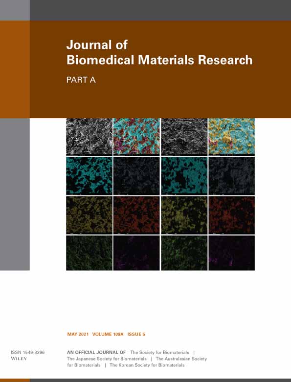
Influence of nano-hydroxyapatite coating implants on gene expression of osteogenic markers and micro-CT parameters. An in vivo study in diabetic rats
Paula Gabriela Faciola Pessôa de Oliveira
Department of Oral and Maxillofacial Surgery and Periodontology, FORP/USP, University of São Paulo, Ribeirão Preto, São Paulo, Brazil
Search for more papers by this authorMariana Sales de Melo Soares
Department of Oral and Maxillofacial Surgery and Periodontology, FORP/USP, University of São Paulo, Ribeirão Preto, São Paulo, Brazil
Search for more papers by this authorAdriana Maria Mariano Silveira e Souza
Department of Oral and Maxillofacial Surgery and Periodontology, FORP/USP, University of São Paulo, Ribeirão Preto, São Paulo, Brazil
Search for more papers by this authorMário Taba Jr
Department of Oral and Maxillofacial Surgery and Periodontology, FORP/USP, University of São Paulo, Ribeirão Preto, São Paulo, Brazil
Search for more papers by this authorDaniela Bazan Palioto
Department of Oral and Maxillofacial Surgery and Periodontology, FORP/USP, University of São Paulo, Ribeirão Preto, São Paulo, Brazil
Search for more papers by this authorMichel Reis Messora
Department of Oral and Maxillofacial Surgery and Periodontology, FORP/USP, University of São Paulo, Ribeirão Preto, São Paulo, Brazil
Search for more papers by this authorBruna Ghiraldini
Paulista University, School of Dentistry, São Paulo, São Paulo, Brazil
Search for more papers by this authorFelipe Anderson de Sousa Nunes
Department of Oral and Maxillofacial Surgery and Periodontology, FORP/USP, University of São Paulo, Ribeirão Preto, São Paulo, Brazil
Search for more papers by this authorCorresponding Author
Sérgio Luís Scombatti de Souza
Department of Oral and Maxillofacial Surgery and Periodontology, FORP/USP, University of São Paulo, Ribeirão Preto, São Paulo, Brazil
Correspondence
Sérgio Luís Scombatti de Souza Departament of Oral & Maxillofacial Surgery and Periodontology School of Dentistry of Ribeirão Preto University of São Paulo Av do Café, s/n. 14040-904 Ribeirão Preto, SP, Brazil.
Email: [email protected]
Search for more papers by this authorPaula Gabriela Faciola Pessôa de Oliveira
Department of Oral and Maxillofacial Surgery and Periodontology, FORP/USP, University of São Paulo, Ribeirão Preto, São Paulo, Brazil
Search for more papers by this authorMariana Sales de Melo Soares
Department of Oral and Maxillofacial Surgery and Periodontology, FORP/USP, University of São Paulo, Ribeirão Preto, São Paulo, Brazil
Search for more papers by this authorAdriana Maria Mariano Silveira e Souza
Department of Oral and Maxillofacial Surgery and Periodontology, FORP/USP, University of São Paulo, Ribeirão Preto, São Paulo, Brazil
Search for more papers by this authorMário Taba Jr
Department of Oral and Maxillofacial Surgery and Periodontology, FORP/USP, University of São Paulo, Ribeirão Preto, São Paulo, Brazil
Search for more papers by this authorDaniela Bazan Palioto
Department of Oral and Maxillofacial Surgery and Periodontology, FORP/USP, University of São Paulo, Ribeirão Preto, São Paulo, Brazil
Search for more papers by this authorMichel Reis Messora
Department of Oral and Maxillofacial Surgery and Periodontology, FORP/USP, University of São Paulo, Ribeirão Preto, São Paulo, Brazil
Search for more papers by this authorBruna Ghiraldini
Paulista University, School of Dentistry, São Paulo, São Paulo, Brazil
Search for more papers by this authorFelipe Anderson de Sousa Nunes
Department of Oral and Maxillofacial Surgery and Periodontology, FORP/USP, University of São Paulo, Ribeirão Preto, São Paulo, Brazil
Search for more papers by this authorCorresponding Author
Sérgio Luís Scombatti de Souza
Department of Oral and Maxillofacial Surgery and Periodontology, FORP/USP, University of São Paulo, Ribeirão Preto, São Paulo, Brazil
Correspondence
Sérgio Luís Scombatti de Souza Departament of Oral & Maxillofacial Surgery and Periodontology School of Dentistry of Ribeirão Preto University of São Paulo Av do Café, s/n. 14040-904 Ribeirão Preto, SP, Brazil.
Email: [email protected]
Search for more papers by this authorThe authors have complete independence in the results and did not receive any kind of aid.
Funding information: National Council of Scientific and Technological Development (CNPq, Brazil), Grant/Award Number: Grant number: 446840/2014-9; State of Sao Paulo Research Foundation (FAPESP, Brazil), Grant/Award Numbers: 2015/09879-0, 2016/14584-2
Abstract
This study evaluated the response of a nano-hydroxyapatite coating implant through gene expression analysis (runt-related transcription factor 2 (Runx2), alkaline phosphatase (Alp), osteopontin (Opn), osteocalcin (Oc), receptor activator of nuclear factor-kappa B (Rank), receptor activator of nuclear factor-kappa B ligand (Rank-L), and osteoprotegerin (Opg)). Three-dimensional evaluation (percent bone volume (BV/TV); percent intersection surface (BIC); bone surface/volume ratio (BS/BV); and total porosity (To.Po)) were also analyzed. Mini implants were surgically placed in tibias of both healthy and diabetic rats. The animals were euthanized at 7 and 30 days. Evaluating all factors the relative expression of Rank showed that NANO surface presented the best results at 7 days (diabetic rats). Furthermore the levels of Runx2, Alp, Oc, and Opn suggest an increase in osteoblasts proliferation, especially in early stages of osseointegration. %BIC in healthy and diabetic (7 days) depicted statistically significant differences for NANO group. BV/TV, BS/BV and To.Po demonstrated higher values for NANO group in all evaluated time point and irrespective of systemic condition, but BS/BV 30 days (healthy rat) and 7 and 30 days (diabetic rat). Microtomographic and gene expression analyses have shown the benefits of nano-hydroxyapatite coated implants in promoting new bone formation in diabetic rats.
REFERENCES
- Ajami, E., Bell, S., Liddell, R. S., & Davies, J. E. (2016). Early bone anchorage to micro- and nano-topographically complex implant surfaces in hyperglycemia. Acta Biomaterialia, 39, 169–179.
- Ajami, E., Mahno, E., Mendes, V. C., Bell, S., Moineddin, R., & Davies, J. E. (2014). Bone healing and the effect of implant surface topography on osteoconduction in hyperglycemia. Acta Biomaterialia, 10(1), 394–405.
- Albrektsson, T., Branemark, P. I., Hansson, H. A., & Lindstrom, J. (1981). Osseointegrated titanium implants. Requirements for ensuring a long-lasting, direct bone-to-implant anchorage in man. Acta Orthopaedica Scandinavica, 52(2), 155–170.
- Buser, D., Schenk, R. K., Steinemann, S., Fiorellini, J. P., Fox, C. H., & Stich, H. (1991). Influence of surface characteristics on bone integration of titanium implants. A histomorphometric study in miniature pigs. Journal of Biomedical Materials Research, 25(7), 889–902.
- Chen, Z., Bachhuka, A., Wei, F., Wang, X., Liu, G., Vasilev, K., & Xiao, Y. (2017). Nanotopography-based strategy for the precise manipulation of osteoimmunomodulation in bone regeneration. Nanoscale, 9(46), 18129–18152.
- Chuang, S. K., Tian, L., Wei, L. J., & Dodson, T. B. (2001). Kaplan-Meier analysis of dental implant survival: A strategy for estimating survival with clustered observations. Journal of Dental Research, 80(11), 2016–2020.
- Coelho, P. G., Granjeiro, J. M., Romanos, G. E., Suzuki, M., Silva, N. R., Cardaropoli, G., … Lemons, J. E. (2009). Basic research methods and current trends of dental implant surfaces. Journal of Biomedical Materials Research. Part B, Applied Biomaterials, 88(2), 579–596.
- Courtney, M. W., Jr., Snider, T. N., & Cottrell, D. A. (2010). Dental implant placement in type II diabetics: A review of the literature. Journal of the Massachusetts Dental Society, 59(1), 12–14.
- Curtis, A., & Wilkinson, C. (1999). New depths in cell behaviour: Reactions of cells to nanotopography. Biochemical Society Symposium, 65, 15–26.
- Dang, Y., Zhang, L., Song, W., Chang, B., Han, T., Zhang, Y., & Zhao, L. (2016). In vivo osseointegration of Ti implants with a strontium-containing nanotubular coating. International Journal of Nanomedicine, 11, 1003–1011.
- de Groot, K., Geesink, R., Klein, C. P., & Serekian, P. (1987). Plasma sprayed coatings of hydroxylapatite. Journal of Biomedical Materials Research, 21(12), 1375–1381.
- de Lange, G. L., & Donath, K. (1989). Interface between bone tissue and implants of solid hydroxyapatite or hydroxyapatite-coated titanium implants. Biomaterials, 10(2), 121–125.
- de Oliveira, P. T., Zalzal, S. F., Beloti, M. M., Rosa, A. L., & Nanci, A. (2007). Enhancement of in vitro osteogenesis on titanium by chemically produced nanotopography. Journal of Biomedical Materials Research. Part A, 80(3), 554–564.
- Denissen, H. W., Kalk, W., de Nieuport, H. M., Maltha, J. C., & van de Hooff, A. (1990). Mandibular bone response to plasma-sprayed coatings of hydroxyapatite. The International Journal of Prosthodontics, 3(1), 53–58.
- Ducheyne, P. (1987). Bioceramics: Material characteristics versus in vivo behavior. Journal of Biomedical Materials Research, 21(A2 Suppl), 219–236.
- Expert Committee on the Diagnosis and Classification of Diabetes Mellitus. (2003). Report of the expert committee on the diagnosis and classification of diabetes mellitus. Diabetes Care, 23(Suppl 1), S5–S20.
- Feldkamp, L. A., Goldstein, S. A., Parfitt, A. M., Jesion, G., & Kleerekoper, M. (1989). The direct examination of three-dimensional bone architecture in vitro by computed tomography. Journal of Bone and Mineral Research, 4(1), 3–11.
- Filiaggi, M. J., Coombs, N. A., & Pilliar, R. M. (1991). Characterization of the interface in the plasma-sprayed HA coating/Ti-6Al-4V implant system. Journal of Biomedical Materials Research, 25(10), 1211–1229.
- Gittens, R. A., McLachlan, T., Olivares-Navarrete, R., Cai, Y., Berner, S., Tannenbaum, R., … Boyan, B. D. (2011). The effects of combined micron−/submicron-scale surface roughness and nanoscale features on cell proliferation and differentiation. Biomaterials, 32(13), 3395–3403.
- Globus, R. K., Moursi, A., Zimmerman, D., Lull, J., & Damsky, C. (1995). Integrin-extracellular matrix interactions in connective tissue remodeling and osteoblast differentiation. ASGSB Bulletin, 8(2), 19–28.
- Goodman, W. G., & Hori, M. T. (1984). Diminished bone formation in experimental diabetes. Relationship to osteoid maturation and mineralization. Diabetes, 33(9), 825–831.
- Hamann, C., Goettsch, C., Mettelsiefen, J., Henkenjohann, V., Rauner, M., Hempel, U., … Rammelt, S. (2011). Others. Delayed bone regeneration and low bone mass in a rat model of insulin-resistant type 2 diabetes mellitus is due to impaired osteoblast function. American Journal of Physiology. Endocrinology and Metabolism, 301(6), E1220–E1228.
- Jiang, H., Ma, X., Zhou, W., Dong, K., Rausch-Fan, X., Liu, S., & Li, S. (2017). The effects of hierarchical micro/Nano-structured titanium surface on osteoblast proliferation and differentiation under diabetic conditions. Implant Dentistry, 26(2), 263–269.
- Jimbo, R., Coelho, P. G., Vandeweghe, S., Schwartz-Filho, H. O., Hayashi, M., Ono, D., … Wennerberg, A. (2011). Histological and three-dimensional evaluation of osseointegration to nanostructured calcium phosphate-coated implants. Acta Biomaterialia, 7(12), 4229–4234.
- Kay, J. F. (1988). Designing to counteract the effects of initial device instability: Materials and engineering. Journal of Biomedical Materials Research, 22(12), 1127–1136.
- Kay, J. F. (1992). Calcium phosphate coatings for dental implants. Current status and future potential. Dental Clinics of North America, 36(1), 1–18.
- Kjellin P, Andersson M; Synthetic Nano-sized crystalline calcium phosphate and method of production. Patent US patent 8206813. 2012.Alexandria, VA, USA: US Patent and Trademark Office.
- Lemons, J. E. (1988). Hydroxyapatite coatings. Clinical Orthopaedics and Related Research, 235, 220–223.
- Li, Z., Kuhn, G., von Salis-Soglio, M., Cooke, S. J., Schirmer, M., Muller, R., & Ruffoni, D. (2015). In vivo monitoring of bone architecture and remodeling after implant insertion: The different responses of cortical and trabecular bone. Bone, 81, 468–477.
- Livak, K. J., & Schmittgen, T. D. (2001). Analysis of relative gene expression data using real-time quantitative PCR and the 2(−Delta Delta C[T]) method. Methods, 25(4), 402–408.
- Longo, G., Ioannidu, C. A., Scotto d'Abusco, A., Superti, F., Misiano, C., Zanoni, R., … Scandurra, F. (2016). Improving osteoblast response in vitro by a nanostructured thin film with titanium carbide and titanium oxides clustered around graphitic carbon. PLoS One, 11(3), e0152566.
- Lopes, H. B., Freitas, G. P., Elias, C. N., Tye, C., Stein, J. L., Stein, G. S., … Beloti, M. M. (2019b). Participation of integrin beta3 in osteoblast differentiation induced by titanium with nano or microtopography. Journal of Biomedical Materials Research. Part A, 107(6), 1303–1313.
- Lopes, H. B., Freitas, G. P., Fantacini, D. M. C., Picanco-Castro, V., Covas, D. T., Rosa, A. L., & Beloti, M. M. (2019a). Titanium with nanotopography induces osteoblast differentiation through regulation of integrin alphaV. Journal of Cellular Biochemistry, 120(10), 16723–16732.
- Lopes, H. B., Souza, A. T. P., Freitas, G. P., Elias, C. N., Rosa, A. L., & Beloti, M. M. (2020). Effect of focal adhesion kinase inhibition on osteoblastic cells grown on titanium with different topographies. Journal of Applied Oral Science, 28, e20190156.
- Ma, X. Y., Feng, Y. F., Wang, T. S., Lei, W., Li, X., Zhou, D. P., … Wang, L. (2017). Involvement of FAK-mediated BMP-2/Smad pathway in mediating osteoblast adhesion and differentiation on nano-HA/chitosan composite coated titanium implant under diabetic conditions. Biomaterials Science, 6(1), 225–238.
- Margonar, R., Sakakura, C. E., Holzhausen, M., Pepato, M. T., Alba j, R., & Marcantonio j, E. (2003). The influence of diabetes mellitus and insulin therapy on biomechanical retention around dental implants: A study in rabbits. Implant Dentistry, 12(4), 333–339.
- Martinez, E. F., Ishikawa, G. J., de Lemos, A. B., Barbosa Bezerra, F. J., Sperandio, M., & Napimoga, M. H. (2018). Evaluation of a titanium surface treated with hydroxyapatite nanocrystals on osteoblastic cell behavior: An in vitro study. The International Journal of Oral & Maxillofacial Implants, 33(3), 597–602.
- McCracken, M., Lemons, J. E., Rahemtulla, F., Prince, C. W., & Feldman, D. (2000). Bone response to titanium alloy implants placed in diabetic rats. The International Journal of Oral & Maxillofacial Implants, 15(3), 345–354.
- Meirelles, L., Arvidsson, A., Albrektsson, T., & Wennerberg, A. (2007). Increased bone formation to unstable nano rough titanium implants. Clinical Oral Implants Research, 18(3), 326–332.
- Moraschini, V., Barboza, E. S., & Peixoto, G. A. (2016). The impact of diabetes on dental implant failure: A systematic review and meta-analysis. International Journal of Oral and Maxillofacial Surgery, 45(10), 1237–1245.
- Moraschini, V., Poubel, L. A., Ferreira, V. F., & Barboza, E. S. (2015). Evaluation of survival and success rates of dental implants reported in longitudinal studies with a follow-up period of at least 10 years: A systematic review. International Journal of Oral and Maxillofacial Surgery, 44(3), 377–388.
- MSC-R, L., M, S. F., L, N. T., E, P. F., H, B. L., TdO, P., … Beloti, M. M. (2016). Titanium with Nanotopography induces osteoblast differentiation by regulating endogenous Bone morphogenetic protein expression and signaling pathway. Journal of Cellular Biochemistry, 117(7), 1718–1726.
- Napoli, N., Chandran, M., Pierroz, D. D., Abrahamsen, B., Schwartz, A. V., Ferrari, S. L., … Diabetes Working, G. (2017). Mechanisms of diabetes mellitus-induced bone fragility. Nature Reviews. Endocrinology, 13(4), 208–219.
- Nevins, M. L., Karimbux, N. Y., Weber, H. P., Giannobile, W. V., & Fiorellini, J. P. (1998). Wound healing around endosseous implants in experimental diabetes. The International Journal of Oral & Maxillofacial Implants, 13(5), 620–629.
- Odgaard, A. (1997). Three-dimensional methods for quantification of cancellous bone architecture. Bone, 20(4), 315–328.
- Ong, J. L., & Lucas, L. C. (1994). Post-deposition heat treatments for ion beam sputter deposited calcium phosphate coatings. Biomaterials, 15(5), 337–341.
- Ong, J. L., Lucas, L. C., Lacefield, W. R., & Rigney, E. D. (1992). Structure, solubility and bond strength of thin calcium phosphate coatings produced by ion beam sputter deposition. Biomaterials, 13(4), 249–254.
- Qian, C., Zhu, C., Yu, W., Jiang, X., & Zhang, F. (2015). High-fat diet/low-dose Streptozotocin-induced type 2 Diabetes in rats impacts osteogenesis and Wnt signaling in Bone marrow stromal cells. PLoS One, 10(8), e0136390.
- Rabbani, P. S., Soares, M. A., Hameedi, S. G., Kadle, R. L., Mubasher, A., Kowzun, M., & Ceradini, D. J. (2019). Dysregulation of Nrf2/Keap1 redox pathway in Diabetes affects multipotency of stromal cells. Diabetes, 68(1), 141–155.
- Ramenzoni, L. L., Bosch, A., Proksch, S., Attin, T., & Schmidlin, P. R. (2020). Effect of high glucose levels and lipopolysaccharides-induced inflammation on osteoblast mineralization over sandblasted/acid-etched titanium surface. Clinical Implant Dentistry and Related Research, 22(2), 213–219.
- Retzepi, M., & Donos, N. (2010). The effect of diabetes mellitus on osseous healing. Clinical Oral Implants Research, 21(7), 673–681.
- Sarve, H., Lindblad, J., Borgefors, G., & Johansson, C. B. (2011). Extracting 3D information on bone remodeling in the proximity of titanium implants in SRmuCT image volumes. Computer Methods and Programs in Biomedicine, 102(1), 25–34.
- Schlegel, K. A., Prechtl, C., Most, T., Seidl, C., Lutz, R., & von Wilmowsky, C. (2013). Osseointegration of SLActive implants in diabetic pigs. Clinical Oral Implants Research, 24(2), 128–134.
- Schneider, G. B., Zaharias, R., & Stanford, C. (2001). Osteoblast integrin adhesion and signaling regulate mineralization. Journal of Dental Research, 80(6), 1540–1544.
- Schwartz, Z., Olivares-Navarrete, R., Wieland, M., Cochran, D. L., & Boyan, B. D. (2009). Mechanisms regulating increased production of osteoprotegerin by osteoblasts cultured on microstructured titanium surfaces. Biomaterials, 30(20), 3390–3396.
- Shin, L., & Peterson, D. A. (2012). Impaired therapeutic capacity of autologous stem cells in a model of type 2 diabetes. Stem Cells Translational Medicine, 1(2), 125–135.
- Sivaraj, K. K., & Adams, R. H. (2016). Blood vessel formation and function in bone. Development, 143(15), 2706–2715.
- Souza, A. T. P., Bezerra, B. L. S., Oliveira, F. S., Freitas, G. P., Bighetti Trevisan, R. L., Oliveira, P. T., … Beloti, M. M. (2018). Effect of bone morphogenetic protein 9 on osteoblast differentiation of cells grown on titanium with nanotopography. Journal of Cellular Biochemistry, 119, 8441–8449.
- Takeshita, F., Iyama, S., Ayukawa, Y., Kido, M. A., Murai, K., & Suetsugu, T. (1997). The effects of diabetes on the interface between hydroxyapatite implants and bone in rat tibia. Journal of Periodontology, 68(2), 180–185.
- Vandeweghe, S., Coelho, P. G., Vanhove, C., Wennerberg, A., & Jimbo, R. (2013). Utilizing micro-computed tomography to evaluate bone structure surrounding dental implants: A comparison with histomorphometry. Journal of Biomedical Materials Research. Part B, Applied Biomaterials, 101(7), 1259–1266.
- Wai, P. Y., & Kuo, P. C. (2004). The role of Osteopontin in tumor metastasis. The Journal of Surgical Research, 121(2), 228–241.
- Wai, P. Y., & Kuo, P. C. (2008). Osteopontin: Regulation in tumor metastasis. Cancer Metastasis Reviews, 27(1), 103–118.
- Zhou, W., Tangl, S., Reich, K. M., Kirchweger, F., Liu, Z., Zechner, W., … Rausch-Fan, X. (2019). The influence of type 2 Diabetes mellitus on the Osseointegration of titanium implants with different surface modifications-a Histomorphometric study in high-fat diet/low-dose Streptozotocin-treated rats. Implant Dentistry, 28(1), 11–19.


