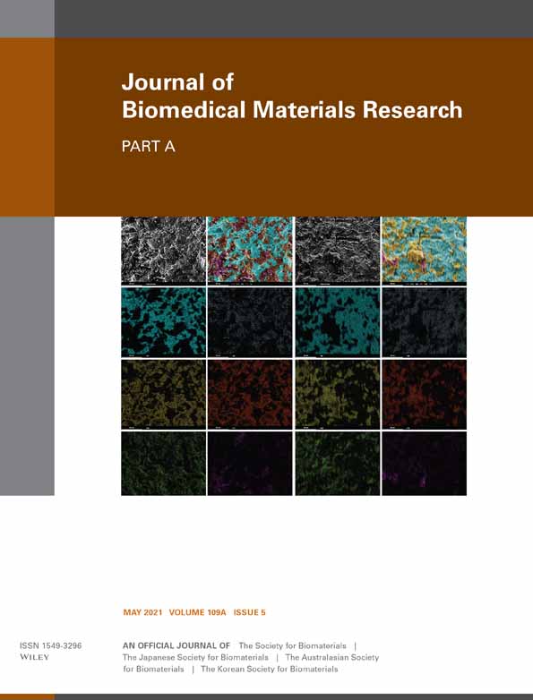
Carbon quantum dots-embedded electrospun antimicrobial and fluorescent scaffold for reepithelialization in albino wistar rats
Sankaralingam Kanagasubbulakshmi
DRDO-BU Center for Life Sciences, Bharathiar University, Coimbatore, India
Search for more papers by this authorKrishnasamy Lakshmi
DRDO-BU Center for Life Sciences, Bharathiar University, Coimbatore, India
Search for more papers by this authorCorresponding Author
Krishna Kadirvelu
DRDO-BU Center for Life Sciences, Bharathiar University, Coimbatore, India
Correspondence
Krishna Kadirvelu, DRDO-BU Center for Life Sciences, Bharathiar University, Coimbatore 641046, Tamil Nadu, India.
Email: [email protected]
Search for more papers by this authorSankaralingam Kanagasubbulakshmi
DRDO-BU Center for Life Sciences, Bharathiar University, Coimbatore, India
Search for more papers by this authorKrishnasamy Lakshmi
DRDO-BU Center for Life Sciences, Bharathiar University, Coimbatore, India
Search for more papers by this authorCorresponding Author
Krishna Kadirvelu
DRDO-BU Center for Life Sciences, Bharathiar University, Coimbatore, India
Correspondence
Krishna Kadirvelu, DRDO-BU Center for Life Sciences, Bharathiar University, Coimbatore 641046, Tamil Nadu, India.
Email: [email protected]
Search for more papers by this authorFunding information: Defence Research and Development Organisation, Grant/Award Number: DRDO-BU CLS Phase-II R&D Programme 2014
Abstract
A prosthetic scaffold development using fluorescent nanofiber is reported for an enhanced reepithelialization in wistar albino rats. In this study, a novel approach was followed to construct the biocompatible fluorescent nanofiber that will be helpful to monitor the tissue regeneration process. Here, a multifunctional carbon quantum dots (CQDs)-embedded electrospun polyacrylonitrile (PAN) nanofiber was fabricated and characterized using standard laboratory techniques. The biodegradation ability was assessed by simulated body fluid thereby analyzing porosity and water absorption capacity of the material. The fluorescent scaffold was tested for cytotoxicity and antimicrobial activity using bacterial and fibroblast cells and fluorescent stability was analyzed by bioimaging of animal and bacterial cells. Tissue regeneration capability of the developed scaffold was evaluated using wistar albino rats. Unlike biomicking scaffolds, the CQDs-embedded PAN-based substrate has given dual support by enhancing reepithelialization without growth factors and acted as an antimicrobial agent to provide contamination free tissue regeneration. Scaffolds were examined by using histostaining techniques and scanning electron microscopy to observe the reepithelialization in the regenerated tissues. The novel approach for developing infection free soft tissue regeneration was found to be phenomenal in scaffold development.
Supporting Information
| Filename | Description |
|---|---|
| jbma37048-sup-0001-AppendixS1.docxWord 2007 document , 649 KB | Appendix S1: Supporting information |
| jbma37048-sup-0002-suppinfoS2.mp4Word 2007 document , 26.5 MB | Supporting information video PAN_Nanofiber-Contact_angle |
| jbma37048-sup-0003-suppinfoS3.mp4Word 2007 document , 26.8 MB | Supporting information video PAN_NanofiberCQDs-Contact_angle |
Please note: The publisher is not responsible for the content or functionality of any supporting information supplied by the authors. Any queries (other than missing content) should be directed to the corresponding author for the article.
REFERENCES
- Alam, A. M., Liu, Y., Park, M., Park, S. J., & Kim, H. Y. (2015). Preparation and characterization of optically transparent and photoluminescent electrospun nanofiber composed of carbon quantum dots and polyacrylonitrile blend with polyacrylic acid. Polymer (Guildf)., 59, 35–41. https://doi.org/10.1016/j.polymer.2014.12.061
- Atiyeh, B. S., Costagliola, M., Hayek, S. N., & Dibo, S. A. (2007). Effect of silver on burn wound infection control and healing: Review of the literature. Burns, 33, 139–148. https://doi.org/10.1016/j.burns.2006.06.010
- Balint, R., Cassidy, N. J., & Cartmell, S. H. (2014). Conductive polymers: Towards a smart biomaterial for tissue engineering. Acta Biomaterialia, 10, 2341–2353. https://doi.org/10.1016/j.actbio.2014.02.015
- Barker, D. A., Bowers, D. T., Hughley, B., Chance, E. W., Klembczyk, K. J., Brayman, K. L., … Botchwey, E. A. (2013). Multilayer cell-seeded polymer nanofiber constructs for soft-tissue reconstruction. JAMA Otolaryngology—Head & Neck Surgery, 139, 914–922. https://doi.org/10.1001/jamaoto.2013.4119
- Bharambe, S. V., Darekar, A. B., & Saudagar, R. B. (2013). Wound healing dressings and drug delivery systems: A review. International Journal of Pharmacy and Technology, 5, 2764–2786. https://doi.org/10.1002/jps.21210
- Biswas, D. P., O'Brien-Simpson, N. M., Reynolds, E. C., O'Connor, A. J., & Tran, P. A. (2018). Comparative study of novel in situ decorated porous chitosan-selenium scaffolds and porous chitosan-silver scaffolds towards antimicrobial wound dressing application. Journal of Colloid and Interface Science, 515, 78–91. https://doi.org/10.1016/j.jcis.2018.01.007
- Choi, J. S., Leong, K. W., & Yoo, H. S. (2008). In vivo wound healing of diabetic ulcers using electrospun nanofibers immobilized with human epidermal growth factor (EGF). Biomaterials, 29, 587–596. https://doi.org/10.1016/j.biomaterials.2007.10.012
- Chun, J. Y., Kang, H. K., Jeong, L., Kang, Y. O., Oh, J. E., Yeo, I. S., … Min, B. M. (2010). Epidermal cellular response to poly(vinyl alcohol) nanofibers containing silver nanoparticles. Colloids Surfaces B Biointerfaces., 78, 334–342. https://doi.org/10.1016/j.colsurfb.2010.03.026
- Contardi, M., Heredia-Guerrero, J. A., Perotto, G., Valentini, P., Pompa, P. P., Spanò, R., … Bayer, I. S. (2017). Transparent ciprofloxacin-povidone antibiotic films and nanofiber mats as potential skin and wound care dressings. European Journal of Pharmaceutical Sciences, 104, 133–144. https://doi.org/10.1016/j.ejps.2017.03.044
- Dong, X., Al Awak, M., Tomlinson, N., Tang, Y., Sun, Y. P., & Yang, L. (2017). Antibacterial effects of carbon dots in combination with other antimicrobial reagents. PLoS One, 12, 1–16. https://doi.org/10.1371/journal.pone.0185324
- Dou, Q., Fang, X., Jiang, S., Chee, P. L., Lee, T. C., & Loh, X. J. (2015). Multi-functional fluorescent carbon dots with antibacterial and gene delivery properties. RSC Advances, 5, 46817–46822. https://doi.org/10.1039/c5ra07968c
- Dozois, M. D., Bahlmann, L. C., Zilberman, Y., & Tang, X. S. (2017). Carbon nanomaterial-enhanced scaffolds for the creation of cardiac tissue constructs: A new frontier in cardiac tissue engineering. Carbon, New York, 120, 338–349. https://doi.org/10.1016/j.carbon.2017.05.050
- Huang, X. J., Xu, Z. K., Wan, L. S., Wang, Z. G., & Wang, J. L. (2005). Surface modification of polyacrylonitrile-based membranes by chemical reactions to generate phospholipid moieties. Langmuir, 21, 2941–2947. https://doi.org/10.1021/la047419d
- Hui, L., Huang, J., Chen, G., Zhu, Y., & Yang, L. (2016). Antibacterial property of graphene quantum dots (both source material and bacterial shape matter). ACS Applied Materials & Interfaces, 8, 20–25. https://doi.org/10.1021/acsami.5b10132
- Ignatova, M., Manolova, N., Markova, N., & Rashkov, I. (2009). Electrospun non-woven nanofibrous hybrid mats based on chitosan and PLA for wound-dressing applications. Macromolecular Bioscience, 9, 102–111. https://doi.org/10.1002/mabi.200800189
- Jhonsi, M. A., Ananth, D. A., Nambirajan, G., Sivasudha, T., Yamini, R., Bera, S., & Kathiravan, A. (2018). Antimicrobial activity, cytotoxicity and DNA binding studies of carbon dots. Spectrochimica Acta, Part A: Molecular and Biomolecular Spectroscopy, 196, 295–302. https://doi.org/10.1016/j.saa.2018.02.030
- Jiang, L., Li, Y., Xiong, C., Su, S., & Ding, H. (2017). Preparation and properties of bamboo fiber/nano-hydroxyapatite/poly(lactic-co-glycolic) composite scaffold for bone tissue engineering. ACS Applied Materials & Interfaces, 9, 4890–4897. https://doi.org/10.1021/acsami.6b15032
- Jijie, R., Barras, A., Bouckaert, J., Dumitrascu, N., Szunerits, S., & Boukherroub, R. (2018). Enhanced antibacterial activity of carbon dots functionalized with ampicillin combined with visible light triggered photodynamic effects. Colloids Surfaces B Biointerfaces., 170, 347–354. https://doi.org/10.1016/j.colsurfb.2018.06.040
- Jithendra, P., Rajam, A. M., Kalaivani, T., Mandal, A. B., & Rose, C. (2013). Preparation and characterization of Aloe vera blended collagen-chitosan composite scaffold for tissue engineering applications. ACS Applied Materials & Interfaces, 5, 7291–7298. https://doi.org/10.1021/am401637c
- Kanagasubbulakshmi, S., & Kadirvelu, K. (2018). Nano interface potential influences in CdTe quantum dots and biolabeling. Applied Nanoscience, 8, 285–295. https://doi.org/10.1007/s13204-018-0774-0
- Kanagasubbulakshmi, S., & Kadirvelu, K. (2019). Photoinduced holes transfer based visual determination of dopamine in human serum. Spectrochimica Acta Part A: Molecular and Biomolecular Spectroscopy, 206, 512–519. https://doi.org/10.1016/j.saa.2018.08.050
- Kandhasamy, S., Perumal, S., Madhan, B., Umamaheswari, N., Banday, J. A., Perumal, P. T., & Santhanakrishnan, V. P. (2017). Synthesis and fabrication of collagen-coated ostholamide electrospun nanofiber scaffold for wound healing. ACS Applied Materials & Interfaces, 9, 8556–8568. https://doi.org/10.1021/acsami.6b16488
- Kokubo, T., & Takadama, H. (2006). How useful is SBF in predicting in vivo bone bioactivity? Biomaterials, 27, 2907–2915. https://doi.org/10.1016/j.biomaterials.2006.01.017
- Lakshmi, K., Kadirvelu, K., & Mohan, P. S. (2018). Photo-decontamination of p-nitrophenol using reusable lanthanum doped ZnO electrospun nanofiber catalyst. Journal of Materials Science: Materials in Electronics, 29, 12109–12117. https://doi.org/10.1007/s10854-018-9317-4
- Lakshmi, K., Mathusalini, S., Arasakumar, T., Kadirvelu, K., & Mohan, P. S. (2017). Highly reactive lanthanum doped zinc oxide nanofiber photocatalyst for effective decontamination of methyl parathion. Journal of Materials Science: Materials in Electronics, 28, 12944–12955. https://doi.org/10.1007/s10854-017-7125-x
- Li, H., He, X., Kang, Z., Huang, H., Liu, Y., Liu, J., … Lee, S. T. (2010). Water-soluble fluorescent carbon quantum dots and photocatalyst design. Angewandte Chemie International Edition, 49, 4430–4434. https://doi.org/10.1002/anie.200906154
- Li, H., Kang, Z., Liu, Y., & Lee, S. T. (2012). Carbon nanodots: Synthesis, properties and applications. Journal of Materials Chemistry, 22, 24230–24253. https://doi.org/10.1039/c2jm34690g
- Long, Q., Li, H., Zhang, Y., & Yao, S. (2015). Upconversion nanoparticle-based fluorescence resonance energy transfer assay for organophosphorus pesticides. Biosensors & Bioelectronics, 68, 168–174. https://doi.org/10.1016/j.bios.2014.12.046
- Neghlani, P. K., Rafizadeh, M., & Taromi, F. A. (2011). Preparation of aminated-polyacrylonitrile nanofiber membranes for the adsorption of metal ions: Comparison with microfibers. Journal of Hazardous Materials, 186, 182–189. https://doi.org/10.1016/j.jhazmat.2010.10.121
- Nithya, S., Selvasekarapandian, S., & Premalatha, M. (2017). Synthesis and characterization of proton-conducting polymer electrolyte based on polyacrylonitrile (PAN). Ionics (Kiel)., 23, 2767–2774. https://doi.org/10.1007/s11581-016-1841-8
- Othman, F. E. C., Yusof, N., Jaafar, J., Ismail, A. F., Hasbullah, H., Abdullah, N., & Ismail, M. S. (2016). Preparation and characterization of polyacrylonitrile/manganese dioxides-based carbon nanofibers via electrospinning process. IOP Conference Series: Earth and Environmental Science, 36, 012006. https://doi.org/10.1088/1755-1315/36/1/012006
10.1088/1755-1315/36/1/012006 Google Scholar
- Payne, B. (2012). Minimise corrosion while maximising distillate. Petroleum Technology Quarterly, 17, 2377–2389. https://doi.org/10.1096/fj.01-0250com
- Ray, G. T., Mandelblatt, J., Habel, L. A., Ramsey, S., Kushi, L. H., Li, Y., & Lieu, T. A. (2016). Breast cancer multigene testing trends and impact on chemotherapy use. The American Journal of Managed Care, 22, e153–e160. https://doi.org/10.1097/OGX.0000000000000256.Prenatal
- Said, S. S., Aloufy, A. K., El-Halfawy, O. M., Boraei, N. A., & El-Khordagui, L. K. (2011). Antimicrobial PLGA ultrafine fibers: Interaction with wound bacteria. European Journal of Pharmaceutics and Biopharmaceutics, 79, 108–118. https://doi.org/10.1016/j.ejpb.2011.03.002
- Thakur, R. A., Florek, C. A., Kohn, J., & Michniak, B. B. (2008). Electrospun nanofibrous polymeric scaffold with targeted drug release profiles for potential application as wound dressing. International Journal of Pharmaceutics, 364, 87–93. https://doi.org/10.1016/j.ijpharm.2008.07.033
- Tissera, N. D., Wijesena, R. N., Sandaruwan, C. S., de Silva, R. M., de Alwis, A., & de Silva, K. M. N. (2018). Photocatalytic activity of ZnO nanoparticle encapsulated poly(acrylonitrile) nanofibers. Materials Chemistry and Physics, 204, 195–206. https://doi.org/10.1016/j.matchemphys.2017.10.035
- Wang, H. M., Chou, Y. T., Wen, Z. H., Wang, Z. R., Chen, C. H., & Ho, M. L. (2013). Novel biodegradable porous scaffold applied to skin regeneration. PLoS One, 8, 2–12. https://doi.org/10.1371/journal.pone.0056330
- Wang, J., Planz, V., Vukosavljevic, B., & Windbergs, M. (2018). Multifunctional electrospun nanofibers for wound application—Novel insights into the control of drug release and antimicrobial activity. European Journal of Pharmaceutics and Biopharmaceutics, 129, 175–183. https://doi.org/10.1016/j.ejpb.2018.05.035
- Xu, F., Dodd, M., Sheardown, H., & Hoare, T. (2018). Single-step reactive electrospinning of cell-loaded nanofibrous scaffolds as ready-to-use tissue patches. Biomacromolecules, 19(11), 4182–4192. https://doi.org/10.1021/acs.biomac.8b00770
- Yao, L. R., Song, X. M., Zhang, G. Y., Xu, S. Q., Jiang, Y. Q., Cheng, D. H., & Lu, Y. H. (2016). Preparation of Ag/HBP/PAN nanofiber web and its antimicrobial and filtration property. Journal of Nanomaterials, 2016, 1–10. https://doi.org/10.1155/2016/4515769
- Yao, Y. Y., Gedda, G., Girma, W. M., Yen, C. L., Ling, Y. C., & Chang, J. Y. (2017). Magnetofluorescent carbon dots derived from crab Shell for targeted dual-modality bioimaging and drug delivery. ACS Applied Materials & Interfaces, 9, 13887–13899. https://doi.org/10.1021/acsami.7b01599
- Zahedi, P., Rezaeian, I., Ranaei-Siadat, S. O., Jafari, S. H., & Supaphol, P. (2010). A review on wound dressings with an emphasis on electrospun nanofibrous polymeric bandages. Polymers for Advanced Technologies, 21, 77–95. https://doi.org/10.1002/pat.1625


